Fundamentals of patient mobilisation
 Call Kai
 Learn
 Practice Test
 Spaced Repetition
 Match
 Flashcards
 Knowt Play
1/65

There's no tags or description
Looks like no tags are added yet.
66 Terms
Body mechanics, Spine biomechanics, pathokineziology
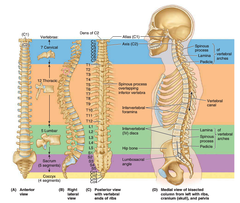
Anatomy of the spine:
32-35 vertebrae
- Cervical (7)
- Thoracic (12)
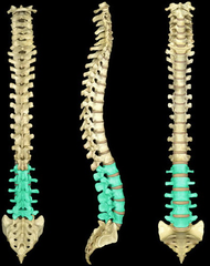
- Lumbar (5)
- Sacral (5)
- Coccygeal (3-6)
Vertebrae verae („true") - Cervical ,Thoracic, Lumbar.
Vertebrae spuriae („false")/fused - Sacral, Coccygeal
Functions:
o Bony axis of the body
o In a standing position, the body weight is directed to the lower limbs
o Absorbs collisions while walking
o Protects the spinal cord
Curvatures of the spine
There are 4 natural curves in vertebral column:
- Cervical LORDOSIS Curvature
- Thoracic KYPHOSIS Curvature
- Lumbar LORDOSIS Curvature
- Sacral KYPHOSIS Curvature
The more elastic curves our spine has, the more resistant it will be to environmental influences.
Curvatures of the spine II
Lateral side: 2x ‚S' curvatures
Thoracic KYPHOSIS and Sacral KYPHOSIS are Primary curvatures.
Cervical LORDOSIS and Lumbar LORDOSIS are Secondary curvatures.
- Thoracic and sacral are formed during fetal development.
- Cervical is formed when infant raises head.
- Lumbar is formed when infant sits up and begins to walk.
Vertebrae and disci of the vertebral column
The vertebrae of the spine are in different sizes and shapes.
- From below to the top: corpus/body vertebrae are smaller
- The Cervical part has to support the head.
- The Thoracic part has to support head, upper limbs and chest.
- The Lumbar part has to support everything...
- The Lumbar part is the most loaded part of the spine.
General structures/parts of the vertebra
Common:
- Vertebral body
- Transverse process
- Spinous process
Differences:
Superior articular process
Inferior articular process
Lamina
Vertebral foramen
Pedicle

CERVICAL spine - 7
C1 - Atlas - no
C2 - Axis
C3
C4
C5
C6
C7
THORACIC spine - 12
Superior view:
spinous process
transverse costal facet
superior articular facet
corpus
superior costal facet
vertebral
Lateral view:
superior vertebral notch
pedicle
transverse costal facet
superior articular facet
superior costal facet
inferior costal facet
inferior vertebral notch
LUMBAR spine -5
Sacrum (5 fused)
from top to bottom - anterior view
sacral promontory
body of first sacral vertebra
transverse ridges (site of vertebral fusion)
anterior sacral foramina
apex
coccyx
from top to bottom - posterior view
body
sacral canal
facet of superior articular process
Auricular surface
median sacral crest
lateral sacral crest
posterior sacral foramina
sacral hiatus
coccyx

Types of vertebrae

Types of vertebrae

Disci of the vertebral column
Between every vertebra (except: atlas-axis)
- Function:
- shock absorption
- maintaining flexibility of the spine
Annulus fibrosus: outer multilayered fibrous bark
Nucleus pulposus: inner gelatinous cores
Elasticity and strong connections between the
vertebral bodies. They take up water when at rest
and release water when under load so their size is
constantly changing.
They are subjected to enormous forces when standing, sitting or lifting when moving. They are constantly being compressed, their water content decreases. The fluid squeezed out is absorbed by the closing plates of the spongy vertebral bodies.
At rest, the pressure is released, the vertebral bodies move away from each other and the discs draw the fluid back out of the vertebral bodies and regain their height.

Connections of the spine
Between vertebra:
- Intervertebral discs
- Ligaments
- Intervertebral joints
- Muscles
Functionally move and stabilize the spine:
• Superficial and deep back muscles
• Abdominal muscles
• Gluteal muscles
The harmonious cooperation of these muscles
is essential for the normal functioning of the
spine.
Muscles of the spine
Superficial back muscles: retraction of the shoulder girdle (scapula adduction)
Deep back muscles: Paravertebral muscles - straighten the spine
Abdominal muscles: spine flexion, stabilize the pelvis and lumbar spine. In case of weakness: increase lumbar lordosis
Gluteal muscles: work with abdominal muscles -
dynamically stabilize the lumbar spine
Posture control
The basic condition for a biomechanically correct posture is a dynamic state of equilibrium of the body parts in relation to each other, which is achieved by the correct position of the pelvis and the muscular balance of the muscles responsible for the posture.
Physiological measures of posture:
• The angle of sacrum: 30°
• Between the horizontal and the line running through the superior aspect of S1
• Lumbosacral angle: 140°
• Lying between the axis of L5 and the sacral axis
• The angle of pelvic tilt: 60°
• Between the horizontal and the line joining the promontory to the superior border of the pubic symphysis
Posture
Biomechanically correct posture also requires that the body's line of gravity, when standing upright on two legs, touches physiological points.
Physiological points affected by the line of gravity:
The spine - found posterior of the body, behind the center of gravity.
center of gravity lies:
Through the atlanto - occipital joint
Tragus of the ear
Anterior humeral head
Anterior - inferior edge of T11
Greater trochanter
behind the patella
Through the lateral malleoli
Posture control II
With correct posture - the tension on the articular
ligament and capsule is physiological, the muscular effort is minimal, and the load on the articular surfaces and intervertebral disc is uniform.
with incorrect posture - the physiological angles and biomechanical parameters are altered, changing the forces acting on the body, the position of the centre of gravity, which overloads the muscles used for correction, increasing the load on the articular surfaces and the intervertebral discs.
Elements necessary for correct posture
• Muscle strength
• Endurance
• Muscle tone balance
• Muscle extensibility
• Physiological weight line
• Lumbar motor control
• Postural automation
• Anatomical and biomechanical postural parameters
The basis of the biomechanical approach is the motion segment (functional spinal unit), which is the functional unit of the spine, consisting of two vertebrae and the intervertebral disc between them, as well as the muscles and ligaments connected to them.
Movements of the spine
Movements of spine
• Flexio ventralis/Forward bending: C>L>Th
• Flexio dorsalis/extension/Backward bending: C>L>Th
• Flexio lateralis/Lateral bending: C>L>Th
• Torsio (rotation): C>Th>L
The load on the intervertebral discs and its changes in postures and work positions
Supine laying - 25%
Lateral laying - 75%
Standing - 100%
Standing slightly leaning forward - 150%
Standing bending forward - 220%
Sitting straight - 140%
Sitting slightly bending forward - 185%
Sitting bending forward - 275%
sitting with protracted shoulders and flexed spine 275% of the load on the body's spine. This is exactly 175% more than standing upright and 250% more than lying supine.
The load on the intervertebral discs and its changes in postures and work positions II
The least load on the spine is when the body is in supine position (spending 20 minutes a day in this position can do a lot for the regeneration of the discs). Gravitational load can be reduced by supporting or lifting the legs. In this position, the
lumbar spine is straightened, the vertebrae are released and the discs are given the opportunity to drain fluid back, increasing their height.
Unfortunately, the load of sitting, whether correctly or incorrectly, is almost equivalent to bending, holding and lifting all day.
The load on the intervertebral discs and its changes in postures and work positions III
According to studies, high forces applied for short
periods of time cause the most damage to the motion segment, and the discus is subjected to twice as much load in dynamic positions as in static positions.
Based on biomechanical assessments, it is recommended to avoid trunk flexion and rotation in order to prevent discus injury.
Biomechanically correct posture
Biomechanically correct posture is achieved when the tension of the joint capsules and ligaments is
physiologically correct, the harmonious cooperation of the muscles responsible for posture results in minimal muscular effort and, as a consequence, the load on the joint surfaces is uniform.
Low back pain (LBP) for health professionals
Among health workers the most common musculoskeletal disorder is low back pain (LBP) syndrome
- According to studies the reason is: overload caused by improperly performed patient transfer tasks
The overload is caused by:
- a high number of patient transfers, mobilisation
- incorrect postures
- prolonged asymmetric postures
- lack of muscle strength
- inadequate equipment
- low beds that cannot be raised.
What is LBP
Pain between the lower ribs and the lower gluteal folds.
All the components of the functional unit of the spine (the motion segment) have pain sensory innervation:
- the vertebrae
- muscles
- ligaments
- fasciae
- joint capsules
- nerve elements
- the anulus fibrosus of the intervertebral discs.
The sensation of pain in the lower back can therefore come from many different
tissue structures.
Classification based on the biomechanical aetiology of low back pain
Spinal origin, mechanical LBP:
- More than 90 %
- symptoms typically change with posture and exercise
- disc degeneration, arthrosis of the facet joint, bony stenosis, osteoporosis, compression fracture, any traumatic fracture of the vertebrae, scoliosis, increased lordosis or kyphosis, spondylolysis or spondylolisthesis...
- associated with lower extremity radiating pain in 7-10% of cases, the most common cause: disc herniation
Classification based on the biomechanical aetiology of low back pain II
Spinal origin, non-mechanical LBP:
- 1 %
- Neoplasms (metastasis, lymphoma, leukemia, tumours..) Infections (osteomyelitis), inflammatory
disease, Scheuermann disease
Classification based on the biomechanical aetiology of low back pain III
Non-Spinal origin:
- 1-2 %
- diseases of the internal organs, these are typically the abdominal organs (kidneys, pancreas, gallbladder), pelvic organs (prostate, uterus) or even aneurysms of the aorta.
Risk factors for the development of low back pain
Ø Genetics
Ø Mechanical (Over)load
Ø Physical activity, sport
Ø Smoking
Ø Psychological factors
Ø Decreased muscle strength (trunk)
Ø Posture
Gnetics
Age-specific studies suggest that LBP is genetically determined, with an estimated heritability of between 30 and 46%.
Based on the scientific evidence to date, the intergenerational transmission of predisposition to LBP is likely to be a multifactorial mechanism involving multiple genes.
Mechanical (Over)load
The different forces acting on the body, which
differ in direction and amount from the physiological load.
But there are large individual differences in the
effects of mechanical loading on the spine.
Physical activity, sport
The risk of LBP as a function of physical activity shows a correlation similar to a "U" shape, i.e. little, as well as significant physical effort, increases the prevalence of symptoms.
Smoking
Both current and former smokers have a higher prevalence and incidence of LBP than non-smokers data from a study among adolescents provide the best evidence that smoking and the
presence of LBP are associated.
Psychological factors
Stress, depression, anxiety, emotional and cognitive behavioural patterns, negative attitudes to pain.
Fear of movement and catastrophising increase the ineffectiveness of therapeutic interventions and are considered as major risk factors for the development of chronic complaints.
Decreased muscle strength (trunk)
Weak muscle strength around the spine has been
identified as a significant risk factor for LBP.
However, it was not clear whether weak muscle
strength was a cause or a consequence of chronic
pain.
Posture
Disorders of the frontal or sagittal spinal harmony,
as well as abnormal physiological positions of the
spine, pelvis and head, may also increase the risk
of developing LBP.
Physiotherapy of the LBP syndrome
Active physiotherapy methods are the first to be proposed, including:
• movement therapy
• patient education.
Long-term effective movement therapy methods:
• Elongation exercises (axial stretching)
• Stabilization therapy
• Progressive strength training
• Sports therapy
Long-term effective movement therapy methods
- Elongation exercises (axial stretching) decrease the pressure within the disc, the disc becomes higher, the intervertebral space increases, the disc base becomes smaller.
- Stabilization therapy, which helps maintain or restore spinal health by strengthening the small, deep muscles of the spine.
- Progressive strength training with increasing loads improves physical endurance.
- Sports therapy uses movement materials from physical education, sports psychology, sport
and conductive pedagogy. It improves physical skills and movement culture through group
sessions with increasing workload.
McKenzie-method
A physiotherapist has the patient repeat specific postures and movements and deduces the cause of the condition from the change in pain. On this basis, PT will draw up a personalised McKenzie
treatment programme consisting of a few simple movements that can be easily done at home or at
work. Through active exercises, the patient is able to relieve the pain on their own, so that they can
influence the outcome of the problem and relieve any recurring pain themselves.
Patient education
is not effective on its own, but it is the most effective conservative treatment when combined with movement therapy. Effective patient education includes the development of correct posture and spinal alignment. An essential element is the automatic development of daily forms of movement that do not increase discus load.
Therapeutic methods that include patient education and movement therapy:
- Back School (BS) programs
- Cesar therapie
- Mensendick therapie
- Multidisciplinary rehabilitation programme (MRDP)
Back School (BS) programs
An essential element of the spinal school programme is:
• raise awareness of the current body position
• learn new postures
• recognise and eliminate harmful postures and movements.
The programme consists of patient education, lifestyle advice and a movement programme.
Cesar Therapy
is a postural therapy, is a specific form of movement based on the cause and effect relationship between posture, lack of movement and low back pain.
The therapy is a learning process in which posture is corrected and movement therapy is used to counteract the lack of movement.
Mensendieck therapy
has been used in the Netherlands and Scandinavia for decades, consists of posture therapy, posture exercises and patient education. Mensendieck therapy emphasises learning by doing, health awareness and skill development to help the patient recognise and avoid functional disability. The sessions are conducted in groups.
A multidisciplinary rehabilitation programme (MDRP)
in MDPR a rehabilitation team plans and implements therapy. The team includes:
o rheumatologist
o rehabilitation specialist
o physiotherapist
o nurse
o occupational therapist
o orthopaedic technician
o psychologist
o social worker
o the patient.
Passive physiotherapy methods
• massage
• electrotherapy
• thermotherapy
• external lumbar support (lumbar support belt, spinal brace)
The main aspects of spine protection
1. Pay attention to correct posture in every position, check and remind yourself several
times during the day!
2. Avoid static positions, prolonged sitting, prolonged standing, change your posture often!
3. Take care of your body weight - it should be appropriate to your height, age and build.
Keep our fitness levels in balance with regular exercise and a balanced diet!
4. Get enough sleep and rest in the right place and in the right position to keep the metabolism of our discs in balance every day.
5. Don't make sudden movements or changes of position, be deliberate!
6. Take time to relax and unwind to relieve muscle tension caused by stress and overwork.
7. Protect your spine at work and in your daily activities, learn and apply the rules of correct
patient movement.
8. When lifting, use the muscles around the hips and knees, and when carrying, minimise and balance the load on the spine.
9. In the early morning, after waking up, avoid sports activities that cause more strain.
10. Exercise regularly, strengthen the deep abdominal and back muscles with targeted
exercises, and build a strong, active "corset" around the spinal column.
Tips to protect and secure your spine
At home:
Sitting puts more strain on the spine than standing.
- Sit up straight in your chair
- Sit all the way back in the chair, with your waist pressed against the backrest.
- Position the top of the computer screen at eye level.
- The desk or keyboard should be at elbow height.
- Put a lumbar pillow or rolled-up towel behind your waist to support your spine.
- Keep your thighs parallel to the floor.
- Knees should be fist distance apart.
- The feet should rest on the floor, using a footrest if the soles do not touch the floor.
- Even sitting in a perfect posture is tiring for the spine. Keep a small break every hour, stand up, stretch, walk.
Lifting and carrying
• Stand close to the object to be lifted.
• Bend your knees to lower your centre of gravity to a squatting position while keeping your
• spine straight.
• Lift slowly to standing position by stretching the knees.
• Pull the load close to your body, elbows in.
• Divide the load into two or more smaller loads (if possible), or ask
• assistance when lifting a large object.
• Push, rather than pull.
• Pushing puts less stress on the spine than pulling,
• twice the energy can be exerted.
• Stay close to the objects.
• Use both arms.
Reaching for an object
• Use a strong chair or ladder.
• Keep your shoulders, hips and legs pointed towards the object, avoiding objects
• When reaching objects, use arms, legs and shoulders to reach the object.
• Before lifting, test the weight of the object to be lifted by tilting one of the arms
• towards one heel.
• Wear flat-heeled shoes.
• If you have a sore back, lie on your back with a
pillow/roll towel under your knees and head.
Travelling
• When carrying suitcases or heavy bags, distribute the load evenly between the two hands - carrying them sideways puts an uneven load on the spine.
• Lift larger, heavy objects close to the body
using the thigh muscles with a straight back.
• Avoid heavy shoulder bags, which can cause
shoulder, neck, waist and leg pain - a backpack is better.
Long-lasting seat
• Frequent changes in posture will change the location of the monotonous, static load on the spine.
• As a driver, we should also stop for a few minutes as often as possible, move our limbs and perform trunk rotation.
• Support your neck and waist with a suitable cushion that is not too thick, which will compensate for the natural physiological curvatures of the spine.
• A "horseshoe cushion" to protect our cervical spine relieves strain on the region and absorbs damaging vibrations during travel.
Sport
• When playing sport, you should not only pay attention to warming up, but also to the correct form of movement.
• Never start exercising with stiff joints! This applies to all sports, a few minutes of warm-up (before sport) and stretching (after sport) is very important!
• Swimming is recommended for everyone, it
strengthens, stretches and relaxes the back muscles. Backstroke is best when both the cervical, back and lumbar spine are well relieved. Extended head swimming are not ideal.
Running and Cycling
• Avoid hard or asphalt roads for running or jogging - due to the increased axial load, choose grass, forest or dirt roads, comfortable, flexible, soft-soled sports shoes and a comfortable pace.
• Cycling is also recommended with adequate lumbar support. Make sure the seat and handlebar height is correctly adjusted, racing handlebars that require excessive forward lean can be harmful in the long term.
Ball and riding sports
• Tennis, racquetball, football and other ball games often involve sudden movements and twisting. It is advisable for non-experienced athletes to learn spine sparing techniques and move with increased caution beforehand in these sports.
• Riding practised with the appropriate technique, or trying it out during the learning phase, under the guidance of a qualified instructor, is beneficial for patients with chronic low back pain.
Patient mobilisation
It has already been established that musculoskeletal disorders are a common
complaint among nurses. This risk may also be increased by the working conditions of nurses working in patient care:
• inadequate patient beds,
• inadequate aids,
• lack of knowledge or inadequate knowledge of movement techniques
Many risk factors affect correct patient movement and increase the risk of injury.
Risks associated with patients
• Patients cannot be lifted like objects, the activity may have to be performed in an unusual posture, and the rules for safe lifting may not always be fully applicable.
• During lifting, patients cannot always be held close to the body, it is not possible to predict what will happen during patient handling.
• Overweight patients who are unable or only partially able to assist, place a particularly heavy physical burden on carers.
• The risks associated with patient weight.
Environmental risks
• Environmental factors that can cause a slip, trip or fall e.g.: electrical wiring, hospital equipment,
etc...
• Irregular working surfaces e.g.: floors...
• Spatial constraints e.g.: toilets, difficult to reach
narrow places, beds that are not accessible from
several sides
Other risks:
• No help is available,
• Lack of appropriate equipment,
• Inappropriate clothing or footwear,
• Lack or inappropriate use of knowledge.
To determine the appropriate patient handling technique, the needs and capabilities of the patient must first be assessed
- The level of assistance required by the patient should not exceed the level of assistance provided.
- Example: For an uncooperative inpatient (bedridden elderly patient, comatose patient, patient resistant to mobilisation, etc.), a device or assistance may be required, whereas a partially able and cooperative inpatient can transfer from bed to chair with a little assistance, even with the help of a scaffold.
Sometimes patients are agitated, aggressive, passive or have limited cooperation from the caregiver
- which increases the risk of injury to the caregiver
- Musculoskeletal injuries can still occur with the correct patient handling techniques, but the likelihood of their occurrence is significantly reduced.
Risk of loss of patient safety due to inappropriate patient movement
Poorly assessed and implemented patient movements can compromise the quality of
patient care and patient safety. Unintended side effects associated with patient movement:
- Patient fear of the procedure,
- pain,
- shoulder injury from manual lifting techniques,
- hip fractures from dropping the patient,
- bruising
- skin damage and further damage to decubitus areas.
Our common (nurse + physiotherapy) goal is
patient safety!
Bobath-method
Dr. Karel and Bertha Bobath - 1940 Examination and treatment of the hemiparetic patient. (stroke)
The Bobath approach is holistic, covering all
aspects of everyday life and including advice and training for carers on treatment, equipment and other modalities. According to the Bobath method, every time we change the patient's position and positioning, we also improve the patient's mobility.
Dotte-method
Paul Dotte - France physiotherapist (1986)
Basic of the method: Personal experience
Aim: Have the caregiver adopt controlled
movement patterns that facilitate care activities,
do not endanger the health of the caregiver and
put the patient first. This will contribute to
improving the quality of patient care. The aim is
not to increase muscular strength, but merely to
acquire and constantly develop a specific skill.
Patient lifting with mechanical aids
• Low-friction mattress covers (30)
• Patient moving sheet (31)
• Sliding sheets (32)
• Moving belts (33)
• Patient turning discs (34)
Electric patient handling devices and overhead lifting devices:
• Electric patient lifting device (35)
• Standing up support device (36)
• Overhead lifting device (37)
Communication during patient mobilisation
An integral part of safe patient handling is communication (nurse + patient)
Verbal:
Find a common language: the carer need to take into account the patient's ability to absorb information and other gender, age and socio-economic characteristics.
Non-verbal:
(which may include the wearing of a "white coat") can play an important role in building trust, among other things.
Emphatic communication has a number of positive effects
• it can reduce feelings of loneliness and isolation
• it can create hope
• increases the patient's sense of importance
• can create positive feelings and change the patient's behaviour
• aggressive behaviour and conflict can be more easily avoided and managed
• patient can be more cooperative
Before starting patient mobilisation
• Always explain the procedure
• the purpose of the movement
• the sequence of movements
• how the patient can help, if he/she can
+ encourage as much cooperation as possible during patient mobilisation
Communication between helpers
• Discuss in advance the method and the way in which the task will be carried out.
• Make sure that all the helpers involved and the helper who will be conducting the movement use a common vocabulary.