SA State C
 Call Kai
 Learn
Learn Practice Test
 Spaced Repetition
 Match
Match Flashcards
 Knowt Play
1/41

There's no tags or description
Looks like no tags are added yet.
Name | Mastery | Learn | Test | Matching | Spaced | Call with Kai | Chat |
|---|
No analytics yet
Send a link to your students to track their progress
42 Terms
1. Disorders of oestrous cycle of the bitch - abnormal oestrus cycle length
Abnormal Estrous Cycles in bitches include variations in cycle length, silent heat, rapid return to estrus, and split heats.
1. Physiological Variation & Diagnostic Markers
Normal Windows: Proestrus and estrus can each last 3 to 21 days. Bleeding for up to 40 days can be entirely physiological. The combined length of Proestrus (P), Estrus (E), and Diestrus (D) normally spans 63 to 142 days.
Silent Heat: Ovaries function normally, but the bitch shows no vulvar edema, vaginal discharge, or attraction to males. Common in young, small-breed dogs before their first true estrus.
Verification: Measure serum progesterone once a month. A level >6.4 nmol/L confirms functional luteal tissue. Alternatively, look for rising superficial epithelial cells on vaginal cytology.
2. Rapid Return to Estrus & Split Heats
An estrous cycle returning in <7 months is classified as a rapid return.
The Mechanism: Often caused by an anovulatory cycle (failure to ovulate). The follicles regress prematurely without ovulating, prompting a rapid new follicular wave. The subsequent return to proestrus frequently results in normal ovulation.
Pubertal Variations: The very first cycle in young bitches often fails to ovulate despite normal proestrus signs. A new proestrus cycle will appear within a few weeks, which may finally result in ovulation. No treatment is necessary.
Split Heat / Split Estrus: A short, 2 to 12-week interval between clinical signs of proestrus.
Demographics: Common at puberty and in bitches <4 years old.
Pathology: Follicles grow and produce estrogen but regress before ovulation occurs. If split heats happen continuously or frequently in an animal, it suggests hypothyroidism or chronic premature luteolysis. Treatment is generally not necessary unless secondary to an underlying endocrine disease.
Abnormal Cycle Intervals & Prolonged Estrus- hormonal imbalances, ovarian pathology, or environmental influences.
1. Shortened Intervals (<7 months)
Ovulation Failure: Proestrus returns ~2 months earlier than expected due to an absent luteal phase, often caused by inadequate LH secretion.
Therapy: 500 IU hCG daily for 3 days at the next oestrus to induce ovulation.
Short Luteal Phase: Progesterone drops to basal levels prematurely (20–40 days post-ovulation instead of the normal 60–65 days) due to corpus luteum insufficiency. Common in German Shepherds and often detected via early pregnancy loss.
Therapy: Progesterone (P4) supplementation during pregnancy (Note: carries risk of male puppy cryptorchidism and female masculinization).
Short Anoestrus: Driven by the "dormitory effect" where housing a bitch with other females in oestrus accelerates cycle return.
2. Lengthened Intervals (>11 months)
Normal Physiology: Normal cycles average 7 months but can stretch to 11 months physiologically. The interval naturally increases as the bitch ages.
Prolonged Luteal Phase: Rare extensions beyond the normal 65-day diestrus window, typically caused by progesterone-producing ovarian tumors or luteal cysts (occasionally seen with pyometra).
3. Prolonged Proestrus or Oestrus (>30–35 days)
Defined as active vulvar bleeding and male attraction lasting >30–35 days, with >80–90% superficial cells on vaginal cytology.
Delayed Ovulation: Normal ovulation can range from day 5 to 25. If delayed past 30 days, it indicates inadequate LH secretion or poor receptor response. Some cases respond to hCG.
Follicular Cysts: Rare, thin-walled, anechoic structures measuring 8–12 mm on ultrasound. All bitches displaying oestral behavior for >30 days should undergo ovarian ultrasound to screen for these cysts or rare estrogen-producing ovarian neoplasms (treated via spay/OHE).
Fra state
Talk about Q38 and 39. Asked about primary and secondary anoestrous as well as split oestrus.
2. Induction of oestrus in the bitch and queens
Estrus induction involves stimulating the estrus phase in an animal that cannot enter it naturally. While inducing the external behavioral signs of estrus using estrogenic compounds is straightforward, achieving a fertile estrus followed by ovulation and successful embryo implantation remains highly challenging in both the bitch and the queen.
1. Indications & Pre-Induction Protocol
Clinical Indications:
Pathological anestrus.
Concurrent endocrine conditions like Hyperadrenocorticism (Cushing's) or Hypothyroidism.
Hunting or show bitches, where extreme alterations in body fat lead to decreased gonadotropin secretion.
Pre-Induction Screening Requirements: Before initiating any protocol, a comprehensive physical and reproductive evaluation is mandatory:
Baseline blood work and full health history (tracking management and activity levels).
Palpation of the mammary glands.
Structural evaluation of the uterus.
Diagnostic vaginal cytology.
2. Induction Methods
Non-Pharmacological (The Dormitory Effect): Involves housing multiple bitches together. Pheromonal exposure naturally stimulates the animals to synchronize and begin cycling together.
Pharmacological Methods: Synthetic drug protocols carry a significant failure rate and distinct clinical drawbacks, including smaller litter sizes and prolonged subsequent cycles. Because of these risks, medical induction is generally considered inappropriate for healthy, normally cycling bitches
Induction Method | Primary Mechanism | Success & Timeline | Key Clinical Notes |
|---|---|---|---|
GnRH Agonists (e.g., Deslorelin implants) | Triggers pituitary release of endogenous LH and FSH, inducing a normal, fertile proestrus. | Estrus occurs in 2–15 days. Ovulation typically at 12 ± 2 days. Pregnancy rate: 50–100%. | Implant is placed near the umbilicus. Keep in place until ovulation (P4 >16–19 nmol/L). Withdraw if no ovulation by day 20. |
Dopamine Agonists (e.g., Cabergoline, Bromocriptine) | Suppresses prolactin secretion, triggering a premature but otherwise natural, fertile proestrus. | Proestrus induction: 80–100%. Pregnancy rate: 60–100%. | Provides high success rates with a very natural cycle progression. |
Gonadotropins (e.g., FSH, LH, eCG) | Delivers direct exogenous FSH & LH activity to physically stimulate the ovaries. | Can induce external proestrus signs, but ovulation usually fails. | Spontaneous ovulation is rare; often requires additional intervention to force ovulation. |
Estrogens | Employs positive feedback on GnRH to induce an LH surge. | Induces a "false" proestrus, which must be followed by a real cycle or paired with gonadotropins. | Primarily used to prime the hypothalamic-pituitary-ovarian axis. |
Estrus induction in the queen focuses on prioritizing natural environmental management, as artificial hormonal stimulation is rarely indicated and carries a high risk of reproductive complications.
1. Non-Pharmacological Methods (Preferred)
Light Manipulation:
The most natural method to trigger estrus. Requires a windowless room to precisely increase and control the daily light cycle.
Social Stimuli: Physical contact or proximity with a tomcat or other actively cycling females provides a natural stimulatory effect.
2. Pharmacological Induction Protocols
Clinical Warnings: There are no authorized compounds for estrus induction in cats. Exogenous hormones frequently create an abnormal endocrine environment, cause ovarian hyperstimulation, or trigger delayed secondary follicular waves with accessory corpus luteum (CL) formation.
Timing: Treatments must only be administered during anoestrus or inter-oestrus to prevent dangerous hyperstimulation. Never treat prepubertal queens, as it routinely induces cystic follicles.
3. Hormonal Selections & Ovulation Induction
eCG (Equine Chorionic Gonadotropin): The first-choice hormone if chemical induction is absolutely necessary. The queen must be mated within 3–4 days of estrus onset.
FSH (Follicle-Stimulating Hormone): Associated with very low conception rates and high rates of embryonic death.
Ovulation Induction: Best performed during mid-oestrus. Ovulation can be triggered pharmacologically using LH, GnRH, or hCG, or naturally via mechanical vaginal stimulation that mimics natural mating.
3. Prevention of the breeding in the female (medical approach)
Female dogs come into oestrus/heat twice a year on average, and it is only at that time that they can conceive.
Indications for birth control
Stop hormone-related behaviours: escaping, roaming, aggression, disobedience
Stop reduced performance in racing animals
Stop attractiveness to male
Prevention of unwanted mating
Stop physiological changes during pro-oestrus and oestrus: vulval bleeding, hypervocalisation (in queens)
Reduce risk of later medical problems: pseudopregnancy, pyometra, mammary tumours (only for OHE)
Methods: 2 methods to prevent oestrus
a) Surgery- ovariohysterectomy/ovariectomy (spaying)
b) Medical management using hormonal drugs
Progesterone and androgens have negative feedback on the H-P-O axis by suppression of ovarian activity (block production/release of GnRH from the hypothalamus). Androgens also decrease functional response of tissues to estrogen. Both - administered first half of anestrus
Medical control of oestrus
Synthetic analogues of progesterone (progestins or progestogens)
Synthetic analogues of testosterone (Androgens). Not much used.
Long-acting GnRH agonists.
1. Progestogens
postponement of estrus - mimicking luteal phase. given in anestrus.
Megestrol Acetate (MA): short-acting synthetic progestogen.
commonly used choice for temporary postponement or quick estrus suppression.
Medroxyprogesterone Acetate (MPA): A long-acting option. Use never exceed 2 years (max of 4 injections at 6-month intervals). Spontaneous cycle recovery takes 1.5 to 26 months.
Delmadinone Acetate (DMA) & Chlormadinone Acetate (CMA): DMA mimics MPA but requires injections every 4 months, with an 8-month recovery. CMA recovery is highly variable (up to 2 years). Post-treatment fertility remains normal.
Proligestone (PGS): A newer generation progestogen with lower progestational activity, though a wide range of side effects are documented. Recovery takes 3 to 9 months.
Severe Class Side Effects: cystic endometrial hyperplasia, pyometra, Diabetes Mellitus (via insulin suppression), acromegaly, mammary neoplasia, and the masculinization of female fetuses.
2. Androgens
suppress gonadotropins via negative feedback on the hypothalamic-pituitary-ovarian (HPO) axis and blunt the tissues' response to estrogen. started 30 days before proestrus.
Testosterone: Side effects include clitoral hypertrophy, vaginitis/discharge, increased aggression, mounting behavior, and a musky body odor.
Mibolerone: A synthetic oral daily androgen used for long-term prevention up to 5 years (though clinically not recommended for >2 years). Estrus returns within 7 to 200 days.
Strict Contraindications: Strictly prohibited in cats (effective dose is near toxic levels). Do not use in future breeding animals, pregnant animals, dogs under 7 months old (causes premature epiphyseal growth plate closure), liver/kidney disease history, or Bedlington Terriers (genetic predisposition to progressive hepatitis).
3. GnRH Agonists
These agents cause a temporary down-regulation of the cycle and inhibit ovulation. They are also clinically used to treat urinary incontinence following an ovariohysterectomy (OHE).
Clinical Note: They may carry an increased risk of mammary neoplasia.
Mandatory Pre-Screening: Requires a complete clinical exam, reproductive history checking for recent cycles within 1–2 months, mammary palpation, vaginal cytology (to verify the dog is truly out of estrus), and a serum biochemistry panel to evaluate liver function and glucose metabolism.
Deslorelin – long-acting GnRH agonist Indications:
Suppression of cyclicity without gonadectomy.
Preserved further reproductive performance.
Implanted SC, repeated every 5 months. Best to implant in dioestrus to prevent heat.
Mechanism of action: stimulates the pituitary to release all its reserves of gonadotropin until downregulation occurs.
Cause oestrus induction if treated in anoestrus.
Fra state:
I briefly talked about the Oestrus cycle and what hormones dominate in each section. Then I went into the three categories of drugs that are used to prevent breeding. Names of the actual drugs was not as important as what types they are and how they act.
4. Unwanted mating and pregnancy termination
Pregnancy termination
Unplanned and unwanted mating of cats and dogs is a common concern.
Indications of pregnancy termination
Unwanted pregnancy
Mis-mating small bitch to large male (single pup syndrome)
Very young or very old bitch (under 1 year, and over 8 years)
High risk of dystocia
Endocrine issues
Metabolic issues (Pregnancy ketosis)
(Told at the clinic):
After fertilization, within 10-11 days, embryo reaches uterine horn, and the next few days up to 18-20 days of pregnancy it moves around to find the best place for implantation. Both implantation and placentation occur around 18-20 days of pregnancy.
Embryo = up to 30 days of pregnancy
Fetus = after 30 days of pregnancy
After 30-35 days of pregnancy = abortion (typical discharge is present)
Before 30-35 days = resorption of embryo and no clinical signs (no behavioral changes, no discharge)
Two stages of pregnancy:
1st stage – embryonal stage. Divided into early and late. Early – till placentation (18-20 days). Late – till the end of organogenesis (30-35 days).
2nd stage – fetal stage from day 35.
Surgical treatment
Ovariohysterectomy
The best stage of oestrous cycle to do gonadectomy → anoestrus.
Best is 2.5-5.5 months following the onset of proestrus.During oestrus: higher risk of bleeding due to vascularity of uterus
Dioestrus: can lead to lactation & pseudopregnancy due to sudden removal of progesterone supply (ovary)
Medical termination of pregnancy (mismating protocols) utilizes target-specific medications selected based on the precise phase of gestation.
Phases of Pregnancy Termination
Gestational Phase | Primary Medication Class | Mechanism of Action & Target Organ | Key Clinical Notes & Comorbidities |
|---|---|---|---|
Early Embryonal (Until Day 9–11) | Estrogens (e.g., Estradiol benzoate, cypionate, valerate) | Alters zygote transit time in the oviduct, alters the local biochemical environment, and acts as a direct embryotoxin. Acts on the uterus. | Not recommended due to severe, high-risk side effects: pyometra and potentially fatal bone marrow suppression. Must be given immediately post-copulation. |
Late Embryonal (Day 9–11 to Day 30) | Antiprogesterones (e.g., Aglepristone / Alizin) | Most commonly used choice. Competes with progesterone (P4) at the receptor level as a true antagonist. Blocks P4 uterine effects without dropping serum P4 levels. Acts on the uterus. | No external clinical signs or discharge are observed when administered during this early embryonal window. |
Fetal Phase (Day 30–35 to Parturition) | Antiprogesterones (e.g., Aglepristone / Alizin) | Competes with progesterone (P4) at the receptor level as a true antagonist. Acts on the uterus. | Administration during this late phase will cause clinical signs, such as vaginal discharge and fetal expulsion. |
Fetal Phase (Day 30–35 to Parturition) | Prostaglandin F2α | Causes luteolysis (lyses the corpus luteum) to drop serum P4 levels, while simultaneously driving uterine contractions and cervical dilation. Acts on the ovaries. | Use extreme caution in patients with pre-existing cardiac or respiratory distress, and in brachycephalic breeds. |
Fetal Phase (Day 30–35 to Parturition) | Antiprolactin Drugs (e.g., Cabergoline / Galastop) | Functions as a dopamine agonist to block prolactin secretion from the pituitary. Because prolactin is required to maintain luteal function in the second half of gestation, its removal forces luteolysis and abortion. Acts on the pituitary. | Provides a reliable, centrally mediated method for mid-to-late term termination. |
Other drugs:
GnRH antagonist, corticosteroids(dexamethasone). Act on pituitary gland. Has side effects, not recommended.
5. Pregnancy loss (embryonal and foetal mortality)
Embryo: an early stage in development that does not yet have morphological features for species recognition. Up to day 35 in bitches, and day 27 in queens.
Embryonal mortality: death of the embryo(s) during the embryonic period of intrauterine development
Foetus: unborn foetus with morphological characteristics typical for the species. From day 36 in dogs, 28 in cats.
Foetal mortality: death of the foetus(es) during foetal period of intrauterine development
Deadborn foetus: stillborn at the time of spontaneous delivery or death occurred within 24 hours after birth.
Abortion: termination of pregnancy by the delivery of a dead foetus, or its expulsion from the uterus before the normal term of delivery which is incapable of survival.
Pregnancy losses may manifest by:
Death of embryo with its resorption
Foetal death with its mummification
Abortion
Stillborn at the time of normal parturition
Early embryonic death
Loss of embryos can occur before a diagnosis of pregnancy can be made.
seen in healthy bitches with cystic endometrial hyperplasia (abnormal uterine development allowing bacteria to enter uterus and interfere with normal embryo development)
It is also thought that individual embryos may fail due to genetic abnormalities.
Resorption:
Resorption of one or more embryos can occur with continuation of the rest of the pregnancy (due to abnormal development of the embryo or competition of placental attachment sites)
Resorption of entire litter may occur in the case of infectious agents – check for canine herpes virus if this occurs.
Resorption only occurs when an embryo fails before day 35
Foetal loss
Early foetal death is Dx by USG by the presence of foetal anatomy without a heartbeat. There are many causes for why a bitch would abort all or part of a litter. These causes are categorized into foetal defects, abnormal maternal environment, and infectious agents.
Aetiology:
Infectious | Non-infectious |
Bacterial – Brucella canis, other bacteria (E. coli, Campylobacter, Pseudomonas, Salmonella, haemolytic streptococci, Clostridium sp.)
Protozoa – Toxoplasma gondii
Viral – Canine herpesvirus, distemper virus, parvovirus, adenovirus, FLV, FIV. | - Congenital/hereditary defects - Endocrine disbalance (hypoluteinism, hypothyroidism) - Exogenous hormones administered during gestation (oestrogens, glucocorticoids, prostaglandins, dopamine agonists – cabergoline, bromocriptine) - Fetotoxic or teratogenic drugs (chemotherapy, antimycotic drugs, some ATB (trimethoprim-sulfonamide, tetracycline, gentamicin], modified live vaccines) - Injury/physical trauma - malnutrition |
Clinicalsigns:
Embryonic death goes unnoticed when the embryos are reabsorbed before pregnancy can be detected.
Abnormal vaginal bleeding, expelled foetuses. Foetal death can be seen as abortion or not seen (foetal mummification - seen on RTG, USG or when uterus is opened surgically)
Diagnosis: Can be difficult.
History (vaccination [time, type], brucellosis test, administration of medicines during pregnancy, trauma, information about breeding environment, information about previous parturition…)
Foetal necropsy
Serology
Treatment: Supportive, casual, etc. ATB (ampicillin or amoxicillin are safe during pregnancy), vaccination, tocolytics
Hypoluteoidism =
The insufficient secretion of progesterone by the corpora lutea during pregnancy, resulting in failure to maintain progesterone concentration above a critical level (>6nmol/l), leading to resorption or abortion.
Caused by primary luteal failure occurring before term, or any toxic or hormonal substance that may stimulate release of prostaglandins & subsequent luteolysis or block the action of progesterone in the uterus.
6. Abort (aetiology, dam examination, necropsy of foetus)
death of a foetus any time during pregnancy.
Embryonic death - very early, such as during the passage of the embryo through the oviduct, or during the early pre-implantation or free-floating phase of embryonic life.
Foetal deaths occur after day 30 when organogenesis is complete
Types
i. Abortion
expulsion of foetuses before full term pregnancy, i.e., before the conceptus is capable of independent life. Abortion is uncommon in the bitch.
ii. Partial abortion
Most commonly in bitch with uterine body pregnancy. There is presence of both live and dead pups.
iii. Foetal Reabsorption
Foetuses will normally only be reabsorbed if they die during the first half of pregnancy. The most common causes are inadequate hormonal support, endometrial or placental disorders or intoxications, and infections, such as canine herpesvirus or brucellosis.
iv. Mummification
The death of foetuses after the calcification of the skeleton, i.e., by day 50 in the dog, may often lead to the mummification of foetuses. The foetuses are maintained within the uterus, and degradation occurs to a certain extent, but the skin dries around the foetus forming a membrane that may delay complete foetal decay and reabsorption for prolonged periods of time. Mummified foetuses may be retained in non-infected uteri, and the general health of the dam is usually not affected.
v. Premature Birth and Stillborn Puppies
The expulsion of foetuses after the foetus is capable of independent life. Stillborn puppies are full-term foetuses that are born dead. Death may occur a few days, hours or minutes before parturition.
Aetiology
Non- infectious causes: endocrine disbalance, exogenous hormones, foetotxic or teratogenic drugs, physical injury
Infectious causes: Brucella canis, miscellaneous bacteria, toxoplasma gondi, neospora, canine herpes virus, canine distemper virus, canine parvovirus, canine adenovirus, etc.
Clinical signs
Often, the pregnancy loss proceeds without any clinical symptoms. There may be fever, colic, discharge
Vaginal discharge containing blood or pus, brown, greenish or black-tinged or with a foul odour, can imply serious pregnancy complications.
Diagnosis: Determination of pregnancy, vaginal smear, ultrasound, relaxing test, radiography
Foetal necropsy
Necropsy is the pathological examination of a corpse, particularly to determine cause of death.
Consist of gross examination, histopathology, bacteriology, virology, parasitology, mycology and serology
Clinical findings
It is also thought that individual embryos may fail due to genetic abnormalities.
Moderate anasarca and severe hemoperitoneum: the subcutis is markedly expanded with oedema (anasarca). The abdominal cavity is severely dilated with blood, most likely because of septicaemia.
Bilateral hindlimb deformity and unilateral hydroureter.
Multifocal haemorrhage in lungs
Systemic canine herpesvirus 1-infections: severe acute interstitial pneumonia, lungs being poorly collapsed and mottled red-beige. Multifocal, severe, acute renal necrosis and haemorrhage with petechia on cut and surface.
Necrotizing hepatitis: toxoplasma
Severe subacute bronchopneumonia: Bordetella bronchisepica
Segmental enteritis: panleukopenia
From state: important to mention embryonal period vs fetal period (up to 35 days in dogs, 27 in cats) and divide causes for embryonal/fetal death (was less interested in the infectious causes - I only mentioned them), wanted to hear about Hypoluteoidism. Asked “when there is fetal death but we don’t see anything?” - what she meant was when you do surgery and find a mummified fetus inside the uterus (but it’s not really seeing something on the bitch if she is not operated… so tricky question)
7. Dystocia of the bitch and queen (assessment of vital functions of foetuses during and after parturition)
Dystocia is the difficulty in passing the foetus through the pelvic canal (difficult/slow/abnormal parturition).
Must be diagnosed in a timely fashion for better outcome of medical or surgical intervention.
Incidence in bitch can be up to 100% in some breeds, while it 5-20% in queens
Assessment of vital functions of foetuses intrapartum
Intrapartum monitoring of the foetus & uterine activity help medically manage labour & identify early surgical intervention. Current monitoring tools:
B-mode ultrasonography
Doppler (foetal heart rate) ultrasonography,
Tocodynamometry: detect and record uterine activity.
The use of Doppler USG & tocodynamometry - accurately identify the current stage of labour, adjust oxytocin & calcium Tx with accuracy, & avoid the adverse effects of excessive oxytocin (uterine tetany, foetal hypoxia & uterine rupture).
Detect if there is any stress in the foetuses or the mother, to help decide if we should perform an emergency C-section or if we can wait.
Dystocia evaluation parameters:
Foetal heart rate can give an indication of whether or not intervention is necessary.
Normal heart rate 180-240 bpm
Distress: 150-160 bpm
<130 bpm: low survival rate if the foetuses are not delivered within 2-3 hours
<100 bpm: immediate intervention is necessary
Maximum time between each foetus is 2 hours. If there are no problems, then it is ok to wait more than 2 hours.
Assessment of vital functions of foetuses after parturition (puppy, about the same in kittens)
We can use Apgar score to evaluate neonatal viability; HR, BR, Grimace, motion, mmc.
Parameter | Score 0 | Score 1 | Score 2 |
HR/min Spontaneous parturition: Caesarean section: |
< 180/ min < 120/ min |
180-220/ min 120-180/ min |
> 220/min > 180/min |
BR/ min | Without vocalization < 6/min | Mild vocalization 6-15/min | Strong vocalization > 15/min |
Grimace | Missing | Grimace | Strong response |
Motion | Weak | Rarely flexions | Active motion |
MM Colour | Cyanotic | Pale | Pink |
Evaluation of the Apgar Score total value:
Distress-free puppy/kitten: 7-10 points (does not need first aid. Only dry them and keep them warm.)
Mild distress: 4-6 points
Serious distress: 0-3 points
A low score → neonate requires medical attention. Continue doing the Apgar score to monitor during treatment. 0 for 10 minutes represents an important risk factor for subsequent death or disability.
Treatment of puppies with AS 4-6
Chest rubbing, GV-26 pressure, umbilical rubbing, clear ventilation
Artificial ventilation and/or O2 through a face mask
Majority of puppies are recovered within short time
Treatment of puppies < 3
50% of puppies with severe distress are dead within 20 minutes
After 20 min resuscitation – 50% puppies can improve to 4-6 and survive
Neonatal hypothermia slows gut motility, leading to functional gastrointestinal stasis (ileus) and a life-threatening systemic cascade if managed incorrectly.
1. Pathophysiology: The Fatal Cascade
The Feeding Danger: Tube-feeding a hypothermic neonate causes the milk replacer to either be regurgitated and aspirated (resulting in aspiration pneumonia) or to ferment in the stagnant gut.
The Vicious Cycle: Fermentation causes severe abdominal bloating, which exerts upward pressure on the thorax and induces labored breathing. The resulting pain and respiratory distress cause the neonate to swallow air, worsening the bloat and triggering circulatory collapse and death.
Strict Feeding Rule: A hypothermic neonate must never be fed if its body temperature is <34.4°C and/or if no bowel sounds are heard via auscultation.
2. Temperature Thresholds by Age
At Birth:
<34.4°C1–3 Days Old:
<35.6°C1 Week Old:
<37.2°C
3. Reheating & Stabilization Protocol
Rewarming Rate: Reheat the neonate slowly at a maximum rate of 1°C per hour while keeping the skin completely dry.
Caloric Support: Warming accelerates metabolic demand. Dextrose must be added to fluid supplementation to prevent hypoglycemic crash and assist stabilization.
External Heating: Heating pads, heat lamps, or warm water bottles may be used cautiously. Crucial: Neonates cannot crawl away from excessive heat; temperatures must be checked frequently to avoid thermal burns.
Severe Cases (Internal Fluids): Administer warm fluids via IV, intraperitoneal (IP), or intraosseous (IO) routes. The fluid temperature must never be more than 1°C higher than the patient's current core body temperature to prevent thermal shock.
Neonatal hypoglycemia glucose drop below 30 mg/dL. While neonates naturally maintain lower baseline glucose levels than adults and tolerate these lower levels better, their reserves are highly limited.
1. Clinical Presentation & Breed Vulnerability
Common Symptoms: Tremors, crying, irritability, increased appetite, dullness, lethargy, stupor, seizures, and coma.
Toy Breed Vulnerability: vulnerable because have a Higher brain mass-to-body weight ratio, requiring significantly more glucose for their size to maintain normal neurologic function.
Underlying Triggers: Beyond direct starvation and hypoxia, causes include sepsis, sub-optimal environmental conditions, congenital metabolic defects, portosystemic shunts (PSS), and hypopituitary dwarfism.
2. Treatment & Monitoring Protocol
Intravenous Therapy: dextrose slowly IV as a 5–10% solution diluted in standard Ringer's Lactate or normal saline.
Emergency Alternative: if IV cannot be placed, higher concentrations of dextrose can be smeared directly onto the mucous membranes of the mouth for rapid absorption.
Post-Stabilization Care: Recheck blood glucose frequently after intervention; aggressive supplementation carries a distinct risk of triggering rebound hyperglycemia.
Neonatal dehydration is a rapid, life-threatening emergency caused by a high total body water content, an inability to concentrate urine, and an immature renal system that cannot autoregulate blood flow, resulting in a low glomerular filtration rate (GFR).
1. Assessment & Diagnostic Keys
Mucous Membranes: The best diagnostic indicator for assessing hydration.
Skin Turgor: Inaccurate and unreliable in neonates due to distinct differences in their skin and subcutaneous tissue.
Urine Color: A highly effective tool; any yellow coloration confirms dehydration, as normal neonatal urine should be nearly clear. Even mild dehydration severely compromises a neonate and requires immediate intervention.
2. Fluid Administration Routes
Oral (PO): Restricted to very mild cases, and only after ensuring gut motility is fully intact with audible bowel sounds.
Subcutaneous (SC): Used in mild cases to allow slow absorption without overloading the homeostatic system.
Intravenous (IV) / Intraosseous: The ideal route for moderate-to-severe cases, typically utilizing the cephalic or jugular vein.
3. Fluid Volume & Rates
Parameter | Clinical Protocol / Metric |
|---|---|
Maintenance Requirements | High baseline needs of 80–100 mL/kg/day. |
Standard Infusion Rate | Restricted to 3–4 mL/kg/hour to prevent rapid volume overload. |
Shock / Severe Bolus | 30–40 mL/kg of warm isotonic crystalloids administered over 5–10 minutes. |
Fluid Temperature | Must be warm, but never exceed 1°C above the neonate's current core body temperature to prevent thermal shock. |
Monitoring | Frequent weight checks and continuous reassessment of the oral mucous membranes. |
Hypoxia
Clinical Stealth: difficult to recognize clinically because neonates tend not to hyperventilate in response to low oxygen levels until they are several days old.
The Normal Acidosis Window: Respiratory and metabolic acidosis is entirely normal during the first 1–2 hours after birth, with most neonates recovering spontaneously within 45 minutes. Medical intervention is only indicated if acidosis persists for more than 3 hours.
1. The Hypoxic Fatal Cascade
Systemic Impact: Hypoxia triggers severe systemic stress, causing respiratory depression, the translocation of harmful intestinal bacteria, and systemic chilling.
The Vicious Cycle: Just like hypothermia, hypoxia halts gut motility. This induces functional stasis, leading directly to a systemic cascade of circulatory collapse and death.
Clinical Markers: Common physical findings in hypoxic neonates include bradycardia (decreased heart rate) and severe hypotension (decreased blood pressure).
2. Treatment & Oxygen Therapy
Primary Intervention: Immediate oxygen supplementation is mandatory.
Delivery Methods:
Use a commercially available incubator.
Create a temporary oxygen cage.
Feed an oxygen line directly into a closed box covered with a blanket (which functions simultaneously to retain vital core warmth and protect the patient from cool drafts).
From state: Know the exact definition of dystocia
8. Dystocia of the bitch (criteria of dystocia, primary and secondary atony of uterus)
Dystocia is the difficulty in passing the foetus through the pelvic canal (difficult/slow/abnormal parturition)
Causes of dystocia is separated into maternal or foetal causes.
Maternal: lack of expulsive forces/uterine inertia. Soft-tissue and bony sections of birth canal
Foetal: Initiation, presentation, position, posture, size
Criteria of dystocia- bitch
a) Before delivery of first puppy
If pregnancy takes longer than 68-72 days with no signs of parturition
24-36 h past drop of rectal temperature with no signs of parturition
Abdominal straining for 30 min or 4 hour (intermittent) without delivery
Foetal fluid = initiation of 2nd stage - without any puppies coming after 2-4 h
Green or black discharge – placenta is separated – puppy cannot breath.
b) After delivery of first puppy
If there is 2-4 hours since first delivery or if there has been persistent straining for 30min.
Abnormal, foul smelling/haemorrhagic discharge
Dam has been in second stage of labour for more than 12 h
Stuck or dead puppy
Maternal signs: apathy, tremor, dyspnoea, whining/crying, abnormal posture
Uterine inertia (atony) refers to abnormal, ineffectual uterine contractions or improper relaxation of the uterus during or after labor. It stands as the primary cause of dystocia in the bitch.
- Hypotonic atony; primary uterine atony & secondary uterine atony
- Hypertonic atony; colicky uterus & hyperactive lower uterine segment
1. Classification Matrix
Type | Definition | Common Causes / Triggers |
|---|---|---|
Complete Primary Inertia | Stage 2 labor fails to initiate entirely; zero puppies are delivered. | Lack of fetal signals, systemic disease, or profound calcium/glucose deficits. |
Partial Primary Inertia | Normal labor initiates and some puppies are delivered, but contractions cease before the entire litter is born. | Large litters (uterine overstretching), small/dead litters, or maternal exhaustion. |
Secondary Inertia | Exhaustion of the uterine musculature after repeatedly contracting against a physical tract obstruction. | Narrow pelvis, pelvic trauma, vaginal strictures, uterine torsion, or vaginal hyperplasia. |
Hypertonic Atony | Pathological, uncoordinated, or hyperactive contractions. | Classically manifests as a colicky uterus or a hyperactive lower uterine segment. |
2. Multifactorial Etiology of Primary Inertia
Metabolic Defects: The myometrium cannot contract due to a failure to respond to fetal endocrine signals, or the bitch is unable to mobilize sufficient ionized calcium or glucose for muscle fiber activation.
Litter Size Anomalies:
Too few puppies (1–2) or dead fetuses: Inadequate hormonal signaling to trigger labor.
Too many puppies or excessive fetal fluids: Pathological overstretching of the myometrium, rendering it mechanically unable to contract.
Systemic & Environmental Factors: Poor nutrition/starvation, genetic predisposition, environmental stress, and acute anxiety/distraction.
3. Clinical Management & Treatment
Medical Management: Indicated ONLY after physical obstruction has been strictly ruled out via digital palpation and imaging.
Therapy: Administration of IV calcium gluconate and glucose maximizes myometrial contractility. Oxytocin may then be carefully utilized if appropriate.
Surgical Intervention: An emergency C-section
is mandatory if a physical obstruction is identified, if secondary uterine inertia has set in, or if medical therapy fails to restore progressive labor.
From state:
Why is discharge green? - Because the placenta is green. Specify difference between primary and secondary atony
9. Dystocia of the bitch (obstructive dystocia, forceps delivery)
will ask for kiispeptin role for reproduction
2 center in hypothalamus remember the difference
Dystocia is the difficulty in passing the foetus through the pelvic canal (difficult/slow/abnormal parturition)
Obstructive dystocia
when foetus does not exit pelvis during parturition due -physically blocked, despite uterus contracting normally.
Aetiology
Bone abnormalities: fracture, breed (bulldog, pug), diet, immaturity, neoplasia, disease
Soft tissue abno.: congenital defect, fibrosis, immaturity, prolapse, neoplasia, abscess, deviation, herniation, adhesion, stenosis, torsion, rupture
Foetal malpresentation/position/posture (most common brachycephalic breed)
.
Disposition of head and limbs of foetus.
Presentation: should be cranial longitudinal, but can be caudal longitudinal or transverse
Position: should dorsal, but can ventral, left lateral or right lateral
Posture: should extended. Deviation of head and limbs.
d) Dystocia due to abnormal size of foetus
Absolute oversize: pelvis normal , -foetus large
Relative oversize: foetus normal dimensions, - maternal pelvis small
e) Dystocia due to fatal monsters
deformed foetus- abnormal number of limbs, absence of body parts, etc.
Conjoined/Siamese twins- common type - from incomplete division of fertilised ovum.
Hydrocephalus - excess of cerebrospinal fluid that has leaked inside the dog´s skull
Schistosomus reflexus (exposed abdominal and thoracic organs, limb malformations, abnormal spinal column, liver and diaphragm hypoplasia, genitourinary and GIT changes. Cannot survive.)
Anasarca (generalized oedema of the whole body)
Management
Manual or forceps delivery & administration of medications to aid in uterine contractions
Manual fetal extraction is performed when a fetus is physically lodged or detected within the vaginal canal and can be safely delivered without immediate surgical intervention.
1. Direction of Traction & Lubrication
2. Gripping Techniques & Alignment
3. Adjunctive Delivery Techniques
Direction & Lube: Pull the puppy backward and downward (caudoventral) to match the natural curve of the pelvis. Apply generous sterile lubricant inside the canal first.
Where to Grip: Pull only by the head/neck (head-first) or pelvis (tail-first). Never pull the limbs, as this causes severe injuries.
Realignment Rules:
Rotate: Gently turn the shoulders or pelvis sideways to fit the widest part of the birth canal.
Reposition: Only push a puppy back in between contractions. Never push against active straining.
Delivery Tricks: Use steady transabdominal massaging to guide the puppy down, and use a gentle side-to-side rocking motion while pulling to slip past tight spots.
Forceps delivery (ONLY IN CASE OF DEAD OR LAST FOETUS)
Forceps should be used with care as they can traumatise vaginal wall via pinching or can cause serious damage to foetus.
Forceps delivery are used as a last resort when it is clear that removal of the affected foetus will enable completion of parturition relatively unaided, otherwise, & in the cases of large litters, it is best to resort to a C-section.
Manipulation is performed between periods of straining & requires adequate lubrication with forceps grasped around head & neck.
10. Dystocia of the bitch (medical and surgical management)
Medical management of dystocia
Is only indicated when the birth canal is fully dilated
No foetal malposition or malformation
No absolute or relative foetal oversize
No birth canal constrictions
General condition of the dam, offspring is still good (HR >180 bpm)
<2 foetuses remain to be delivered
Methods
1) Fluids (lactated ringer)
provided with IV fluids to correct hydration, electrolyte, & blood glucose abnormalities.
2) Tocolytic agents (Calcium gluconate)
Calcium solutions increase the strength of uterine contractions.
IV calcium gluconate may augment the oxytocin’s effect.
Monitor heartrate!! Give slowly. If no effect within 30 min, then also give oxytocin.
3) Oxytocin
Oxytocin increases the frequency of the uterine contractions.
Oxytocin has direct action on rate of calcium influx into the myometrial cell, which is essential for myometrial contraction.
Give 1 dose oxytocin IM – wait 30 min. If straining begins, the treatment can be repeated (MAX 2 doses oxy!) if necessary, although each additional administration will elicit a weaker response.
If nothing happens in30 minutes, it is not likely that further treatment will be successful à proceed with Forceps or C-section.
Oxytocin:
can ONLY be given when the puppy is free to come out à NO OBSTRUCTION!!
IM or SC – NEVER IV! à Rupture of uterus, spasms that can kill the puppy.
Surgical management (C-section) for dystocia is critical when medical or manual extraction is impossible. The timing of the procedure depends directly on fetal lung maturity (surfactant develops at day 58 in puppies to prevent lung collapse/atelectasis), maternal progesterone levels, and core body temperature (BT).
1. Indications for C-Section
Emergency Indications: Dystocia, fetal or maternal distress, prolonged labor, systemic sepsis, uterine torsion, abnormal vaginal discharge, complete primary uterine inertia, and maternal tract obstruction/secondary uterine inertia.
Elective / Voluntary Indications: History of previous C-section, advanced maternal age at first pregnancy, single-pup syndrome (or large fetuses), brachycephalic breeds, and historical maternal pelvic trauma.
2. Preoperative Stabilization & Physiological Rules
Cardiovascular Stabilization: Patients in prolonged stage 2 labor are frequently hypovolemic and hypotensive. Preoperative blood work must be performed, and fluid boluses must be given to correct perfusion deficits prior to induction.
The "Conscious Prep" Rule: To drastically minimize anesthesia time (as inhalants like isoflurane potentiate hypotension), perform all surgical clipping and preliminary site preparation while the patient is still conscious.
Pre-Oxygenation Requirement: Pregnant dams have an increased oxygen demand paired with a decreased functional residual capacity and total lung volume due to the heavy, gravid uterus compressing the diaphragm. This compression worsens in dorsal recumbency. Always pre-oxygenate the patient prior to induction to prevent severe hypoxemia, and keep the time from induction to puppy delivery as short as possible
3. Anesthetic Protocol Guidelines
Pregnant animals have heightened sensitivity to anesthetics. Because any drug that crosses the blood-brain barrier will cross the placental barrier, always use the lowest effective dose of short-acting, rapidly metabolized, or reversible agents.
Protocol Phase | Preferred Drugs | Avoid / Contraindicated Drugs | Clinical Justification |
|---|---|---|---|
Premedication | Glycopyrrolate, Alfaxalone (low dose), or Atropine. | Acepromazine, Diazepam, Midazolam, Medetomidine, Xylazine, and Opioids. | Glycopyrrolate does not cross the placenta, protecting fetal vitals. Phenothiazines and alpha-2 agonists cause severe fetal depression and maternal hypotension. |
Regional (Optional) | Lidocaine epidural or local line block along the abdominal midline. | — | Minimizes the requirement for systemic induction and maintenance agents. |
Induction | Propofol or Alfaxalone. | Ketamine. | Propofol and Alfaxalone are rapidly metabolized by the dam and neonates, ensuring high Apgar scores. Ketamine causes severe neonatal depression. |
Maintenance | Isoflurane or Sevoflurane. | — | Offers rapid adjustment of anesthetic depth and swift elimination upon turning off the gas. |
Procedure
Ventral midline approach (incise linea alba), taking care to avoid uterine or neonatal lacerations
The uterus should be isolated and packed off with surgical laparotomy sponges.
Make a single incision in the body of uterus. All foetuses are extracted through this incision.
Puppy with placenta is removed, or placenta is broken open and the umbilicus is ligated and divided before the puppy is transferred to an assistant, then placenta is removed.
After removal of all neonates, uterus should be palpated & visually inspected from cervix to ovaries
The uterine incision may be closed with absorbable suture in a single-layer suture pattern (inverting)
Trenger sikkert ikke å nevne den her, men kanskje lurt å kunne om de spør
Dogs and cats have endotheliochorial placentation, i.e. the endothelium of endometrial blood vessels is in direct contact with the chorion. They are not able to absorb transferring-bound iron directly from the maternal compartment like humans. Most domestic animals phagocytose RBCs that have been extravasated by endometrial capillaries. This occurs across the areas of placental attachment. It is only in canids and felids in which phagocytosis of RBC occur predominantly at the margins of their zonary placentas.
The special villi in the marginal area have intricate surfaces that lie within the marginal hematomas on the endometrium forming the co-called hematophagous zones. Heme from maternal blood on the endometrium is immediately broken down by hemoxidase into biliverdin. Biliverdin is bright in colour, accounting for the appearance of the hematophagous zone. The postpartum discharge in bitches is green in colour.
Heme is broken down in the same manner in felids, but the hematophagous zone is not green. In cats, biliverdin may be catabolized to bilirubin very rapidly in this area, so that its presence never becomes obvious. The postpartum discharge in a queen is brownish red.
11. Dystocia of the queen (criteria of dystocia)
Most cats have no problems during parturition.
4 points indicating that the cat is in phase 2 of parturition:
Temperature has risen to normal – not normally measured in cats
Contractions
The first water bag (allantois) bursts and the foetal fluids are passed
One or more kittens are born, but there are more kittens left
Dystocia evaluation parameters:
General health status of the animal
Vaginal discharge – colour
Strength of contractions including abdominal contractions
Duration of foetal expulsion, time between two foetuses, expulsion of placenta, duration of parturition
Foetal heart rate:
Normal: 180-240/min
Distress: 150-160/min
<130/min – low survival rate if the foetuses are not delivered within 2-3 hours
Before delivery of the first kitten:
More than 68 days from the last mating, no signs of impending parturition
Intermittent abdominal straining for more than 2 hours without delivery of a kitten
Haemorrhagic vaginal discharge
Passage of the foetal fluids before onset of abdominal straining
Persistent abdominal straining for more than 5 minutes without delivery of a kitten
Passage of dark brown vaginal discharge is a sign of placental detachment (same as in the case of green discharge in dogs). Kitten will die within a few hours.
If no kitten has arrived 2-3 hours after the water bag (allantois) has burst.
Weak, irregular contractions for 2-4 hours
After delivery of the first kitten:
More than 2 hours since the birth of the last kitten
Persistent abdominal straining for more than 5 minutes without delivery of a kitten
Abnormal, foul smelling, haemorrhagic vaginal discharge
Presence of a kitten stuck in the birth canal (a kitten which protrudes from the vulva should be delivered within 3-5 minutes)
The birth of a dead foetus
Maternal compromise: crying, abnormal posture, apathy, tremor, dyspnoea
From state:
Only asked how long the parturition could take in queen, which is several days apparently. Because they sometimes can start the parturition but then can stop and wait for some days before finishing it she said.
12. Indication for voluntary and urgent caesarean section
progesteron oic know function 4 of them
Surgical management (caesarean section) of dystocia should only be performed in cases of atony, oversize, maternal abnormalities, obstruction and foetal malformations.
Indications of surgical management
Reasons for urgent/emergency C-section:
Complete primary atony
Partial primary uterine atony where large nr. of foetuses remain and the response is unsatisfactory to drugs, or secondary uterine atony/obstruction
Gross abnormal maternal pelvis
Maternal illness
Suspicion of uterine torsion, rupture, prolapse, herniation
Evidence of foetal distress with poor response to medical intervention = HR <150 bpm
Foetal oversize
Absolute oversize: maternal pelvis is of normal size, but foetus is too large
Relative oversize: foetus is of normal dimensions, but maternal pelvis is too small
Malpresentation that is not able to manipulate
Foetal putrefaction
Reasons for voluntary C- section:
Dystocia in previous parturition,
Urgent C-section in previous parturition,
First parturition at old age > 6y,
Small number of foetuses/large foetuses,
Brachycephalic breeds, due to disproportion between the size of the pelvic canal of the dam and the size of the neonatal head resulting in a physical blockage to delivery.
Previous pelvic trauma.
Single puppy syndrome
Criteria and time for C- section
Foetal maturity: lungs should be completely developed (58 days post ovulation in puppies)
Determination of date of mating/insemination à because foetuses must be mature, otherwise they won´t survive in external environment (lungs surfactant develops at day 58)
Progesterone content: <6nmol/l at end of pregnancy
Body temperature at the end of pregnancy – there is a decrease in body temperature before delivery. Measure temp. regularly the last week of pregnancy, when it suddenly drops from 38°C to 36°C it means the delivery is on the way. If nothing happens within 24h – dystocia
Clinically recorded onset of parturition
Some researchers suggest that C section should be done after 20-24 hours after Aglepristone (termination of pregnancy) has been administrated on 59th-60th day from ovulation.
From state
· Not too worried about emergency, as long as you can say something. Give aglepristone before voluntary C-section! What day the surfactant is formed (can’t do C-section before this).
· First and foremost, her definitions of voluntary and urgent is that anything you can see beforehand (malproportion, single pup etc) is planned, while sick mother, smelly discharge are emergencies (she corrected me on this a few times). She was happy with the reasons that Ingrid has in her document. Important to talk about lung surfactant - why it is important that puppies are not taken out before it is developed and on what day that is (day 57, 58). I said that you can use aglepristone to induce abortus before c-section, but apparently it’s not used in both planned and emergency, only in one of them – VOLUNTARY.
Got this November 2021: I only listed all the reasons for urgent and for voluntary c-section listed in Vildes document. Hornakova didn't ask me anything about aglepristone or time of voluntary c-section. But she was super happy with my list
13. Disorders of the puerperium (uterine prolapse, retention of placenta)
diestrus no folucle only corpura loteum with progesterone
diestrus finished by birth or lutelizes lutheal cells undergo apoptosis
Uterine prolapse protrusion of a portion or the entirety of the uterine horns and body through the vulvar lips. It occurs as a post-parturition complication rare in cats.
1. Classification & Pathophysiology
Morphology:
Incomplete Prolapse: Appears cylindrical; the tissue may reside within the cranial vagina or just begin to evert.
Complete Prolapse: Appears Y-shaped (reflecting both horns), presenting outside the vulva as a doughnut-shaped, congested mass darkened by debris and venous stagnation.
The Fatal Risk: Prolapse can tear the broad ligament and rupture the uterine artery. The resulting internal hemorrhage can cause rapid hypovolemic shock and death if left uncontrolled. Prolonged exposure leads to tissue drying, severe edema, and necrosis.
Etiology: severe abdominal straining during delivery, improper or forced manual fetal extraction, excessive intrapartum bleeding, and hypocalcemia.
Diagnosis: Identified by physical palpation of a firm, tubular mass protruding from the vulva postpartum, paired with the inability to locate the uterus on abdominal ultrasound.
Difference between vaginal and uterine prolapse? = The time it happens
- Uterine prolapse during parturition (prior, during or post-parturition)
- Vaginal prolapse during estrus (hormonal indicated by estrogens)
2. Treatment Options
Conservative Management (Fresh, Viable Cases Only):
Perform under general sedation or anesthesia.
Lavage the mass with warm saline and gently massage it to reduce edema.
Lavage with a hypertonic dextrose solution to rapidly draw fluid out and shrink the tissue swelling.
Lubricate with a water-soluble gel and manually replace the tissue using steady external pressure, combined with flushing sterile fluid under pressure into the horn to help unfold it.
Post-reduction: Administer oxytocin to promote rapid uterine involution, which closes the cervix and prevents a recurrence.
Surgical Management:
Indications: Mandated if the uterine tissue is devitalized/necrotic, mechanically irreducible, or if the broad ligament vessels have ruptured.
Approach: Perform a laparotomy to either facilitate manual reduction from the inside or proceed directly to an ovariohysterectomy (OHE/spay). Post-operatively, administer antibiotics and oxytocin.
Retention of placenta
failure to expel foetal membranes together with the puppy/kitten during parturition. The placentas are not always expelled after each foetus and several times may be delivered together some time later during parturition.
Causes: The larger the litter, the more likely is the placenta to be retained. More common in toy breeds.
Clinical signs: Green discharge from vulva, restlessness, depressed, fever. In general, queens have similar clinical signs, although in some cases there are no signs of vulvar discharge until several weeks after parturition.
Consequences if not treated: Atony of the uterus, placentitis, placental necrosis, septicaemia, toxaemia.
Dx: USG, vaginal examination & abdominal palpation. The vaginal speculum shows dark green, sometimes friable membranes in the cervical canal, body & horns of uterus, & large quantities of dark black-green watery discharge.
Treatment
a) Conservative:
In early stages (within 24 hours of parturition), repeated administration of oxytocin may be sufficient to cause expulsion of the retained placenta. This results in resolution of the clinical signs, although the dam should be treated with broad-spectrum antimicrobial agents safe for nursing neonates (e.g. amoxicillin clavulanate) to prevent development of secondary metritis.
In later stages: Removal of the retained placenta(s) with forceps via an inserted vaginal speculum.
Broad spectrum antibiotics (amoxicillin). Repeated treatment with low-dose prostaglandin.
b) Surgical: it may be difficult to remove the placenta, in which case hysterectomy may be indicated. In animals that are not going to be used in breeding, OHE is the treatment of choice.
14. Disorders of the puerperium (subinvolution of placental sites, disorders of maternal behaviour)
Subinvolution of placental sites (SIPS)
Subinvolution of Placental Sites (SIPS) represents an abnormal repair process of the endometrial layers where the placentas were attached during pregnancy.
Target Demographics: It is most commonly diagnosed in young bitches under 3 years of age following the delivery of their very first litter.
Clinical signs: Bitch appears normal except for haemorrhagic uterine discharge from vulva for more than 16 weeks postpartum.
Diagnosis: Diagnosis is by exclusion; differentials include metritis, vaginitis, and cystitis.
Treatment
a) Conservative: supportive, recovery is spontaneous.
b) Surgical: ovariohysterectomy – recommended in bitches that become anaemic.
Disorders of maternal behaviour
Maternal bonding is a vital, pheromone-mediated event initiated at parturition. Appropriate maternal behavior—including attentiveness, grooming, protecting neonates, and facilitating nursing—is absolutely critical to neonatal survival.
1. Etiology & Environmental Triggers
Instinct vs. Interference: maternal behavior instinctual, however sensitive to negative external influences. Anesthetic drugs, acute pain, environmental stress, and excessive human interference can disrupt bonding.
Management Rule: Whelping and nursing should always take place in quiet, familiar surroundings with minimal human disruption to allow natural pheromonal and behavioral pathways to establish.
2. Pathogenesis of Behavioral Anomalies
Neonatal Neglect: Dams that demonstrate little to no interest in resuscitating their neonates immediately after birth are highly likely to exhibit poor maternal behavior throughout the entire postnatal period.
Maternal Aggression: Stress or fear can occasionally manifest pathologically as excessive protective behavior or dangerous, fear-induced maternal aggression directed at the neonates or handlers.
3. The Physiological Mechanism of Stress-Induced Agalactia
Stress directly induces a physiological block on milk letdown through the following pathway:
[Environmental Stress / Anxiety]
                 │
                 ▼
     [Epinephrine Release]
                 │
                 ▼
    [Mammary Vasoconstriction]
                 │
                 ▼
[Blocks Oxytocin Entry to Mammary Glands]
                 │
                 ▼
      [Failure of Milk Ejection]
Normal Physiology: Neonatal suckling triggers the release of oxytocin, which binds to mammary receptors to drive milk ejection.
The Stress Block: High stress triggers the release of epinephrine (adrenaline). Epinephrine induces acute vasoconstriction of the mammary vasculature, physically blocking oxytocin from entering the mammary gland. This prevents milk ejection, leaving the nervous or agitated dam with poor milk availability despite normal milk production
Clinical signs: Little interest in neonates, aggression, infantophagia
Diagnosis: Clinical signs
Treatment: Dopamine antagonist tranquilizers (acepromazine), benzodiazepines, anti-anxiety pharmaceuticals.
15. Disorders of the puerperium (metritis, eclampsia)

Metritis
inflammation of the wall of uterus due to a bacterial infection, usually occurring within a week after a dog has given birth. not the same as pyometra.
Aetiology:
Predisposing factors: dystocia, contaminated obstetrical manipulations, or retained foetuses and/or placenta.
E. coli - most common - Other streptococcus, staphylococcus, Proteus. May spread into the blood, causing a blood infection.
Clinical signs: Lethargy, anorexia, decreased lactation, fever and malodorous vaginal discharge. Pu/Pd.
Diagnosis:
Physical exam, chemical blood profile, a complete blood count, an electrolyte panel, urinalysis (bacteriuria)
Hematologic and biochemical: show septicaemia, systemic inflammation reaction and endotoxemia.
Vaginal cytology - haemorrhagic to purulent septic discharge.
USG
Treatment
Conservative: IV & electrolyte support, appropriate ATB, uterine evacuation (prostaglandin F2 alpha, ergometrine)
Surgical: ovariohysterectomy may be indicated if the bitch's condition permits
Eclampsia (puerperal tetany)
Eclampsia - criitical drop in blood calcium levels. It most commonly affects small-breed dams during peak lactation, roughly 2 to 4 weeks after whelping.
1. Pathophysiology & Clinical Signs
Body depletes its natural calcium - meet the immense demand for milk production.
Giving oral calcium supplements during pregnancy can cause eclampsia because it down-regulates the parathyroid gland, making unable to rapidly mobilize calcium when lactation begins.
Early Signs: Restlessness, pacing, panting, whining, and a stiff gait.
Advanced Signs: Muscle tremors, muscle rigidity (tetany), hyperthermia (>105°F / 40.5°C), tachycardia, seizures, and circulatory collapse.
2. Emergency Calcium Treatment & Dosages
Treatment must be initiated immediately upon diagnosis. The goal is to safely restore serum calcium and halt neuromuscular symptoms.
Acute Emergency Intravenous Dose: 10% Calcium Gluconate is administered at 0.5–1.5 mL/kg IV
Elemental Calcium Equivalent: This translates to a dose of 5–15 mg/kg of elemental calcium.
Administration Protocol: This dose must be given slowly over 10 to 30 minutes. Critical Monitoring: You must continuously monitor the heart via ECG or direct auscultation during the infusion. If bradycardia (slow heart rate), arrhythmias, or premature ventricular complexes (PVCs) develop, the infusion must be stopped immediately.
Contraindication: Corticosteroids are strictly contraindicated because they further lower serum calcium levels.
Maintenance & Follow-Up: Once stabilized, the dam can be transitioned to oral elemental calcium at 25–50 mg/kg/day divided into 3 to 4 doses for the remainder of lactation, alongside rapid weaning of the puppies.
From state: She wanted to ONLY talk about eclampsia in detail - learn everything about this. Esp. learn the dose of Ca used in treating. We didn’t even talk 1 sentence about metritis (but maybe learn a sentence to know what it is at least).
16. Disorders of the ovaries (cysts, tumours, remnant syndrome)
Ovarian cysts → fluid-filled structures that develop within the ovary
Pathogenesis: Two types of ovarian cysts have been described in dogs.
Follicular cyst: most common. Follicle that develops to pre-ovulation size - does not rupture or regress. single or multiple - seem to be unilateral in the majority of cases.
Persistent production of oestrogens by granulosa cells (no luteinisation). Often after termination of pregnancy by oestrogens (used till 9-11 days of pregnancy). Continuous production of E2 à persistent pro-oestrus/oestrus. Follicular cysts are often discovered after pyometra has occurred and the average age at diagnosis is 8 years.
Luteal cyst: form from CL that do not regress at the end of dioestrus. Continuous production of P4 à Persistent anoestrus.
Cystic corpora lutea: Rarely -usually are asymptomatic.
Cystic subepithelial structures (SES): folds of the external ovarian epithelium that protrude into the ovarian stroma. The cells there undergo hyperplasia and cystic distension. can also undergo neoplastic changes → cystadenomas or cystadenocarcinomas.
Cystic rete ovarii: They are cystic enlarged tubules, derived from mesonephric tubules, around the ovary. They may sometimes replace the surrounding normal ovarian tissue, without resulting in any overt disease.
Paraovarian cysts: They develop from remnants of the mesonephric and paramesonephric structures surrounding the ovaries. They do not impair ovarian function. It is not always easy to differentiate them from ovarian follicular cysts using USG.
.
Can occur at any age. Nullipara. Accidentally during castration of older bitches
Clinical signs:
Follicular cyst: prolonged proestrus or oestrus - prolonged E2 production, vaginal bloody discharge, swelling of the vulva, attractiveness for male dogs. Pyometra, increased mammary gland tissue, symmetrical alopecia, anaemia.
Luteal cysts: prolonged anoestrus or infertility - prolonged P4 production. The long-term effect of P4 may lead to CEH/pyometra. Pseudopregnancy.
Dx: Vaginal cytology, USG.
Other:
Abnormal hormone production, cystic mass in the ovary, series of vaginal swabs, long duration of proestrus-oestrus (>40 days).
E2 in the blood
P4 in the blood (easily measurable, increased levels up to 60 days after oestrus, if longer à luteal cysts). ‘
History, clinical examination, abdominal palpation (large cysts), USG (repeated; no change in image. Normally it should change.), surgically,
Treatment
a) Possible spontaneous cyst destruction.
b) OVH
c) Follicular cysts: GnRH, hCG à luteinization, ovulation
d) Luteal cysts: PGF2a à loss of luteal tissue and cessation of P4 production
e) WARNING! All treated bitches must be subsequently monitored, due to the risk of developing CEH/pyometra from long-term effect of E2/P4 on the uterus
f) In cats (copulation or mechanical vaginal irritation?)
Ovarian remnant syndrome
persistence of ovarian activity in a neutered bitch, which usually becomes obvious during proestrus and oestrus.
happens when a fragment or an entire ovary (rare) is left at the time of neutering.
happens most on right side because located more cranially and the suspensory ligament connecting right ovary to right kidney is shorter than on the left side. It may be evident 3 months to 7 years after ovariectomy. More often in cats than in bitches.
Aetiology:
A fragment or an entire ovary remaining after neutering
Ectopic tissue – referred to as ‘extraovarian tissue’. Very uncommon.
Excessive administration of estrogenic drugs (for urinary incontinence)
Consumption of owner’s estrogenic medication
no bread no age
Clinical signs: onset of CS from the date of castration is different (days to years). Oestrus signs (vulval oedema, attraction of male, vulval discharge, oestrus behaviour), perineal alopecia, lactation.
Reasons for ORS:
Surgical errors – incorrectly established ligature, leaving part of the ovarian tissue in the abdominal cavity à unlikely, because it is not difficult to perform OVH correctly & the rest of the ovary is almost always in the ovary area.
Hypothesis: it is assumed that some animals have small accessory pieces of ovarian tissue under the ovary itself & they become functional after the removal of the ovary.
Diagnosis:
E2 secretion followed by P4 secretion (ovarian tissue only),
History, CS, vaginal cytology (vaginal swab show cornified cells à E2),
Determination of P4 (after in dioestrus).
DD: adrenal diseases (E2 secretion, but without subsequent P4 secretion).
Treatment:
Make sure that there is no iatrogenic origin.
Ovarian tissue is removed surgically. Check the other side too!
Laparotomy is recommended during cyclical change (easier identification).
Prognosis: good.
Ovarian tumours
Occurrence in older dogs. Usually not hormonally active & no CS.
Most benign. Some can be hormonally active & metastasize.
3 groups of ovarian tumours:
Epithelial cell tumour: benign; adenomas, fibromas. Malignant; adenocarcinoma – Often bilaterally.
Stromal tumours: Granulosa cell tumour. They arise from the granulosa cells of the follicle that normally produce E2.
50% are malignant, but they are rare in general.Germ cell tumours: teratoma & dysgerminoma (and mixed tumours). Teratomas arise from germ cells that began to divide, but developed into a tumour mass instead of differentiating into a foetus. All are malignant
Non-specific soft tissue tumours: Haemangiomas, hemangiosarcoma’s, leiomyomas and leiomyosarcomas
Clinical signs:
Enlargement of the abdomen, ascites in case of obstruction of lymphatic vessels, persistent bloody or purulent vaginal discharge, weight gain, vomiting, diarrhoea (as a manifestation of GIT disorders), oestrogen-related alopecia may develop. Production of E2 or P4 (symptoms of oestrus or infertility).
- DD: ovarian cyst), onset or worsening of CEH/pyometra, spread to other organs (symptoms according to place of metastasis.
Diagnosis:
History, CS,
Palpation – large ovarian tumours
X-ray – soft tissue mass. Mineralization in case of teratoma
USG – best for ovarian tumours (rounded or irregular structures with heterogenous density large mass with a cystic structure caudal to the kidney), and can be used to check uterine changes such as CEH.
Vaginal cytology – can detect presence of oestrogen (in cases of GCT)
Treatment: OHE. During surgery tumours should be handled with extreme care because shedding of cells from the tumour into the abdominal cavity must be avoided. Thorough lavage of the abdomen at the end of surgery is recommended. In breeding bitches where only one ovary is affected, unilateral ovariectomy may be considered. In case of teratoma, both ovaries and the uterus should be removed because of the high risk of metastasis.
Prognosis: it is estimated that no more than 30% of neoplasms metastasize. OHE usually resolves the problem.
Prevention: Routine USG is recommended yearly for elderly bitches to detect ovarian tumours early.
17. Disorders of tubular part of reproductive apparatus - uterus - cystic endometrial hyperplasia/pyometra, tumours
all dystokia criteria before deliveri 1 puppu or after dthe dylivery
Cystic endometrial hyperplasia/Pyometra
CEH is hyperplasia of the endometrium and abnormal dilatation and proliferation of the endometrial glands.
Pyometra is:
presence of purulent contents within the uterus.
A hormonally mediated disorder characterized by cystic endometrial hyperplasia (CEH) with secondary bacterial infection.
Always connected with dioestrus – common in older bitches - usually occur 2 weeks to 4 months after oestrus.
Pathogenesis:
CEH/pyometra involves estrogenic stimulation of the uterus, followed by periods of progesterone influence, which results in endometrial proliferation, uterine glandular secretions, cervical closure and decreased myometrial contractions.
The effects of these are often cumulative during several oestrus cycles.
The mechanism of CEH is mediated by hormones.
The mechanism of pyometra is mediated by infection.
Forms of pyometra:
- Open form – in many cases less systemically ill than closed form
- Closed form
- Stump pyometra – when there is failed to remove all ovarian tissue. Does not depend on the length of the endometrial tissue left, but rather remnants of ovarian tissue in addition to leaving too much endometrial tissue.
Pyometra develops this way:
Pyometra - secondary infection - result of hormonal changes in reproductive tract.
Following oestrus (heat), progesterone elevated up to 2 months → causes lining of uterus thicken in preparation for pregnancy.
If no pregnancy for many oestrus cycles, uterine lining continues to increase in thickness until cysts form within the uterine tissues (= CEH). Thickening lining secretes fluids and mucus → bacterial can grow.
muscles of uterus cannot contract either due thickening of wall or high levels of progesterone. → bacteria that enters the uterus and mucus that have accumulated cannot be expelled.
During oestrus, WBCs normally protect against infection, are unable to go to uterus.→ this normally allows sperm to enter.
cervix tightly closed except during oestrus, relaxes - sperm to enter. If cervix is open/relaxed, bacteria easily enter the uterus.
- 3 important effects of P4 (secreted during dioestrus):
a. Negative effect on the immune protection of Leu (to prevent rejection of embryo if pregnant)
b. Suppress uterine contractility
c. Increased secretion of endometrial glands
Aetiology: Escherichia coli is the most common - Staphylococcus, Streptococcus, Pseudomonas and Klebsiella have also been recovered. In cats we can also find Moraxella spp., and Acinetobacter spp.
Development of endotoxemia à septic shock, kidney/liver damage, immunosuppression.
Clinical signs:
often asymptomatic, except for infertility
Closed pyometra – dangerous! Open pyometra - less dangerous
Vaginal discharge if open (brownish red, smelly), PU/PD (several litres!), enlargement of the abdomen, lethargy, inappetence, anorexia, vomitus, diarrhoea, weakness of hind legs, polyarthritis (from bacteraemia).
In progressive conditions; dehydration, shock, coma and death.
Diagnosis:
History – intact female, recent oestrus, CS
Exclusion of pregnancy must be done
USG – best way to diagnose pyometra. Anechoic or hypoechoic fluid-filled organ with variable thickness, and also proliferative or cystic changes of the wall of the uterus
Haematology – increased Leu (left shift), anaemia
Cytology - large numbers of degenerating PMN leukocytes
Palpation – should be avoided due to risk of rupture
Treatment
Surgical: OHE (best choice! Prevent recurrence.) Stabilize patient before surgery; ATB, fluid therapy.
Conservative/medical:
Antibiotics (amoxicillin with clavulanic acid, enrofloxacin) – broad spectrum should be used!
Intravenous fluids
Antiprogesterone: Aglepristone; Alizin 1st, 2nd & 8th/14th day to block effect of P4 to end dioestrus.
Prostaglandin F2a (stimulate contraction of uterus. OBS! Rupture of uterus! Especially if closed pyometra.
Prognosis and prevention:
Recurrence after successful medical treatment is up to 25% - during dioestrus of the next season.
Pregnancy can reduce risk of pyometra.
Uterine tumours
Rare. Mostly benign tumours (leiomyoma and fibroma), sometimes malignant (squamous cell carcinoma, adenocarcinoma).
most common in dog over 10 y → leiomyomas. 3 forms:
intramural,
intraluminal or
expanding from the serosal surface into the peritoneum.
Mixed tumours that resemble angiolipoleiomyomas have also been found.
CS: Most uterine tumours - no clinical signs unless large + compress the GIT or urinary tract.
abnormal oestrus and/or a mucoid or haemorrhagic vaginal discharge -tumour irritation and vascular erosion.
obstruct of cervix → pyometra, therefore CS include purulent vaginal discharge, pyrexia, anorexia, vomiting, PU/PD.
compress colon, bladder or urethra, causing straining or obstruction. Other signs may include abdominal distention, dysuria, haematuria, dyspnoea and/or loss of consciousness.
Diagnosis:
Radiography and USG may show a mass in the uterine area. The echogenicity of uterine masses is variable. Ultrasound-guided biopsies may provide information regarding tumour type.
Abdominal x-rays, CT and MRI images should be evaluated for evidence of lymph node enlargement or visceral metastasis and thoracic x-rays (three views) should be evaluated for metastasis.
Palpation – abdominal mass, larger vulva (in case of increased oestrogen)
Definitive diagnosis requires histopathology. ¨
Treatment: OHE
18. Disorders of vagina and vulva - vaginal prolapse, vaginitis, tumours
oxy only subcutan in dystocia never intra venous
Difference between vaginal and uterine prolapse? = The time it happens
- Look clinically the same, clinical symptoms are the same
- Uterine prolapse during parturition (prior, during or post-parturition)
- Vaginal prolapse during estrus (hormonal indicated by estrogens)
Vaginal prolapse (Vaginal hyperplasia)
excessive oedema and prolapse of the vagina within the vaginal lumen, - more severe cases prolapse through the vulvar lips. Occur under normal oestrogen stimulation.
Prolapse may occur with hyperoestrogenism or weakness of vaginal connective tissue. Young dog around 2 or 3 oestrus seem to be most frequently affected. genetic trait is strongly suspected but unknown.
Predisposed breeds: large brachycephalic breeds. rare in cats.
Pathogenesis:
IVaginal hyperplasia happens when high estrogen levels cause the vaginal lining to swell during a dog's heat cycle. This swelling can push out of the body (vaginal prolapse), starting from the vaginal floor just ahead of the bladder opening.
While the exposed tissue starts out smooth and shiny, staying outside makes it dry, cracked, and prone to vaginitis or bladder infections. Fortunately, the condition heals on its own as soon as estrogen levels drop. resolves spontaneously once Oestrogen declines.
Clinical signs:
The most common sign is a mass protruding from the vulva. A slight vaginal discharge may be present.
Reluctance to breed or failure of intromission may be the only clinical sign if the hyperplastic tissue is contained within vagina. Clinically, 3 different stages:
Stage 1 - slight protrusion of vaginal wall within the vestibule lumen, without any protrusion outside the vulvar lips
Stage 2 à moderate protrusion of wall and parts of the lateral vagina, forming a pear-shaped mass between the vulvar lips
Stage 3 à protrusion of the wall, the lateral and dorsal part of the vagina, forming a huge oedematous mass at the vulvar opening
Diagnosis:
History (stage of the oestrous cycle) and examination of the vagina. Vaginal cytology should confirm estrogen stimulation. Aspiration cytology helps differentiate prolapse from neoplasia.
Tx:
If the hyperplastic tissue is not causing problems, Tx is not indicated. If it protrudes from the vulva, it should be kept clean & moist & ATB ointment & sugar applied. OHE permanently corrects this condition.
True vaginal prolapse (not hormone-dependent!)
Happen following parturition, due to excessive force during labour, or after dystocia.
During natural mating when the dogs have been separated by force by their owners.
you will see- vaginal mucosa everted in a conical structure outside the vulvar lips. In mild cases, the vagina may be cleaned and replaced using lubricant. Severe cases or traumatized tissue, vaginal pexia under laparotomy and/or surgical amputation may be required.
Juvenile vaginitis
older than 8 weeks of age, before first oestrus. dog generally in good health. aetiology not identified, and bacterial swabs usually negative. Spontaneous resolution. The first oestrus will normally fix the condition – Therefore, do not spay before first oestrus!
Vaginitis in the intact adult
Primary vaginitis: Can be viral (canine herpesvirus) often bacteriological. The bacteria of the normal flora may proliferate for unknown reasons and become pathogenic.
Secondary vaginitis: most common form. underlying vaginal problems including hermaphroditism, anatomical problems of the vulva (e.g. vaginal septum), urinary problems, trauma, foreign bodies, uterine diseases (e.g. pyometra), vaginal neoplasia and endocrine diseases (e.g. DM)
Clinical signs:
Common: Vulvar discharge (mucoid to purulent), recurrent cystitis, perivulvar pruritus and attention from males (outside oestrus). Less common: heavy purulent vulvar discharge, pain, discomfort, excessive licking of vulva
Diagnosis:
Other sources of vaginal discharge (proestrus, pyometra) must be excluded!
Vaginal cytology – large number of PMN cells
Vaginal endoscopy – confirm diagnosis and help identify the underlying problem. Will show local or diffuse areas of hyperaemia of the mucosa, and possibly anatomical abnormalities.
Bacteriological swab – taken as cranial as possible. More than 15% of normal bitches show a heavy growth of one or more bacteria in the cranial vagina without any problems and this doesn’t justify the diagnosis.
Serological test for Brucella canis – All bitches with vulvar discharge should be tested for this in countries where this is possible.
Treatment:
Secondary vaginitis – treat underlying cause first.
Primary vaginitis – ATB in case of bacteria (sensitivity testing). Should be careful not to destroy the normal vaginal flora, as this might give rise to other pathogens.
Vaginitis in neutered adult
Vaginitis can become chronic and is often difficult to cure – Avoid neutering before first oestrus!!
It is suggested that the lack of oestrogen can cause changes in the normal vaginal flora.
Administration of oestrogens may be helpful. Corticosteroids in severe cases.
Tumours
Vaginal & vulvar tumours.
Usually seen in bitches >10 years of age
Aetiology
Benign tumours (90%): leiomyomas, fibroleiomyomas, fibromas, polyps, lipomas, sebaceous adenomas, fibrous histiocytomas, benign melanomas, myxomas and myxofibromas. Often hormone-dependent à intact females.
Malignant tumours: leiomyosarcoma, transmissible venereal tumours (TVTs), adenocarcinoma, squamous cell carcinoma, hemangiosarcoma, osteosarcoma, mast cell tumour and epidermoid carcinoma.
Transmissible venereal tumour (TVT): highly contagious tumour transmitted during coitus.
Cause swelling, serosanguineous vulvar discharge and protrusion of cauliflower-like masses.
Can be seen in nasal or oral mucosa.
Clinical signs: Vulvar bleeding, discharge, enlarged vulvar mass, dysuria, haematuria, tenesmus, excessive vulvar licking & dystocia
Diagnosis: clinical exam with digital palpation of the rectum and vagina, vaginoscopic examination, FNA cytology, radiographs, USG. Vaginal prolapses are sometimes mistaken for tumours, but have a much softer consistency on palpation.
Treatment: Ovariohysterectomy is the treatment of choice. Chemotherapy may be given as an adjuvant treatment.
From state:
Asked about the treatment of juvenile vaginitis. Apparently, you give nothing, and allow the mucopurulent discharge go away on its own.
19. Disorders of the mammary gland - macro morphological irregularity, pseudopregnancy, mastitis
The mammary glands are highly modified and enlarged sweat glands. The gland itself consists of epithelial glandular tissue and the supporting connective tissue. The number - location varies, but most - 5 bilaterally symmetric pairs - queen 4 pairs. Are numerated from cranial to caudal as M1 to M4/M5.
Macro-morphological irregularities - Physical abnormalities:
Inverted nipples and unusually shaped nipples: pruritic, waxy discharge
Nipple hyperplasia and hypoplasia: overgrowth/undergrowth of tissue
Supernumerary nipples: may be complete and functional, partially functional or non-functional. Usually smaller than original nipple.
Accessory mammary gland
Proliferative conditions:
Non-neoplastic – mammary fibroadenomatous hyperplasia
Neoplastic – mammary gland neoplasia is the second most frequent type of tumour seen in bitches (after skin tumours)
Clinical signs:
In non-breeding females abnormalities may insignificant and may go unnoticed.
In breeding females there is decreased milk production and a build- up of milk à mastitis
Diagnosis: Careful examination to establish number of nipples or detect abnormalities.
Treatment: May not be necessary.
Inverted nipples – Evert them to clean them. Put on ointment if necessary (anti-inflammatory/ATB)
Nipple hyperplasia – surgical removal, topical creams to prevent irritation
Mammary gland asymmetry – supplemental feeding to pups in case of more neonates than nipples
Pseudopregnancy in dog
“Phantom pregnancy” - A normal physiological event with physical, psychological or behavioural manifestations in bitches. rare in queens because they are induced ovulatory.
Aetiology:
All bitches have a luteal phase (dioestrus) for two months following ovulation regardless of whether they are pregnant or not. There is no difference in the plasma concentration of progesterone in pregnant and non-pregnant bitches.
P4 has a negative feedback on prolactin production, meaning that there will be an increased prolactin production when there is decreased plasma P4. A sudden decrease in P4, with subsequent increase in prolactin production causes pseudopregnancy. This can be caused by for example OHE carried out in second half of dioestrus (should be done first 2 weeks of dioestrus or during anoestrus) or abortion in late pregnancy.
Clinical signs
Commonly observed in the bitch 6-8 weeks after the end of heat/oestrus.
Development of mammary glands: with secretion of minute quantities of milk
Licking behaviour: the bitch licks her abdomen, sometimes even suckles on herself
Maternal behaviour: nest building, adoption of inanimate objects (dolls, small toys)
Behavioural signs: nervousness, mild anorexia, PU/PD, aggressiveness
CS in Queens: absence of oestrus. May have enlargement of mammary gland.
Treatment:
No Tx is recommended, because the condition resolves spontaneously in 1–3 wk.
Tx only required in females who show marked behavioural changes, excessive milk secretion or develop mastitis.
Tranquilizers (diazepam) may be considered for bitches with significant behavioural changes.
Androgens (mibolerone) may decrease CS of false pregnancy.
Avert maternal behaviour: remove “adopted” toys & items. Elisabeth collar to prevent self-nursing or licking behaviour. Give smaller food portions and increase physical activity to distract the dog from nursing behaviour.
Galastop (cabergoline: prolactin inhibitor) for 5-7 days to stop milk production.
Permanent prevention – OHE
Pseudopregnancy in queens
Because queens are primarily induced ovulators (though spontaneous ovulation occurs), pseudopregnancy triggers when a queen ovulates without fertilization due to:
Infertile mating: Breeding with a sterile male.
Spontaneous ovulation: Occurring without mating, which can be triggered by housing with other cats (35–58% of cases) due to visual, odor, or mechanical stimuli, or very rarely by owner handling.
The queen's endocrine system cannot initially differentiate between a pregnant and non-pregnant luteal phase. However, unlike dogs, pseudopregnant cats rarely show mammary enlargement or behavioral signs, and the condition rarely requires treatment.
Mastitis
inflammation of the mammary gland associated with bacterial infection (E.coli, staphylococcus, streptococcus). Occurs in postpartum period or in pseudopregnancy bitches & less commonly postpartum queens.
Risk factors for developing mastitis include poor sanitary conditions, trauma, systemic infection, no suckling, pseudopregnancy.
Types of mastitis:
Acute: affected gland(s) is hot and painful. The secretion can be brownish, purulent, or haemorrhagic. Fever, lethargy. Death of puppies or kittens - “toxic milk syndrome”.
Septic/gangrenous: often progression of severe acute mastitis:
abscesses and necrosis, gland- dark, cold, ulcerated. Systemic illness – Septicaemia.Chronic/subclinical: the gland(s) show minimal inflammatory changes. May appear swollen with palpable nodules.
Main CS is failure of offspring to thrive, does not gain weight, neonatal mortality.
Diagnosis:
History and physical examination – usually enough to Dx.
Microscopic examination of milk may reveal inflammatory cells, haematology (neutrophilia), cytology, pH of milk (<7,3)
Treatment:
Treatment of mastitis is based on the use of broad-spectrum ATB according to sensitivity tests & effect on neonates.
Should always take into consideration the pH of the milk.
Broad-spectrum ATB:
Erythromycin, lincomycin pH <7.3
Ampicillin, cefalexin pH >7.4
Some ATB not recommended - stain tooth enamel (chloramphenicol, doxycycline, tetracycline)
It is generally recommended to continue natural feeding except in abscesses or gangrenous mastitis
Adjuvant therapies include manual emptying of the milk (every 6h) to avoid accumulation within the mammary gland.
Mastectomy is the last choice that might be necessary in some cases – necrosis.
From state:
Asked me to talk about what causes pseudopregnancy, focusing on hormones and time in the cycle, also the treatment. Didn’t ask about macromorph at all. Causes and treatment of mastitis
20. Disorders of the mammary gland – Agalactia, fibroadenomatous hyperplasia
Absence of milk ejection must be differentiated from agalactia. This may occur when mammary glands are congested (preventing milk from passing through the glands) or inhibition of milk letdown à adrenalin response to pain and stress. This is treated with oxytocin or immediately before feeding the litter.
Agalactia → absence of milk in female that should be lactating.
Primary (total stop of milk production): gland malformation, dam malnutrition. Progesterone supplements during gestation can disrupt of pituitary-ovarian-mammary gland axis.
Secondary (temporary stop): Occur in primiparous bitch (first time pregnancy) or after C-section - due to lack of synchronization of milk & birth. Premature delivery of neonates, severe stress or illness, eclampsia, metritis or mastitis
Tx: difficult and not always effective. Whatever the cause is, attempts are made to stimulate prolactin secretion indirectly using drugs. At the same time it is important to encourage the suckling reflex and the release of oxytocin by placing the puppies or kittens with the mother, even if they are fed artificially.
Stimulate prolactin release by:
Metoclopramide à recommended treatment; used to treat nausea, vomiting and acid reflux in dogs, by helping food pass quickly through the upper GIT and promotes prolactin release.
Acepromazine à tranquilizer/sedative and prolactin (enables milk production)
Domperidone à used as antiemetic, gastroprokinetic agent and galactagogue (stimulation of prolactin)
Because Oestrogen promotes lactogenesis, the adequacy of mammary development should be assessed before a C-section. OHE should not have a negative effect on bitches and queens with adequate lactogenesis at term.
Fibroadenomatous hyperplasia
benign condition characterized by rapid abnormal growth of one or more mammary glands. Non-neoplastic. MFH mainly affects young queens and bitches after their first oestrus.
Cause:
MFH appears to be related to P4 as a hypersensitivity to either endogenous progesterone or administration of exogenous progesterone. The affected tissue has a high number of progesterone receptors and low number of oestrogen receptors (treatment with antiprogesterone). Local activation of progesterone receptors triggers a cascade of specific and sequential series of molecules, specific for each glandular element, which stimulates mammary gland proliferation.
Pathogenesis:
A hormone-dependent dysplastic change in the mammary gland. Hyperplasia occurs within 1–2 wk after oestrus or 2–6 wk after progestin Tx.
There are 2 basic types of hyperplasia of the feline mammary gland:
· Lobular hyperplasia: palpable masses in one or more mammary glands in intact cat
· Fibroepithelial hyperplasia: occurs in young cycling, or pregnant cats and males.
Clinical signs:
Vary from just one simple cyst in the mammary gland to multiple enlarged, uniform, firm, non-painful masses.
Enlarged glands may appear erythematous, and some of the skin may be necrotic.
Oedema of the skin and both hindlegs is common, and the condition can easily be confused with acute mastitis.
The animal may become anorexic, lethargic and pyretic.
The condition may evolve quickly resulting in a very painful, reddish lesion sometimes with skin ulceration with or without discharge
The condition in the bitch is not as common nor severe as in the queen
May end in death in queen due to acute ischemic necrosis or thrombosis.
Diagnosis
Based on symptoms, signalment and history with a careful diff. diagnosis of other mammary gland tumours. Important criterion is the rapid onset of the mammary swelling. Biopsy, USG are also helpful.
Treatment
a) Conservative: stop progesterone- Aglepristone (antiprogesterone), antibiotics (if there is ulceration or infection present), NSAIDs (to diminish inflammation and fever along with pain control).
b) Surgical: ovariohysterectomy or mastectomy is curative, although spontaneous remissions occur.
21. Disorders of the mammary gland – tumours (diagnosis of tumours, classification in bitch and queen - staging)
Mammary gland - blood supply
Mammary supply: Epigastric arteries
Cranial and caudal epigastric arteries connect in the umbilical region.
Venous drainage pathways closely mirror the arterial structure and cross midline more often than arteries .
Cancer metastasis: Frequent midline venous connections allow malignancy to spread easily to the opposite side.
Mammary gland tumours
dog- 25-50% of all tumours. 50% of MGT are benign.
Queen – 3rd most common tumour (after lymphosarcoma, skin). 80% adenocarcinomas.
Aetiology:
Hormones - important role in hyperplasia +neoplasia
Oestrogen or progesterone influence hormone-induced mammary neoplasia
Risk factors
Age Risk: typical 10–12 years.
Tumors heavily rely on hormones; spaying after the 1st estrus is recommended.
Late Spaying: Spaying after 3–4 estruses fails to reduce malignant tumors, but lowers benign risk.
Intact Risk: Unspayed bitches face a 7-fold higher risk of developing mammary tumors.
Caudal Predilection: Caudal glands are most affected due to higher fat, gland volume, and lactation secretion.
Male Incidence: Males can develop these tumors, though it is highly uncommon.
Early Obesity: Acts as a distinct risk factor and can negatively impact post-surgery survival times.
Breed Malignancy: Approximately 25% in small breeds are histologically malignant compared to 58% in large breeds
Histological classification
Dog Carcinomas (with six types and additional subtypes) Sarcomas (four types) Carcinosarcomas (mixed mammary tumours) Benign adenomas | Queen Carcinomas Sarcomas Mixed mammary tumours Adenocarcinomas (80%) |
CS:
Benign (small, circumscribed) or malignant (rapid, ill-deformed boundaries), associated with nipple or gland.
M4 & M5 are involved more often than M1-M3. Grossly, tumours appear as single or multiple nodules (1–25 cm) in one or more glands. The cut surface is usually lobulated, grey-tan, & firm, often with fluid-filled cysts.
Inflammatory mammary carcinoma: rapid, multiple, firm, warm, oedema, erythema (NO SURGERY-does not improve survival rate)
Diagnosis:
Clinical history – previous progestogen treatments, episodes of oestrus and pseudopregnancy, age when OHE.
Palpation – most important step. All glands + regional lymph nodes.
X-ray – thoracic radiography (metastases)
USG – eliminates other causes, metastases
Blood analysis – liver, kidney, blood calcium, WBC, RBC
FNA – ddx between inflammation and neoplastic lesions
Biopsy – definite dx
Therapy
Conservative: chemotherapy (doxorubicin, vincristine, cyclophosphamide, cisplatin), radiation, hormonal
Surgical: (OHE-preventative)
lumpectomy (nodulectomy- small firm, superficial benign)
Mammectomy; removal of one gland, tumour >1 cm, fixation to skin
Regional mastectomy: 1-3 or 3-5 should be removed together
Unilateral or bilateral mastectomy (1-5 + LN)
LN removal
Clinical staging of mammary tumours
Based on TNM- system: Tumour size (T). Lymph node status (N). Metastasis (M).
The most important requirement to staging are to:
Evaluate the primary tumour
Evaluate the regional lymph nodes
Attempt to identify any distant metastatic sites (including distant lymph nodes and lungs)
Recent rapid growth, size
Clinical evidence of invasiveness (fixation to skin or fascia)
Ulceration
Clinical evidence of inflammatory carcinoma
T – primary tumour size in dog: - T1 <3 cm max diameter - T2 3-5 cm - T3 >5 cm | Primary tumour size in cats: - T1 <2 cm - T2 2-3 cm - T3 >3 cm |
N – regional lymph node metastasis (nodal metastasis) - N0 – histologic or cytologic – no metastasis - N1 – histologic or cytologic – metastasis present | |
M – distant metastasis - M0 – No metastasis detected - M1 – distant metastasis detected. | |
Stages | Size | Regional metastasis | Distant metastasis |
I | T1 | N0 | M0 |
II | T2 | N0 | M0 |
III | T3 | N0 | M0 |
IV | Any T | N1 | M0 |
V | Any T | Any N | M1 |
Stage I-III = Surgery. Stage IV-V = death
From state:
- Lumpectomy, nodulectomy, mastectomy
- Mastectomy 3 types: simple, regional, chain
- Blood supply to the mammary gland!!
- Non-surgical therapy: chemo, radiation, hormonal, NSAIDS + Atb.
- Asked what region is more prone to tumours and why
- Asked for what the sizes are in each stage for both dogs and cats!
· Didn’t ask me anything about mastectomy etc, wanted to know predispositions for mammary gland
· tumours (obesity, intact, age)
· Wanted to know the most common place for metastasis of mammary gland tumour - lungs
· Examples of the most common malignant and benign tumours
22. Treatment of the mammary gland neoplasia – surgical (nodulectomy, mastectomy, mammectomy) and non-surgical therapy
Surgical treatment
Surgery is the treatment of choice for all tumours except inflammatory mammary carcinomas or tumours with presence of distant metastasis. The surgical margin is based on tumour characteristics (size and invasiveness). If benign (only a marginal rim is removed), malignant (2-3 cm), very malignant (5-7 cm); in all directions.
It is usually recommended to do OHE simultaneously with any mastectomy.
i. Nodulectomy (Lumpectomy)
Local removal of small (<0,5cm), encapsulated, non-invasive tumours known to be benign. Skin is incised and the tumour is excised, including a marginal rim of normal tissue. Malignant tumours should not be removed by nodulectomy.
ii. Mammectomy
The removal of the one gland. This can be used for larger, more or less fixated tumours centrally located in the gland.
Used If mild or moderate fixation to the underlying muscle fascia or skin. Nb! In many cases glands 1-3 and 4-5 are merged, and single mammectomy may be more complicated than removal of the mentioned gland as a unit
iii. Regional mastectomy
Based on the venous and lymphatic anatomy of the canine mammary glands and usually implies altogether removal of glands 1-3 for tumours in the thoracic gland or 3-5 for tumours in the caudal abdominal or inguinal gland, including the closely related superficial inguinal lymph node.
iv. Chain mastectomy – unilateral or bilateral/radical mastectomy
Removal of glands 1-5 is indicated in multiple nodules, tumours in the third gland or large masses(>1cm)
with fixation or suspicion of malignancy. Bilateral mastectomy removes more than 95% of all mammary tissue. Unilateral mastectomy 4-6 weeks apart is usually tolerated the best and is preferred to bilateral mastectomy in most dogs. In young animals with multiple tumours, tumour recurrence or formation of new primaries is best prevented by chain mastectomy. Recommended to maybe also perform OHE and remove lnn.
Non- surgical therapy
With tumours that are not resectable.
Chemotherapy: When malignant or metastatic tumour is suspected. Doxorubicin, vincristine (good for TVT), cyclophosphamide, cisplatin. Doxorubicin and cyclophosphamide in combination is more effective.
Radiation therapy – not much used in dogs. Only considered in dogs with tumours that are too extensive for surgery.
Hormonal treatment: Used in benign tumours as they have a higher concentration of hormone receptors.
- Tamoxifen (antioestrogen) for 4-8 weeks
- Oestrogen antagonists: nb side effects- ovarian disease, pyometra, urinary incontinence, alopecia, and some estrogenic signs such as increase in vulva size.
- Aglepristone (antiprogesterone): 1 week prior to surgery à reduction in tumour size and progesterone receptors – prevents binding
- Aromatase inhibitors: Oestrogen synthesis inhibitor, therefore slows growth of Oestrogen responsive tumours
NSAIDs: control pain and inflammation, potentially anti-neoplastic
- Firocoxib – decreased tumour size and decreased risk of metastasis
- Others – meloxicam, piroxicam, carprofen
Antibiotics - Systemic ATBs are useful when ulceration or secondary infection
23. Neonatal disorders (physical examination, neonatal resuscitation)
Physical examination of the neonatal (till 2 weeks age)
At birth - examined for a cleft palate or other malformations.
Puppies and kittens - observed and weighted 2x during first 24 h see colostrum has been ingested.
history of breed, sex + age + relatives may give insight to possible underlying genetic diseases.
born with closed eyes and ears.
Eyes open - day 10-14
Ear canal - day 14-16.
Equipment: paediatric stethoscope, digital thermometer (read temp as low as 32°C)
Oral Cavity & Reflexes
Membrane Color: Hyperaemic for days 2–4, then pink; pale or cyanotic indicates distress.
Hydration Status: Tacky/dry means 5–7% dehydrated; very dry with poor skin elasticity means 10%.
Suckling Reflex: Strong at birth; disappears by 3 weeks of age.
Umbilicus & Discharges
Umbilical Inspection: Check for infection, trauma, hernias, or abdominal wall abnormalities.
Cord Length: Cut at exactly 2 cm; shorter causes hernias, longer invites maternal biting.
Cord Ligation: Tie off securely using thick suture material or thread.
Body Discharges: Only feces and urine are healthy; all other orifice discharges are abnormal.
Conformation & Head-to-Tail Checks
Head Defects: Inspect for open fontanelles, cleft palate, hydrocephalus, or bulging eyelids.
Chest & Neck: Watch for flat chest (swimmer syndrome), esophageal gas, ectopic heart, or goiters.
Abdominal Shape: Mildly rounded is normal; bloated indicates waste retention, aerophagia, or pain.
Ventral Defects: Inspect urachus and abdomen for urine scalding, closure defects, or maternal cannibalism.
Perineum & Tail: Verify patent anus/genitals via cotton-ball stimulation; check tail tone for pelvic nerve defects.
Neuro Assessment: Evaluate using postural reactions (righting reflex when placed on the back).
Physiological parameters:
Puppies have the following differences from adults:
Limited ability to control body temperature – should be kept in a warm area.
Insufficient control of water and energy volume in the body
Immature immune system ‘
Irregular breathing
Age | Breathing rate/min | Heart rate/min | Body temperature |
After birth | Up to 15 | 150-220 | 35-37.2 °C |
After 24 hours | 20-30 |
| 35.5-37.5 °C |
1 week |
| 200-220 | 35-36 °C |
4 weeks | Adult frequency | Adult frequency | 37.2-38.3 °C |
Initial Stabilization
Normal Neonate: Characterized by a breathing rate under 15 breaths/minute and vocalization.
Warmth First: Immediately place the neonate on a warm, dry towel.
Airway Clearing: Suction fetal fluids from the upper airway; never swing the puppy (causes cerebral hemorrhage).
Tactile Stimulation: Rub intensively to stimulate breathing; use gentle mouth-to-mouth/nose if needed.
Hypothermia Prevention: Rub with warm towels; successful resuscitation is impossible if the patient remains hypothermic.
Energy Supply: Provide early energy to prevent the hypothermia-hypoglycemia-dehydration syndrome.
CPR: No Breathing (Heartbeat Present)
Ventilation Rate: Provide controlled breathing at 25 breaths/minute until spontaneous breathing starts.
Equipment Options: Utilize an ambu bag, intubation, oxygen mask, incubator, doxapram, or Jen Chung point stimulation.
Duration Rule:
Continue respiratory support for at least 30 minutes if a heartbeat is detected.
CPR: No Heartbeat
Compression Technique: Compress the chest directly behind the bent elbow using a thumb and forefinger.
Compression Rate: Perform rapidly at a minimum of 120 compressions/minute.
Ventilation Ratio: Deliver a puff of air every 15–20 seconds; check vitals every minute.
Timeline Threshold: Continue for 20 minutes; if the heart does not restart, give up to 5 more minutes before stopping.
Tactile & Reflex Stimulation
Regional Rubbing
: Massaging the umbilical or genital regions during the first 3 days postpartum directly increases breathing frequency.
Acupuncture Point: Stimulating the Jen Chung (GV 26) point triggers sympathetic activation and releases catecholamines to jumpstart the heart and lungs.
Medical & Mechanical Intervention
Doxapram Administration: Acts as a central breath stimulator, but fails during hypoxia; always pair with an oxygenated incubator.
Mechanical Ventilation: Use a pediatric Ambu bag or endotracheal intubation to maintain a controlled rate of 25 breaths/minute until independent breathing begins.
Assessment of vital functions of foetuses after parturition (puppy, about the same in kittens)
We can use Apgar score to evaluate neonatal viability; Heart rate, Breath rate, Grimace, motion, mmc.
Parameter | Score 0 | Score 1 | Score 2 |
HR/min Spontaneous parturition: Caesarean section: |
< 180/ min < 120/ min |
180-220/ min 120-180/ min |
> 220/min > 180/min |
BR/ min | Without vocalization < 6/min | Mild vocalization 6-15/min | Strong vocalization > 15/min |
Grimace | Missing | Grimace | Strong response |
Motion | Weak | Rarely flexions | Active motion |
MMC | Cyanotic | Pale | Pink |
Evaluation of the Apgar Score total value:
Distress-free puppy/kitten: 7-10 points (does not need first aid. Only dry them, and keep them warm.)
Mild distress: 4-6 points
Serious distress: 0-3 points
A low score means that the neonate requires medical attention. Continue doing the Apgar score to monitor during treatment. An Apgar score of 0 at 10 minutes represents an important risk factor for subsequent death or disability.
Treatment of puppies with Apgar Score 4-6
Chest rubbing, GV-26 pressure, umbilical rubbing, clear ventilation
Artificial ventilation and/or O2 through a face mask
Majority of puppies are recovered within short time
Treatment of puppies with Apgar Score < 3
50% of puppies with severe distress are dead within 20 minutes
After 20 min resuscitation – 50% puppies can improve to 4-6 and survive
24. Neonatal disorders (bradycardia, hypothermia)
Bradycardia
Definition:
Kittens and puppies– in the first 4-5 days if HR <150 bpm
Adults – HR less than 80 for cats and small dogs, medium size dogs <50 bpm, and for large breeds <40 bpm
The most important causes:
Hypoxia (indicative for myocardial hypoxia)
Hypoglycaemia
Hypothermia à If the rectal temperature drops to 22°C, the heart rate decreases to 40-50 bpm. The hypothermia protects neonates from ischemic brain injury that accompanies cardiovascular failure.
Clinical signs:
Weakness, lethargy, depression, shivering, muscle stiffness, low heart and respiratory rate, shallow respiration, stupor, pale or blue gums, fixed and dilated pupils, coma
Diagnosis:
Temperature, HR, clinical signs
Treatment:
02 & artificial ventilation & heat.
If not effective perform external chest compressions (1-2/sec). Keep neonate warm, because resuscitation may not be effective in hypothermic neonate.
Stimulate heart rate by epinephrine - drug of choice for cardiac arrest (atropine is not indicated as bradycardia is not vagally mediated)
Neonate puppies do not have thermoregulatory functions, and the reflex of skeletal muscle tremor in hypothermia is not developed.
This occurs first 1 week postpartum.
The pups do have a heat search reflex towards mother and other siblings.
Hypothermia
Puppies & kittens are considered hypothermic if body temperature is less than 34.4°C in the first week after parturition.
Neonates are poikilothermic & are unable to regulate their own body temperature. Following birth, body tem falls quickly, before it recovers. Normal rectal temp is
35-36°C (week 1),
36,4-37,8°C (weeks 2 & 3)
weaning (37.5-39°C).
Newborn metabolism is controlled by metabolic rate & controlled heat loss (newborn has little fat, body surface is wet & heat loss is great).
Pathogenesis:
Suppress metabolism and body functions including respiration.
Clinical Signs:
>31.1°C: restlessness, loss of appetite, continuous crying, red mucous membrane, cold skin
28-29°C: lethargic, uncoordinated, bradycardia, bradypnea, ileus, hypoglycemia
<21°C: the neonate appears dead.
Tx:
Slowly reheating (not more than 1°C per hour) – rapid reheating may cause delayed organ failure.
Dextrose – reheating increases caloric demand due to increased metabolic rate.
If severely hypothermic: warm fluids iv, ip, or inter osseus (temp max 1°C higher than the patient’s body temperature).
Prevention:
Thermophore, infra lamp, heated pads, incubator
Consequences of hypothermia:
Slower gut motility (ultimately ileus)
Suckling reflex is depressed (regurgitation and aspiration of milk replacer, resulting in pneumonia), fermentation of ingesta (leading to bloated neonate) à = NEVER FEED hypothermic puppies!!
Inhibition of cellular immune functions (increased susceptibility to bacterial infection), bradypnea, bradycardia.
From state:
What categorizes bradycardia and hypothermia. She asked why to not feed puppies that are hypothermic, because ileus. She also wants an exact definition with temp below 34,5 and heart rate below 180-240 bpm. Asked if puppies are able to shiver, they are not.
25. Neonatal disorders (hypoxia, dehydration)
Hypoxia
When the blood doesn't carry enough oxygen to the tissues.
Hypoxic conditions are often a result of physical conditions during expulsion.
Constriction of umbilical cord: compression of circulation leading to acidosis
Obstruction or dystocia
Pathogenesis:
During hypoxia in neonates there is a selective reduction in oxygen consumption, with neonates able to redistribute the blood flow to the heart, brain, diaphragm and adrenal glands, and away from the spleen, GIT, skin and kidneys. In severe hypoxaemia (with failure of compensatory redistribution of the blood flow) there will be a decline of heart rate and increase in intestinal motility, amniotic fluid aspiration and damage of the intestinal mucosa and some other tissues of high oxygen requirements (adrenal cortex, heart, brain). Hypoxia during birth can be the origin for the development of bacterial translocation and sepsis.
Clinical signs:
Irregular or absent spontaneous breathing, respiratory noise, cyanotic mucous membranes, low tonus, absence of suckling reflex and decreased heart rate.
Treatment:
Conservative: the emptying of the upper airways by a bulb and emptying of the stomach probably filled with foetal fluid by a feeding tube and a syringe to decrease the pressure against the diaphragm.
Supplementation of oxygen (in a box or by nasal catheter). Maintain normal body temperature.
Dehydration
Neonates need about 130-220 ml/kg/day of water during first 2 weeks of life. 120 ml/kg/day in weanlings.
Neonates are predisposed to dehydration because of high body water content, the ratio of surface area to body weight is large and lower renal capacity to conserve water compared with adults.
Causes:
Common causes are diarrhoea, vomiting, pneumonia, decreased milk intake and excessive ambient temperatures.
Pathogenesis:
Hypovolemia-hypotension – intracranial haemorrhage from water loss in the brain.
Dx:
Urine colour: yellow indicates dehydration. Should be blank. Stimulate genital area with cotton wool to obtain urine sample.
Hydration of MM – Tacky, dry.
Skin elasticity (unsuitable)
Treatment à Fluids:
Very mild cases: oral fluid (ONLY if gut motility is intact – assessed by auscultation) – Recommended if dehydration by diarrhoea
Mild cases: SC fluid administration à slow absorption without overloading the homeostatic system
IV fluids are ideal – jugular or cephalic vein (MUST be aseptic due to immature immune system)
IO fluids – femur (through trochanteric fossa) or humerus (through greater tubercle)
Maintenance fluid requirements of the neonate are high – 80-100 ml/kg/day – because of increased total body water, increased body surface area, higher metabolic rate, reduced renal concentrating ability and increased insensible losses.
Should be given at a rate of 3-4 ml/kg/hr
Should not be more than 1°C warmer than body temperature
Monitor rehydration by assessing mucous membranes and weighing the neonate frequently.
From state:
She wants to know why this happens, so talk about the hypothermia as that is the reason for these problems as well. I talked about how they were connected. And she didn't ask me anything just why they get dehydrated.
She was happy with my answer just explaining the hypoxia asked me about how to treat if in hypoxic state (main importance is to oxygenate). I described the dehydration and she asked about the best way to rehydrate the animal (IO catheterisation because there is a very fast absorption rate from the long bones)
26. Neonatal disorders (septicaemia, diarrhoea, respiratory distress)
Neonatal sepsis
Life threatening condition that occurs when body’s response to infection cause damage to its own tissue and organs.
Bacteriemia: A systemic bacterial infection in bloodstream.
Septicaemia: when bacteria and their toxins enter bloodstream, cause blood poisoning and trigger sepsis.
Aetiology: Bacterial/viral invasion through umbilical cord, umbilical trauma (one pup infected) Ingestion or inhalation (whole litter).
Clinical signs:
SIRS (Systemic Inflammatory Response Syndrome) – is the clinical manifestation of a response to inflammation.
The presence of 2 or more of these CS:
Body temperature (Hypo/hyperthermia),
Heart rate >120bpm,
Respiratory rate
Presence of more than 3%(dog) or 5% (cat) young form of leucocytes/ neutrophils.
Septic shock: complication of sepsis, resulting from the damage to microcirculation due to hypotension. This type is resistant to therapy by fluid substitution and vasopressors.
MODS (Multiple Organ Dysfunction Syndrome):
The impaired microcirculation and inadequate tissue perfusion cause multiple organ dysfunction.
Commonly affect kidney, liver, lung brain, heart. In cats – mainly lungs.
Treatment:
Rapid blood circulation support, ATB (ampillicin), supportive therapy (isotonic crystalloids – NOT TOO FAST! à pulmonary oedema.
Diarrhoea
Diarrhoea may become life threatening in newborns, as it rapidly cause dehydration, followed by collapse, hypothermia and death.
Acute/Chronic – more than 3 weeks or recurrent
Non-infectious – Dietic causes, ingestion of garbage/toxins/foreign bodies. Stress colitis (mostly cats)
Infectious (bacterial – Salmonella, E.coli), Viral: rotavirus, parvo, FIV, FIP, FeLV. Parasites.
Respiratory distress (2 forms –RD and ARDS)
Respiratory distress (RD)
An acute NON-inflammatory disease. Usually collapse of the pulmonary alveoli, especially in premature foetuses – which lack surfactant of the inner lung surface. Premature puppies have difficulty breathing and are prone to pulmonary collapse if born too early.
Acute Respiratory Distress Syndrome
Most severe form. ARDS occur secondary to an inflammatory disease process anywhere in the body, in both adult and newborn.
Causes:
ARDS are caused by Sepsis, pneumonia (infectious), chest trauma, gastric content aspiration.
Adult dog à Pulmonary hypotension (heart diseases).
Newborn à lack of surfactant (premature puppies), aspiration of meconium, excess fluid in airways.
There is damage to the lung endothelium, followed by oedema and lung alveoli collapse.
Inflammation with the release of various inflammatory chemicals leads to the leaking of capillaries within the lungs. These leaky capillaries leak fluid into the lungs; this fluid interferes with the body’s ability to exchange oxygen effectively via the lungs. When the animal can no longer exchange oxygen, tissue damage occurs throughout the body and can lead to death.
Treatment:
Treat underlying cause + support lung function - oxygen therapy, mechanical ventilation.
The surfactant is developed at day 56-58. If born before this then there will be collapse of lung and suffocation of newborn. Puppies should not be born before day 60!
27. Neonatal disorders (fading puppy syndrome, neonatal isoerythrolysis)
Fading puppies
caused by different underlying disease processes, characterised by a pattern of increasing weakness, failure to nurse, weight loss, hypothermia and death prior to weaning, usually within the first 2 weeks of life. 2 types:
1 type: the puppy/kitten is sick at birth
Low birthweight, sickness, congenital defects
The puppy is not able to suck – dehydration, hypoglycaemia, hypothermia = death during the first days postpartum
Usually caused by perinatal bacterial infection, respiratory problems as the cause of organ failure = death
2 type: The puppy/kitten is born healthy, but during the first week p.p. weakness, depression, anorexia – fatal circle – dehydration, hypoglycaemia, hypothermia = death
In some cases, especially in kittens, death occurs in weaning period (6-8th week)
Common causes
In most cases, the primary cause is hypothermia. The secondary causes is usually due to failure of colostrum intake and includes infection, hypoglycaemia, dehydration leading to kidney and heart failure and eventually death.
Other common causes to fading puppy syndrome:
Unsuitable environment
Not receiving colostrum / Inappropriate milk composition
Septicaemia
Low birthweight
Congenital abnormalities
Infectious causes (Canine Herpes Virus-1, Brucella)
Trauma
Pathogenesis: During the first 2 weeks of life, puppies/kittens are very vulnerable to illness & environmental stress since they are unable to regulate their body temperature and their immune system isn´t fully functioning.
Diagnosis: Difficult.
History (vaccination – especially maternal Herpes Virus, time of vaccination, type of vaccine, brucellosis test – in countries with occurrence, nutritional supplements and administration of medicines during pregnancy, trauma during pregnancy, participation in exhibitions, information of the breeding environment, information about previous parturitions, number of affected foetuses …)
Clinical examination (umbilical cord, blood examination, patient stabilization)
In the case of multiple foetus damage – consider the euthanasia of the most affected – necropsy
Infectious causes
Herpesvirus, antibodies
Queens – FeLV, FIV, coronavirus
dog/queen – parvovirus
Parasites – fleas, ticks, Neospora caninum, Toxoplasma gondii
Bacteria – streptococci, E. coli, …
Congenital and genetic causes (DNA test for specific genetic diseases, euthanasia, urinalysis, samplings, kidneys, liver)
Clinical signs: No obvious CS or pathological findings. Fading puppies usually have a low birthweight, are restless and cry, often beginning shortly after birth. Birth defects (genetic & drug/environmental causes) account for a large number of fading puppies.
Treatment: It is important to ensure that the puppy receives adequate fluid and is kept warm
Neonatal isoerythrolysis (NI)
An immune-mediated haemolytic disease caused by an incompatible blood group reaction between the serum antibodies of the mother and the erythrocytes of the new-born. NI is caused by ingestion of maternal colostrum containing antibodies to one of the neonate’s blood group antigens.
Blood groups:
Blood groups of cats: Type A, B, AB
Type A has weak antibodies against type B
Type B has strong antibodies against type A
It is a natural property that does not require previous pregnancies or blood transfusion
Types of blood groups in the cat population
Type A=75-100%
Type B=0-25%
AB=0-10%
Occurs only when a queen with a group B is mated with tomcat with group A or AB
Kittens with blood groups A and AB become infected after receiving colostrum with anti A antibodies.
Pathogenesis:
NI is caused when the neonate inherits erythrocyte antigens from the sire. These Ags pass through the placenta into the dam’s circulation where the dam produces Abs against them. These Abs enter the circulation of the newborn after ingestion of colostrum causing intravascular haemolysis & anaemia.
Clinical signs:
Normal at birth but develop severe haemolytic anaemia within 2–3 days and become weak and icteric. Tail and ear necrosis, tachypnoea, tachycardia, haemoglobinuria, sudden death. High mortality.
Diagnosis:
Screening maternal serum, plasma, or colostrum against the paternal or neonatal RBCs.
Treatment:
Stop colostrum (weaning) while giving supportive care with blood transfusions (not effective in the majority of cases. Prevention is best!).
Prevention:
Determination of blood groups (British breeds) before mating
Do not mate queens with group B with tomcats with group A
Weaning of A/AB kittens from group B mother/if father is unknown for at least 24 hours (prevent colostrum intake – only colostrum is dangerous – intestine closes for immunoglobulins after 24h.)
Prevalence of blood group B in cats (over 10%):
- Abyssinian → 16%
- Japanese Bobtail → 16%
- Birman → 18%
- Persian → 14%
- Scottish Fold → 19%
- Somali → 18%
- Sphinx → 17%
- British SH → 36%
- Cornish Rex → 33%
- Devon Rex → 41%
From state:
Straight forward. Talked about fading puppies and basic neonatal isoerythrolysis. She did not have any questions.
She was happy to hear which cat breeds was most susceptible and which blood group. British cat breeds (eg. British shorthair) when mom is blood type B and kitten A
28. Congenital abnormalities of neonate
abnormalities of structure or function present at birth They arise from disruptive events at one or more stages through embryonic and foetal development. The defects are often of genetic origin.
6 conditions most common and most serious:
Hydrocephalus
Pathophysiology: Build-up of cerebrospinal fluid (CSF) causing damaging pressure on the brain cortex.
Etiology: Caused by CSF drainage blockages, fluid overproduction, or fluid leaking into white matter (causing edema/compression).
Canine Predilection: Most common in toy and brachycephalic breeds, particularly Chihuahuas.
Canine Mechanism: Most commonly caused by fused rostral colliculi, which narrows the CSF ducts.
Congenital Signs: Expanding soft skull creates a characteristic dome-shaped head and open fontanelles.
Adult Transition: Once the skull hardens, fluid expansion stops and intracranial pressure rises sharply.
Feline Causes: Triggered in utero by maternal feline panleukopenia virus or griseofulvin (ringworm treatment) exposure.
Acquired Form: Can develop in adulthood secondary to brain inflammation.
Clinical signs: usually evident by 8-12 weeks of age - Enlargement of head, neurological symptoms (seizures, head pressing, circling, gait abnormalities, blindness, dull behaviour)
Diagnosis: Clinical presentation, CT, MRI. CSF analysis – should identify encephalitis.
Treatment - is directed at limiting CSF production and reducing intracranial pressure
Conservative: corticosteroids (reduce brain swelling), diuretics (reduce fluids), euthanasia
Acetazolemide, Omeprazole - to reduce CSF production
Surgical: Surgical shunt - ventriculoperitoneal shunt placed surgically into one of the ventricles to drain excess fluid away to another area of the body where it does no harm (abdomen). Shunt revision are needed as the animal grow. There are some complications to this; risk of infection, over-/under-drainage.
Atresia ani et recti
Pathophysiology: Congenital embryonal defect where the hindgut fails to communicate with the perineum due to an unruptured dorsal membrane.
Anatomical Impact: Can affect the anus, the rectum, or both structures simultaneously.
Type I (Stenosis): Congenital narrowing of the anal opening.
Type II (Imperforate Anus): Anus is closed; rectum ends immediately cranial to the perineal skin.
Type III (Abdominal Imperforate): Anus is closed; rectum ends further cranially within the abdominal cavity.
Type IV (Atresia Recti): Anus forms normally, but the rectum ends in a blind pouch within the pelvic cavity.
Associated Anomalies: Frequently co-occurs with tail agenesis, shortened colon, absent anal sacs, cleft palate, hydrocephalus, or spina bifida..
Breed disposition – Toy poodles, Boston terriers.
Clinical signs: Swelling of abdomen, progressive abdominal pain, tenesmus, retention of faeces.
Treatment: depends on the individual case. May need surgical intervention or not, with stenosis a balloon dilation can be performed. Anoplasty, anoplasty with rectal pull-through, some may need an abdominal approach to make sure the colon and rectum are manipulated correctly. Complications can occur.
Cleft palate / congenital oronasal fistulas
Failure of fusion of the palatine tissues during gestation. Clefts can be either of the primary palate (involving the lip and incisive bone) causing cleft lip (harelip), or of the secondary palate (involving the hard and soft palate) causing cleft palate. The defect may be so severe that there is a direct connection with the nasal passages.
Brachycephalic dogs are overrepresented. The problems in the puppies/kittens is that they may not be able to latch properly on to a nipple and are not able to eat properly and become malnourished and their health might decline, there is also the risk of aspiration of the feed if there is connection with the nasal passages.
Aetiology: Genetic (main cause) - passed from the mother or father; they should not be bred further.
Nutritional deficiencies, viruses, poisons affecting the mother may also increase the risk of cleft palate.
Drugs presumed to be associated with increased risk for developing cleft palate/lip: vitamin A excess, aspirin, corticosteroids, metronidazole, sulphonamides, griseofulvin, amoxicillin.
teratogenic, and nutritional causes.
Diagnosis: Oral examination, clinical signs – Sneeze food, suckling problem. Neonates usually dies early.
Treatment:
Conservative: feeding via orogastric tube, custom made moulded palate prothesis
Surgical correction: should be done at 8-24 weeks of age.
Umbilical hernia
Protrusion of the abdominal lining, abdominal fat, or a portion of abdominal organs through the area around the umbilicus. In most cases it is the abdominal fat protruding. Caused by the incomplete closure of the umbilical ring after birth. More common in puppies than in kittens. It can be congenital or acquired (if umbilical cord was cut close to the abdomen (< 2 cm).
Clinical signs: bulge where the umbilical cord was attached. CS of strangulating hernias; a large, warm hernia sac.
Dx: physical examination. Can push contents back in with a finger.
Treatment: Small hernias are unproblematic and may spontaneously close by 3-4 months of age. Can be reducible (able to push back in) or non-reducible (not able to push back in). Umbilical hernias that do not close may require surgery, especially if an intestinal organ protrudes through it. Closing of the umbilical hernia can be done simultaneously as neutering/spaying, if it doesn’t cause any problem before. Prognosis is usually excellent.
Anasarca
Foetal anasarca is a condition characterized by massive generalized subcutaneous oedema with or without visceral effusion (intrathoracic and intraperitoneal fluid accumulation). Affected puppies usually cause dystocia and neonatal mortality is very high. It is a congenital condition resulting from imbalance in the homeostasis of the foetal fluids. A higher prevalence can be seen in specific breeds, like English and French Bulldogs and Pugs.
Aetiology:
Not known. Genetic, infectious, traumatic. Cardiac malformations are considered among the most common causes.
Clinical signs: Puppies - bloated or distorted features, rubber looking skin, flat chest, front splayleg. Usually cause dystocia.
Diagnosis: Ultrasound (of dam), clinical signs. Prenatal diagnosis can be useful for the planned c-section, in order to apply treatment as soon as possible at delivery.
Treatment: Low survival rate. Mild cases - furosemide, KCl. encourage urination (every 30-60 min).
If treatment is still ineffective after three cycles of furosemide, it should be stopped.
Schistosoma reflexum
A major congenital anomaly that occurs during embryonal development.
Abdominal organs are on the outside of the abdominal cavity at delivery + the vertebral column is incomplete and bent. This is an unusual congenital disorder most commonly seen in ruminants, especially rare in cats.
Aetiology: often unclear. Majority are related to genetic factors, mutations, chromosomal anomalies, infectious agents. and environmental factors or a combination of these.
Treatment: they often die before or shortly after delivery. If not à Euthanasia. In case of partial closure, the abdominal wall, subcutaneous and cutaneous tissues may be sutured,
From state
· Pretty much only wanted to know about hydrocephalus in detail.
· She asked to pick one and talk about it. And only asked about treatment.
· I chose cleft palate - she wanted to know how often to feed (every 3h), how much feed (think she said 1ml pr 28g puppy or something like that)
· Then asked me to list all congenital diseases into inherited and non-inherited…
Wanted to know if hydrocephalus is inherited in chihuahua (which it is) and if crypto is inherited (yes)
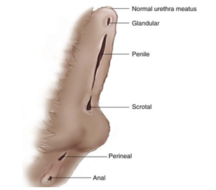
29. Disorders of the penis and prepuce – hypospadias, phimosis, paraphimosis
Hypospadias
Pathophysiology: Congenital external genitalia abnormality characterized by an abnormal ventral and caudal location of the external urethral opening. [1, 2]
Embryonal Cause: Triggered by a fusion failure of the urogenital folds and incomplete development of the penile urethra along the ventral shaft. [1, 2]
Gender Predilection: Observed almost exclusively as a urethral opening defect in male dogs. [1, 2]
Anatomical Classifications: Categorized by localized opening site: glandular, penile, scrotal, perineal, or anal. [1, 2]
Concurrent Condition: Frequently occurs alongside cryptorchidism (undescended testicles)
Aetiology: unclear. The inadequate production of foetal androgen is suspected.
Clinical signs:
Urinary incontinence, periurethral dermatitis or recurrent UTI, excessive licking.
Diagnosis:
During first puppy examination, or owner might report urine output from an unusual orifice (urinary incontinence)
Treatment: Abnormal urethral opening near penile tip may not require surgery. In other cases reconstruction is recommended (scrotal or perineal urethrostomy combined with castration & removal of prepuce & penile tissues). Dogs that have severe irritation associated with abnormally fused prepuce or penile hypoplasia usually require penile and preputial amputation combined with scrotal and perineal urethrostomy.
Phimosis
Inability to normally protrude the penis out of the preputial orifice. Rare in dogs. In tomcats it is predominantly found in young cats with a congenitally small preputial opening.
Aetiology:
Persistent penile frenulum– connective tissue that joins ventrum of glans penis to penile shaft or prepuce)
Congenitally short penis
Intersex condition
Small preputial orifice or preputial stricture resulting from trauma/wound healing
Result of neoplasia of prepuce or penis
Apprehension and/or pain associated with erection, anatomical, chronic inflammation from trauma/balanoposthitis
Temporary – accumulation of debris (smegma, preputial hair)
Inadequate testosterone production at puberty, hormonal imbalances, testicular hypoplasia, testicular agenesis, chromosomal abnormalities, abnormally thick frenulum.
Neoplasia, chronic inflammation.
Clinical signs
Dogs: excessive licking, dermatitis from urine scalding on hind legs or inguinal area, phallocampsis (ventroflexion of the penis), pain at breeding, inability to penetrate bitch
Cats: dysuria, outflow obstruction, haematuria, distended urinary bladder, secondary balanoposthitis.
Diagnosis:
Physical examination - Penis can’t be fully protruded – sedation or General Anaesthesia may be needed to protrude penis. Semen collection in presence of bitch may help to determine if the problem is physical or behavioural.
Treatment: Depending on cause.
Surgery: Transection of persistent frenulum with local anaesthesia, sedation or GA.
Preputial wedge resection – widening of the orifice
Congenitally short penis – do not breed (hereditary)
Adhesions – surgical breakdown, topical ATBs, steroid creams to prevent re-adhesions
Inflammation – culture to find cause and treat causally
Associated with fear – behavioural training
Paraphimosis
Inability to withdraw penis into prepuce. Common in dog - in Toy breeds that frequently masturbate. Testosterone mediated, prepubertal, sexual arousal problem. Bitches in heat.
Cause: Oedema of the penis as a result of chronic balanitis or balanoposthitis. A small preputial orifice, a short prepuce.
Preputial trauma. Neurological deficit caused by encephalitis or an intervertebral disc disease. Tranquilizers of phenothiazine (oedema of penis).
Clinical signs: Once penis becomes entrapped in prepuce, it becomes ischemic, dry and excoriated
The longer the condition continues, the more severe the damage
Self-mutilation may occur due to pain
Stranguria, haematuria, anuria
Prognosis: Prognosis depends upon the promptness of treatment and degree of trauma or necrosis present
Diagnosis: physical exam, neurological exam, x-ray, MRI, CT (IVDD). Spinal tap (encephalitis)
Treatment
Supportive treatment: apply lubrication. Sedate/anesthetise animal. Apply cold, flat compress soaked in hyperosmolar solution to minimize gravitational oedema. Reposition penis, Prevent masturbation
Surgical: opening of the small preputial orifice. Amputation of penis if necrosis is present.
Preventative: Castration is helpful.
From state:
- Hypospadia: Just what it was and where it could occur (opening on penis scrotum etc). Main CS and how to treat = Urethrostomy and she was happy with this. And i mentioned was congenital problem mainly in Boston terriers.
- Phimosis and paraphimosis: Just wanted the difference between them both and if there are paraphimosis what the treatment would be = If can’t get penis back in then do amputation of the penis to prevent trauma.
2021: only wanted me to talk about one of them.
which testicular disease often predispose to phimosis? Cryptorchidism
Difference between phimosis and paraphimosis
Are there any predispositions for phimosis? Apparently not
Otherwise same questions as above
30. Disorders of the penis and prepuce – balanoposthitis, priapism, urethral prolapse
Balanoposthitis
It is an inflammation of the penis (balanitis) & prepuce (posthitis). Balanoposthitis cause stenosis of preputial orifice, adhesions of penis, pain & discomfort. Usually affect dogs older than 4 years of age, intact & neutered, but also young dogs close to puberty which has discharge – related to hormonal changes. Tomcats usually get balanoposthitis after trauma following mating, fights or blunt trauma where adhesions may be formed. It is a rare condition in cats however.
Aetiology: Can be acute/chronic, infectious/non-infectious, ascending/descending.
Infectious: viral (Herpesvirus, Calicivirus)
Non-infectious: trauma (e.g. after fights/mating/blunt trauma), excessive licking (e.g. due to phimosis, paraphimosis),
self-mutilation (anxiety disorders, pruritus)Descending: from prostate, urinary bladder
Ascending: from environment (e.g. poor hygiene).
Clinical signs:
Erythematous and hyperaemic MMs
Green-yellow, purulent, fetid discharge. May be blood
Painful urination, dripping urination, excessive licking,
Diagnosis: physical exam (discharge), swab for bacteria (cultivation + atb sensitivity), cytological evaluation
Treatment
Mild: neck collar (if excessive licking), flushing of prepuce with warm saline/antiseptic, antibiotics.
Severe: frequent antiseptic or ATB ointments, systemic ATB, regular gentle protrusions, sexual rest,
enlarging orifice (if small preputial opening)
Priapism
Persistent penile erection lasting longer than 4 hours, without sexual stimulation/not due to hormones, with subsequent inability to retract the penis into the prepuce. Uncommon in dog, older dogs more prone.
Aetiology:
Neurological disorders, decreased venous outflow form the penis (a coagulation disorder or mass effect), DM, neoplasia, trauma, coagulopathy, reaction to drugs (phenothiazine tranquilizers, hypertensive medications, general anaesthetics).
Pathogenesis:
Priapism is categorized as either nonischaemic (arterial, high flow) or ischemic (veno-occlusive, low flow).
Clinical signs: Dysuria, Stranguria, hematoma, necrosis.
Diagnosis:
Anamnesis (medication administration), physical exam, complete dg work-up – underlying metabolic disease or neoplasia (bloods, coagulation panel, urinalysis, x-ray, USG of abdomen).
Treatment: Flushing with heparinized saline and infuse with sympathomimetic drugs (phenylephrine).
In most cases the penis has become ischemic and excoriated at the time of presentation, and amputation and urethrostomy has to be performed. Castration is NOT helpful – not testosterone mediated!
Urethral prolapse
Urethral prolapse is when the inner lining of the urethra sticks out through the opening of the urethra.
Usually the entire distal end of the urethra is prolapsed and becomes oedematous.
Aetiology: secondary to urethritis, sexual arousal or masturbation.
Seen in younger dogs (< 5 months) and the English bulldog (Hereditary in some lines)
Clinical signs:
A prolapsed urethra often looks similar to a pea-sized mass, and may be red or purple in colour. This can often be observed as a small mass of tissue on the end of the penis. It can become enlarged or inflamed. Intermittent drops of blood from end of penis. Can cause difficulties urinating. Licking. Pollakiuria
Diagnosis: physical exam (blood drops from the tip of penis, doughnut-shape, or pea-shape structure), pollakiuria.
Treatment
Conservative: sexual rest, tranquilization, neck collar (to prevent licking)
Surgical resection: in case of extensive bleeding or pain.
Resection of prolapsed tissue.
Urethral guide to push prolapsed tissue back into urethra. Urethra then sutured to shaft of penis
31. Disorders of the testes – anorchism, monorchism, cryptorchidism
Normal testicular descent in the dog occurs in 2 steps:
Movement through the abdominal cavity into the internal inguinal ring – transabdominal phase
Movement from the inguinal canal to the scrotum – inguinoscrotal phase (movement through and out of the inguinal canal = transinguinal phase)
Initially the testes lie near the kidneys in the abdominal cavity. There is a ligamentous cord (caudal suspensory ligament) called the gubernaculum which attaches to the end of the testis and then runs through the inguinal canal and attaches at the far end to the scrotum. The gubernaculum enlarges under stimulation of certain protein hormones to anchor the testis in place during testicular descent. Testicular descent is complete by 10-42 days of age in dogs. In cats this event appears to be prenatal.
Anorchism/monorchism
Anorchism is the complete absence of both testicles.
Monorchism is the absence of one testicle.
Aetiology: Unknown, but proposed to be connected with interruption of testes blood supply by torsion or vascular occlusion even in utero. Previous orchiectomy. These two conditions are quite rare in dogs.
Diagnosis: Palpation, USG evaluation, CT, MRI, exploratory abdominal surgery.
Treatment: Surgery - First remove scrotal testicle, wait 5-7 days, then perform testosterone stimulation test.
If there is still testosterone production, we know the dog is cryptorchid.
If there is no testosterone production, we know the dog is monorchid.
If no testosterone before castration, we know the dog is anorchid. (Testosterone concentrations can be used to differentiate a bilateral cryptorchid patient from an anorchid or castrated patient)
Cryptorchidism
Failure of one or both testicles to descend into the scrotum (retained testicles). Several breeds of dogs are predisposed, but almost every breed can have it. The retained testicle is predisposed to neoplasia and testicular torsion.
Testicular descent is complete by 10-42 days of age in dogs
Lack of two testes in the scrotum by 8-10 weeks of age is considered to be suspicious for cryptorchism
If an animal reaches 6 months of age without decent testicle/s – considered cryptorchid
In cats, testicular decent appears to be a prenatal event
May be unilateral or bilateral (unilateral is more common)
Right testicle is more likely to be cryptorchid than left – right kidney is more cranial, so starting position of the right testicle is more cranial.
Location
1) Abdominal
2) Ectopic (subcutis).
These are the ONLY 2 forms in dogs, they do NOT have inguinal cryptorchidism!!! Because they have only inguinal ring, no inguinal canal, or very short inguinal canal.
Clinical signs
Testes are very small, soft, and flaccid
No spermatozoa are produced by the retained testis.
Spermatogenesis is completely inhibited by the elevated temperature affecting the testis.
Retained testes predisposes to neoplasms: Sertoli cell tumours (hyperestrogenism) and seminomas tends to be most malignant.
Also predisposes to testicular torsion.
Diagnosis:
Clinical examination, palpation. Testosterone stimulation test, radiography, ultrasound, exploratory laparotomy.
Treatment
Surgical: castration – Best option.
Conservative: hCG/GnRH: stimulate puberty and testicular growth and descent (low effectiveness)
Prevention: To eliminate this condition all parents of cryptorchid animals should be removed as breeders.
From state:
· Asked which are the most common tumors the dog gets from cryptorchidism. Sertoli cell tumors
· Also asked if i knew how anorchism is caused- physiological process
· How you diagnose cryptorchid. Types of cryptorchidism (abdominal etc)
· How do you differentiate between monorchid and cryptorchid
32. Disorders of the testes – orchitis, epididymitis, spermatic cord torsion
Orchitis and epididymitis
Orchitis is the infection of the testicle
Epididymitis is infection of epididymis. Primary or secondary (from orchitis). Tail is most often affected.
Aetiology
Non-infectious: trauma/puncture to scrotum or spermatic cord, auto-immune disorder,
Infectious causes: Brucella canis, B. abortus, B. melitensis (venerally transmitted), streptococcus, staphylococcus, Proteus vulgaris, Bacillus, E. coli, Klebsiella, Pseudomonas, Mycoplasma, Ureaplasma, Blastomyces dermatitidis, fungi,
Canine distemper virus.Descending: Infection
Ascending: infection from the prepuce, urethra, urinary bladder or prostate gland.’
Can be acute or chronic
Hematogenous infection.
Clinical signs: Enlargement, erythema & pain (unilateral or bilateral), swelling may extend up the spermatic cord & involve the vaginal tunic
Acute: fever, lethargy, hindquarter lameness, scrotal pain, erythema, swelling, scrotal oedema, purulent preputial discharge
Chronic: swelling/contraction of all or part of scrotal contents, non-painful and firm
Diagnosis:
Physical examination, CS.
Semen collection – pyospermia. Pain in acute cases can cause inability to ejaculate. Culture of sperm, Brucella testing.
Imaging – USG (heterogenous tissue with inflamed areas being hypoechoic, abscesses)
Cytology – neutrophilic inflammation in cases with infectious bacterial or fungal causes, and lymphoplasmacytic inflammation in association with autoimmune diseases
Brucellosis test – agglutination test, ELISA
Treatment and prognosis:
ATB of choice of testicular, prostate and urinary disease: enrofloxacin (effective against majority of repro pathogens)
Hemicastration or complete castration.
NSAIDs, cold pacing of the scrotum – reduce inflammation and swelling.
Sexual rest.
The prognosis for return to normal function of affected testes is poor. Sperm production and morphology are often affected permanently.
Spermatic cord torsion/testicular torsion
It is defined as a twisting of the testes or testicle on its connective tissue. Seen in middle-aged to older dogs but is quite uncommon condition in dogs, and very rare in cats.
Aetiology
Long caudal ligament of the epididymis or proper ligament of the testis
Retained testes (increased mobility of abdominal testes)
Neoplastic testes: heavier and therefore more mobile
Already enlarged testes: worsening of torsion
Pathogenesis:
Torsion of testicles à venous occlusion à oedema, inflammation, PAIN!
Torsion must be greater than 180 degrees to produce venous occlusion.
Clinical Signs:
Acute abdominal pain, abdominal splinting, depression, lethargy, anorexia, vomiting, fever, haematuria, stranguria, stiff gait on hindlegs.
Diagnosis:
CS, USG (colour Doppler – abnormal venous blood flow of testes), exploratory surgery to confirm diagnosis.
Treatment:
Surgical removal – bilateral castration.

33. Disorders of the testes – Spermatocele, tumours of testes
Spermatocele
A spermatocele is a cystic distension of the epididymis, usually caused by a blockage of the epididymis leading to accumulation of sperm. Some will leak out & result in an inflammation process & the development of a nodule of tissue referred to as a sperm granuloma. Can be unilateral or bilateral.
Spermatocele is seen in intact male dogs of any breed. The risk of developing this disorder increases with age.
Aetiology
Trauma, hyperplasia, adenomyosis (the ingrowth of the epididymis lining into the deeper tissues, which may be associated with excess estrogenic stimulation), congenital disorders causing a blockage of the ducts.
Pathogenesis:
Obstruction cause distension of epididymal tubules, which cause sperm stasis leading to spermatocele formation and the sperm escape into surrounding tissue, which causes an inflammation reaction due to foreign bodies and secondary granuloma formation in the caput region. Nb. This is a gradual process
Clinical signs:
When only one sperm duct is affected there is usually no symptoms. When there is bilateral occurrence, dogs can develop azoospermia. Spermatoceles are usually discovered due to infertility. Sometimes dogs can have a hard palpable mass on the scrotum, but it is rarely associated with pain or visible/palpable lesions.
Diagnosis:
Testicular examination and palpation, semen evaluation (azoospermia), ultrasound, Biopsy of testicle.
Treatment:
Those that have developed insufficient sperm count rarely spontaneously recover. Bilateral blockage is usually only treatable by surgical intervention (castration). If unilateral; hemi-castration. (One case with unilateral spermatocele underwent hemi-castration on the affected testicle and there was collection of excellent-quality semen from the remaining testicle a month later).
Testicular tumours
Most common: Sertoli cell tumors, interstitial cell tumors, seminomas
Less common: undifferentiated carcinomas, fibrosarcoma, lymphomas, hemangiomas, gonadoblastomas, embryonal carcinomas, sarcomas, granulosa cell tumors and teratomas
Epidemiology & Overview
Second most common
Signalment: Typically older, intact male dogs (9–11 years); rare in tomcats.
Breed Predisposition: Boxers, Poodles, and German Shepherds.
I. Sertoli Cell Tumor (SCT)
Prevalence & Retention: Most common (44%); primary tumor of cryptorchid (retained) testicles (State Exam Topic).
Gross Appearance: Solitary, white to pale yellow, large (1–12 cm), firm on cut section. Low metastasis.
Endocrine Profile: Highly active; secretes excess estrogen, causing feminization syndrome (hyperestrogenism).
Clinical Signs: Mimics Cushing's disease; bilateral symmetrical alopecia, hyperpigmentation, dry skin, "red tail", pendulous prepuce, gynaecomastia (enlarged mammary glands/nipples), behavioral changes (squatting to urinate), and lethal bone marrow suppression (anemia).
II. Seminoma (Germ Cell Tumor)
Prevalence & Location: Makes up 31% of tumors; predominantly found in descended testes (75%).
Gross Appearance: Single or multiple, large (1–10 cm), cream to pink-gray, homogenous/lobulated. Higher local metastasis risk.
Endocrine Profile: Not endocrinologically active (no hormone production).
Clinical Signs: Often asymptomatic; may cause local pain from tumor growth pressure.
III. Interstitial / Leydig Cell Tumor
Prevalence & Location: Makes up 25% of tumors; most common tumor of scrotal (descended) testes.
Gross Appearance: Benign, small (<1 cm), single/multiple, soft, rounded, and distinctly tan/yellow/orange. Rarely metastasize.
Endocrine Profile: Endocrinologically active; increases estrogen and testosterone.
Clinical signs: depends on type of tumour. Similar to SCTs, but more symptoms in association with the testosterone production such as prostatic disease, perianal adenoma, perianal gland hyperplasia, and perineal hernia. Dyspnoea – metastasis to lungs. Other signs can be; A single enlarged testicle or asymmetric testicle, soft “swelling” of one or both testicles, generalized scrotal enlargement, infertility/subfertility in breeding stud, penile atrophy, testicular atrophy of the unaffected (noncancerous) testicle – due to hormonally active testicle.
Diagnosis:
Suspicion of a testicular tumour is based on the patient history and physical examination findings – enlarged testis or palpable mass. Exfoliative cytology of the preputial mucosa – cells, number of layers, character of each layer is dependent on hormones (oestrogen). Similar principle as swab of vagina. Will find anuclear cells (same as in heat of bitch). This is valid for SCT where there is increased oestrogen concertation.
Semen evaluation – abnormalities in morphology with primary defects predominating
USG – usually, the neoplastic tissue is hypoechoic compared with normal parenchyma
Testicular aspiration or biopsy
Histopathology – even though macroscopic appearance is very typical for different tumours, they must send for histology for definite diagnosis.
CBC, biochemistry – in case of increased oestrogen, there is suppression of bone marrow à nonregenerative anaemia, leukopenia, thrombocytopenia
X-ray – of lung to find metastasis
Treatment:
Medication in the case of non-operable patients – chemotherapy (cisplatin, vinblastine, cyclophosphamide)
Hemicastration – in unilateral lesion in breeding animals
Complete castration – best option
From state:
Only definition of spermatocele
Tumours and hormones secreted from them. Clinical signs and which are most common
34. Orchiectomy - preventive and therapeutic
Orchiectomy
An orchiectomy is a form of castration in male animals, in which the testicles are removed. Unlike a castration, an orchiectomy can be either unilateral or bilateral, meaning one or both of the testicles can be removed in this type of surgical procedure. Orchiectomy is a relatively quick procedure.
In the UK, castration of tom cats before the age of 6 months is recommended to all owners. The opinions on the routine castration of dogs are more divided. In some countries (e.g. Scandinavia) where stray dogs are not a problem, routine surgical neutering is considered mutilation and is illegal. In Norway only 7% of dogs are neutered and canine overpopulation is not a problem.
Castration is performed far more frequently in tom cats than in dogs.
Indications
Preventative orchiectomy
Decreased roaming and stray animals: important vectors of disease, social problem
Population control - Prevention of unwanted litters
Decreased testosterone induced behaviour: urine marking (cats), territorial aggression (dog)
Increased concentration and obedience
Therapeutic orchiectomy
Cryptorchidism: higher incidence of neoplasia
Prevention/reduction of testosterone- induced disease: benign prostate hyperplasia (BPH), prostatitis, prostatic abscesses, perianal or perineal adenomas, perineal hernias and certain castration-responsive skin disorders (dermatoses).
Castration of a male carrying a harmful genetic trait like hip dysplasia, epilepsy and so on, to prevent the spread of these conditions in the population.
Procedure
Preparation: clip and prepare pre-scrotal area for aseptic surgery. Do not clip scrotum as may result in significant inflammation. Dorsal recumbence, cover with surgical drapes.
Skin and subcutis incision: move first testis from scrotum to pre-scrotal area. Stabilize testes between index finger and incise on midline and incise skin, subcutaneous tissues and spermatic fascia. Facilitate extrusion of first testis
Open method: incise vaginal tunic
Clamp, ligate and transect: apply three clamps across ductus deferens, testicular arteries or venous pempiniform plexus, apply circumferential ligature. One close to patient, another between first suture and next clamp. Transect spermatic cord. Remove clamp and assess for bleeding
Closure: simple interrupted of subcutaneous skin, intradermal of skin. Bury knot
fordel å lese over punkt 6. i tilleggsinformasjonen på slutten av dokumentet.
From state:
Wanted basic therapeutic and preventative indications, did not want to hear about procedure.
Wanted to know names of drugs that could chemically castrate the dog.
35. Disorders of the prostate – benign prostatic hyperplasia, neoplasia
Benign prostatic hyperplasia (BPH)
BPH is common & occurs in ca. 60% of intact males older than 5 years of age. Hormonal cause.
It is usually benign, however when inflamed it may cause problems with urination & defecation. Two forms- with/without follicles.
Aetiology:
Occurs from long-term exposure to dihydrotestosterone (DHT) - the hormone that stimulates prostate growth.
DHT is the active metabolite of testosterone.
Pathogenesis:
As cell numbers & size increase, prostate gland enlarges. Increased vascularity can result in vascular leakage/haemorrhage into the gland, increased swelling causing pressure & cyst formation. Prostate is located under colon & above bladder, so it presses on the colon (constipation) & on the urethra.
Clinical signs:
Sanguineous (red) or serosanguinous (pink) discharge from the penis
Pollakiuria, stranguria, haematuria
Incontinence, inability to urinate
Infertility
Constipation and production of a ribbon-like stool may occur
o Body of the prostate is the first starting to grow. Grow outwards, so problems with defecation can often be the first problem. With longer duration – problems with urination and defecation.
Diagnosis:
Physical examination – digital palpation reveals an enlarged but smooth and symmetrical gland
X-ray – enlarged soft tissue opacity in the caudal abdomen or cranial pelvic canal. Retrograde cystourethrography
USG – uniform hypoechoic to hyperechoic parenchyma with mild heterogenicity
Culture – aerobic bacteria, Mycoplasma, Ureaplasma
Cytological examination – through prostatic massage or semen collection (third fraction of ejaculate is prostatic)
Treatment
Conservative: oestrogen, progestogens.
Finasteride - hormone antagonist à Stop the conversion of testosterone to dihydrotestosterone.
Ypozane (Osaterone-acetate) - antiandrogen + progestogen à inhibit testosterone.
Chemical castration
Surgical: Castration. Prostatectomy (difficult to remove all of it as some is in the urethra)
Prostate neoplasm
Tumours of the prostate is rare.
Adenocarcinoma: main prostate tumour. Characterized by many nodules and asymmetric enlargement that may cause compression of the urethra and colon. Aggressive tumour - Metastasis mainly to lungs, but also lymph nodes, liver, urethra, spleen, rectum, colon, bladder, bone, heart, kidneys and adrenal glands.
Other possible tumours: transitional cell tumour, lymphosarcoma, hemangiosarcoma, squamous cell tumour.
Clinical signs:
Tenesmus, constipation, obstipation, ribbon-like faeces, stranguria, haematuria, weight loss, hindlimb ataxia,
prostate gland can be enlarged and painful on rectal exam
Diagnosis:
Palpation – may be enlarged and painful, and there is typically asymmetry of the gland
Leucocytosis and neutrophilia may be present. Elevations in ALP are common.
X-ray – prostatomegaly. Prostatic mineralization may occur. Contrast studies
USG – heterogenous architecture with neoplastic tissue being hyperechoic
Biopsy or cytology – for confirmation of disease
Treatment:
Chemotherapy & radiation therapy – but metastasis is common by the time it is diagnosed. Low survival rate.
Total prostatectomy – but cannot remove the part that is in the urethra, so might not be very effective + complications with urinary and faecal incontinence, stranguria, haematuria, hindlimb oedema.
Piroxicam – NSAID used to treat some cancers in dogs and cats, including prostate cancer.
Castration is useless.
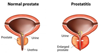
36. Disorders of the prostate – cysts, prostatitis
Prostatic cysts
Prostatic cysts are fluid-filled pockets, commonly associated with Benign prostatic hyperplasia. They are more common in older dogs averaging around 8 years and in large breeds.
Can be intra- or extraprostatic:
Intraprostatic cysts – multiple and smaller in size (<1 mm to 3 cm in diameter), commonly associated with hypertrophy and hyperplasia of the glandular epithelium
Intra- and extraprostatic retention cysts are distinct cysts with a well-defined, thickened wall, small to large, usually single.
Cause: Not completely understood, but maybe arise from blockage of ducts in the prostate, with subsequent expansion of the cyst due to accumulation of prostatic secretions.
Clinical signs:
Dysuria, stranguria, haematuria, constipation, obstipation, tenesmus, ribbon-like faeces, abdominal distention, abdominal pain, anorexia, depression.
May be asymptomatic
Diagnosis:
Palpation – enlarged, asymmetrical.
X-ray – soft tissue opacity caudal or dorsal to the bladder. Retrograde cystourethrography
USG – provides a definite diagnosis, allowing determination of size, location and nature of the cystic structures.
The fluid within the cyst is usually hypoechoic or anechoic
Treatment:
Small cyst associated with BPH alone may decrease in size with medical treatment, but not resolve completely.
Needle aspiration of fluid – temporary solution (fills over time)
Surgery – in case of solitary extraprostatic cysts
Intraprostatic cysts – removal is often difficult, so drainage should be performed.
Large cysts can be marsupialized (formation of new, permanent draining duct)
Prostatitis
= Infection of the prostate gland. Very common, seen in all age groups. Most common in intact than in neutered animals. Predisposing factors include BPH, prostatic cysts, neoplasia or squamous metaplasia.
Aetiology:
Infectious: Ascending bacterial infection from the urinary tract, preputial flora, epididymitis/orchitis
Pathogens: E. coli (most common), Klebsiella, Pseudomonas, Pasteurella, Streptococcus, Staphylococcus, Enterobacter, Brucella, Mycoplasma,
Hematogenous spread
Venereal transmission
Non-infectious: BPH, prostatic cysts, neoplasia or squamous metaplasia.
Chronic prostatitis is more common than acute.
Clinical signs:
Acute: Swelling, oedema, fever, malaise, abdominal pain, vomiting, dysuria, stranguria, haematuria, tenesmus, constipation, obstipation and ribbon-like faeces. Unwillingness to breed. Painful, swollen, soft or firm, asymmetrical prostate gland. Pyuria, haematuria
Chronic: Stranguria, haematuria, tenesmus, constipation, obstipation, ribbon-like faeces. Pyospermia or pyuria
Normal to enlarged symmetrical/asymmetrical, non-painful prostate.
Diagnosis:
Acute:
Physical examination – CS, dehydration, fever, shock, sepsis, multi-organ disease (blood-prostate gland barrier is breached, so hematogenous spread of bacteria can result in severe conditions), elevated hepatic enzymes (particularly ALP).
X-ray – prostatic enlargement. Retrograde cystourethrography
USG – focal to diffuse heterogenous echotexture with or without cavitating lesions filled with hyperechoic fluid.
Chronic:
Physical examination – CS, typically healthy animals, urinary signs/rectal or colonic signs. CBC and chemistry are typically normal.
X-ray – same as acute, but minimal uptake of contrast medium on retrograde cystourethrography
USG – asymmetrical gland with heterogenous architecture.
Cytology – prostatic wash or massage fluid. Marked inflammatory response.
Treatment:
Acute prostatitis requires aggressive therapy.
Supportive: IV fluids on basis of dehydration and electrolyte imbalance.
NSAIDs (careful with kidneys if their function is compromised!)
ATB – based on cytology and Gram stain until culture results are available. Enrofloxacin - Continue with ATB for 4+ weeks (acute) or 12 weeks (chronic cases or abscesses)
Castration of animals with concurrent BPH.
Drainage of prostatic abscesses. Partial prostatectomy
From state:
Cysts: Which are larger - internal or external
Prostitis: What’s the basic clinical sign? Answer: infertility
Treatment? Answer: castration
37. Infertility of the bitch - disorders of the heat (primary and secondary anoestrus)
Primary anoestrus (delayed puberty)
Primary problem – bitch does not show signs of oestrus despite having reached age of puberty/no oestrous activity before 24 months of age (normal: puberty at 7 months, or up to 12 months of age or later).
May indicate a primary endocrinological failure. Clinical investigation is not necessary before 24 months of age.
Causes:
Ø Normal physiological variation
generally reach puberty between 7-12 m.- larger breeds later.
Greyhound may not until 3 or 4 years. Puberty can also have later onset in underweight dogs or those with chronic diseases (e.g. hypothyroidism or Cushing’s).
Ø Inadequate observation by the owner or “silent oestrus”
Frequent cleaning or no male dog can make it more difficult to detect. Elevated plasma progesterone (>0.2 ng/ml) will demonstrate that oestrus has occurred within the last 60 days.
Ø Abnormal sexual differentiation
Chromosomal abnormalities that cause primary anoestrus. Vulva is small, prepuce-like and positioned more cranial.
Will never be fertile – should remove gonads and reproduction tract to prevent pathology later.
Ø Ovarian agenesis/aplasia
Can be difficult to diagnose. It is a rare condition characterized by an absence of ovaries. There is high concentration of FSH and LH, but low concentration of oestrogen. Administration of hCG will not cause increase in plasma oestrogen.
At age 7 or 8 years: decreased quality of estrus, increased interval between estrus, egg quality decreased = decreased fertility + decreased number of live puppies + congenital disorders present
Therapy:
The exclusion of other diseases must be done before induction of the heat cycle and ovulation. The bitch must have reached a suitable age. Options for induction of oestrus:
Induction with oestrogens are not recommended (high toxicity)
eCG (equine chorionic gonadotropin)
20 IU/kg of eCG for 5 days in a row, and 500 IU of hCG on day 5.
Fertility of induced oestrus is variable, but is generally low.
Cabergoline (Deslorelin implant) - inhibit prolactin. Heat in 5-6 days.
Secondary anoestrus (prolonged anoestrus)
Secondary problem – oestrous activity has not occurred within 12 m of previous cycle.
The bitch had a cycle/cycles but is now in a period of an abnormally long anoestrus.
Normal inter-oestrous interval = 7 months. >12-month anoestrus – can be associated with oestrus being unnoticed or pathological conditions. Individual variation (basenji – 1 cycle every 12 months)
Causes:
Ø Inadequate observation by the owner: Inexperienced owner or frequent cleaning. Detection of elevated plasma progesterone tells that oestrus has taken place the last 60 days.
Ø Poor body condition or systemic illness: Rapid return to cyclicity when condition is corrected.
Ø Prolonged luteal phase: Failure to return to cyclical activity. Rare, unless luteal cyst or ovarian neoplasia producing progesterone.
Ø Endocrinopathies: Hypothyroidism and Cushing’s may influence the return to cyclicity.
Ø Premature ovarian failure: rare. Principal problem is thought to be an inadequate ovarian response to pituitary gland gonadotropin stimulation. Elevated FSH and LH but no response of ovaries. No signs of oestrus since low oestrogen.
Ø Exogenous: Ovariectomy or ovariohysterectomy at early age, use of progesterone or glucocorticoids
Diagnosis: Clinical symptoms are faint and unspecific
Measurement of serum progesterone: progesterone levels in the normal reproductive bitch will rise above 2 ng/ml for 2 months after oestrus. Failure to detect increased levels of serum progesterone over a 6-8-month period in the bitch will strongly suggest a state of prolonged anoestrus.
Other blood tests: to detect elevated concentrations of luteinizing hormone (LH) or follicular stimulating hormone (FSH). Primary anoestrus: GnRH stimulation test for FSH/LH or Oestradiol. TT4, FT4, TSH.
Treatment
- Correction of the underlying abnormality
Prolactin inhibitors (cabergoline – 5 ug/kg) – continual daily administration until the first day of vulval swelling and discharge. Bitches that are responding to this therapy usually respond within 30 days, and if administration is stopped once proestrus has begun, there is usually normal fertility
38. Infertility of the bitch - disorders of oestrous cycle (split oestrus, shortened interval between cycles, lengthened period of pro-oestrus or oestrus)
There is variation between individuals in how long the estrous cycle lasts. As proestrus and estrus can last from 3-21 days each (and still be considered normal), bleeding for 40 days can be physiological! That means that the total length of P+E+D can be from 63 to 142 days.
Split oestrus
No or very short oestrus signs develop despite the presence of proestrus signs (bitch will enter proestrus and fail to enter oestrus). Common at puberty and in bitches <4 years of age. Not typically a problem in older bitches. The bitch is observed to enter proestrus again within 3–4 weeks, and the next cycle is usually a normal ovulatory cycle
Aetiology
Once: young bitch. The condition usually recovers spontaneously
Frequent: lack of pre-ovulatory LH or follicle LH receptors à ovulation failure
Treatment: Not necessary during puberty. hCG if repeating problem.
Shortened interval between cycles
Definition: A shortened cycle where the next estrus begins less than 4 months after the previous one ended.
Exclusions: Does not apply if the short interval is caused by a split estrus or a pubertal estrus.
Pathophysiology: Frequent heat cycles and progesterone exposure cause damage; a brief anestrus prevents proper endometrial recovery (uterine lining).
Clinical Consequence: Bitches with this condition characteristically exhibit a higher rate of infertility
Aetiology and Treatment
Ovulation failure:
Proestrus returns earlier than expected (no ovulation – no luteal phase – oestrus occur 2 months earlier than expected).
Can be caused by low LH.
Therapy = 500 IU hCG IM every day for 3 days to ensure that ovulation occurs at next oestrus
Short luteal phase:
Inadequate production of progesterone from CL – the progesterone decreases to basal concentration after 20-40 days after ovulation, rather than the normal 60-65 days
Can be detected when there is loss of a confirmed pregnancy
Most common in German shepherds
Therapy = supplementation with P4 during pregnancy (risk of cryptorchidism in male puppies and masculinization of females).
Short anoestrus:
Anestrums = affects the length of the interval. HUGE variability.
Shortening of anoestrus is commonly seen in bitches that are together with other bitches that are in oestrus (dormitory effect).
Lengthened period of pro-oestrus or oestrus
Normal physiological lengthening: A bitch normally ovulates day 12 after onset of pro-estrus. It is normal that it occurs as late as day 25, or as early as day 5 also.
Pro-oestrus and oestrus usually last approximately 20 days in total. This can be longer either as a normal physiological variant, or as a result of delayed ovulation, ovarian neoplasia or exogenous E2 administration.
Prolonged oestrus: the mean duration is 9 days. If longer than 21 days without ovulation is defined as prolonged oestrus.
Prolonged proestrus: A proestrus that lasts 3 weeks or longer without following oestrus or ovulation.
Aetiology and Treatment
Delayed ovulation: if ovulation hasn’t occurred within 30 days. Failure of adequate LH production or insufficient LH receptors leading to anovulatory follicle/follicular cyst with prolonged Oestrogen production and therefore prolonged oestrus.
Tx: hCG for 3 days. If no response, supply progesterone (OBS pyometra) or OV.Ovarian neoplasia:
Oestrogen-producing ovarian neoplasms, causing signs of persistent oestrus and bone marrow suppression.
Rare. Mainly older bitches. Tx: OHE
Follicular cysts: rare. Image all bitches with proestrus/oestrus behaviour for more than 30 days to see if follicular cysts are present. They are large (8-12 mm), thin-walled, anechoic. Can cause bone marrow suppression (persistent elevated oestrogen levels). Can be treated by inducing ovulation using hCG, or even OHE.
Exogenous oestrogen administration
Dx: Vaginal smear (cells are permanently cornified), blood sample (low progesterone, high oestrogen).
39. Infertility of the bitch - abnormal mating (behavioural problems, abnormality of reproductive tract)
Behavioural problems
Previous poor experience
presented for mating at an inappropriate time and therefore being force-mated, or when the stud dog is too dominant. This can lead to reluctance or refusal of subsequent mating attempts. Correct choice of male is crucial.
monitor vaginal cytology and plasma progesterone to determine best time for mating.
Poor response to the male dog
Marked submission or aggression. It is in most cases caused by poor social skills of the bitch. These cases may be difficult to manage and require careful introduction of the male and prolonged periods of play before attempting mating.
Male preference
Some bitches (more commonly older ones) may show preference for certain males, especially if the bitch has been raised/housed around individual males. If housed away from males, an oestrus female seldom rejects a male based on preference. Also, if a female dog has poor canine social skills from living in a pure human household with little contact with other dogs it may cause poor courting and mating behaviour in the bitch
Behavioural problems during mating can be avoided by proper planning and management of the process. Especially ensuring that the bitch’s first mating is not traumatic is important to establish a normal mating behaviour. Have the male and female meet several times before the actual mating to familiarize with each other and make sure that there is no aggression. For the mating itself, determine the optimal time and have a calm, non-stressful environment.
Abnormality in reproductive tract
Some abnormalities occur within the vagina and are only identifiable upon digital exploration.
Vestibular and vaginal malformations
Error in the formation of the caudal female reproductive tract during foetal development. These are observed most commonly at the junction of the vestibule and vagina.
Vestibulovaginal bands
Fibrous bands or constrictions at the junction between the vestibule and the vagina found just proximal to the external urethral orifice. Not uncommon. Cause pain and failure to achieve intromission.
Can in some cases cause vaginitis (uterine and vaginal discharge is being trapped, causing bacterial overflow).
Treatment: thin bands – broken down manually, larger – episiotomy, ligation and transection.
Vulvovaginal bands
Less common.
A tight ring at the vulval opening that prevents normal intromission.
There is usually an acute pain response at attempted mating, and the bitch will then refuse subsequent mating attempts.
Diagnosis by clinical examination.
Mild to moderate cases can be solved with manual dilatation under general anaesthesia
Vaginal hypoplasia
Rare. The majority of the vagina is affected and is small in diameter and non-dilatable.
Presented most commonly with pain at attempted intromission, or with vaginitis.
- Endoscope can be used to confirm the severity of the condition.
Vaginal hyperplasia:
Hyperplasia of the vaginal wall is a normal response to elevated oestrogen concentrations during oestrus. In some bitches there appears to be an exaggerated response to normal concentration of oestrogen, such that prolific thickening of the vaginal wall occurs especially in the regions just cranial to the external urethral orifice. This can obstruct the vaginal lumen. The hyperplastic tissue develops during proestrus and regresses when oestrogen concentration decreases, and progesterone concentration begins.
Clinical signs:
Mild cases: may go unnoticed, except that it causes a pain response at attempted intromission
More severe cases: hyperplastic tissue may protrude from the vulval lips (ddx: vaginal polyp or neoplasm)
In cases with protruding mass, there may be excessive licking and self-trauma
Diagnosis:
Hyperplasia can be confirmed usually because of the relationship to oestrus, the origin of the tissue (just cranial to the urethral orifice) and the wide base of attachment.
Treatment:
The condition usually regresses fully at the onset of the luteal phase, so most cases can be managed conservatively by preventing self-trauma and using protective creams.
Bitches with protrusion of a small mass can be bred by holding the mass ventrally and allowing the dog’s penis to penetrate dorsal to the mass.
In bitches that become pregnant the hyperplasia will have regressed by the time of parturition and will not contribute to dystocia.
Neutering is recommended if there is progression of severity with each subsequent cycle.
AI for bitches with large protrusions.
Other:
Segmental aplasia - Female is missing part of the tubular genitalia – uterine body or oviduct. very rare.
Uterine tumours or polyps - Uterine masses may interfere with sperm transport or implantation
Cervical stenosis - Stenosis obstructs entry of sperm into the uterus
Cystic endometrial hyperplasia - Due to P4 or E2 treatment, or abnormal reaction to natural P4 or E2
Endometritis - Inflammation of the uterine mucous membrane that prevents implantation and progression of pregnancy.
From state
I first talked about some behavioural problems (wrong timing, dominant male, bad experience, etc.). Then I talked about vestibulovaginal bands, vulvovaginal bands, and vaginal hypoplasia and hyperplasia. She asked about problems with the uterus or ovaries so I mentioned cystic hyperplasia w/ bacteria, ovarian hypoplasia, and follicular cysts. She was happy with that.
November 2021: she was super chill. Mentioned things I remembered about behavior (she was very happy that I mentioned wrong timing of breeding cause it’s most common reason, and aggressivity). Then abnormalities of genital tract - just mentioned what I remembered, vaginitis, cystic endometrial hypoplasia. She also said that sometimes you can have missing uterus or too narrow vagina so that the male can’t penetrate. Was overall happy.
40. Infertility of the bitch - infertility after apparently normal mating
Mating at incorrect time - too long before or after ovulation
Probably the most common cause of infertility. The bitch shows marked variability in the time that ovulation occurs in relation to the onset of pro-oestrus and the signs of standing oestrus.
Methods to determine right day: monitor onset of vulvar softening or the change in the characteristics of vaginal discharge, measurement of plasma progesterone concentrations, exfoliative vaginal cytology and vaginoscopy.
The time of maximum fertility appears to be from the day of ovulation until 4 days after ovulation.
Majority of bitches will ovulate between 9 and 14 days after the onset of proestrus, ovulation may occur as early as 5 days and as late as 30 days after the onset.
Progesterone begins to increase rapidly from baseline levels approximately two days before ovulation, which allows for anticipation of ovulation. Exfoliative vaginal cytology shows stratified squamous epithelium during the fertile period.
Mating at correct time, but there is still pregnancy failure
This is characterized by the bitch being mated at an appropriate time, but not conceived.
Normally involves abnormalities of the reproductive tract that prevents sperm transport, embryo implantation or interfere with the development of a fertilized embryo. Includes uterine diseases and male infertility.
Uterine diseases and male infertility:
Infertile male /Poor male fertility
Stress – transport, alteration of social structure, poor nutrition, parasites à leading to PGF2a release
Missing parts of uterus or oviduct
Uterine tumours – interfere with transport of spermatozoa or with implantation.
Cervical stenosis – obstruct entry of sperm
Congenital defects of oviduct
Failure of ovulation – Oestrus is normal, but proestrus interval is shorter à failure of P4 level to increase. May be due to preovulatory increase in LH.
Early embryonic loss – Endometritis, embryonic defects, interbreeding, inadequate luteal function. Embryonic death may be followed by pseudopregnancy.
Hypoluteoidism – Result in inadequate concentrations of progesterone.
Cystic endometrial hyperplasia – pyometra
Endometritis
Infections
Infections by various agents may also interfere with successful conception including Canine Herpes Virus, Brucellosis, mycoplasma or Ureaplasma.
From state:
- Dominant/ aggressive male
- Failed detection
- Wrong time
- Male problems - she asked about types of ejaculation 1) retrograde 2) premature 3) delayed
Other things
- Endoscope can be used to confirm the severity of the condition.
- Asked about other causes - cysts, uterus/endometrium
- Follicular cysts
- Degen diseases of testicles – tumour
Azootospermia vs Aspermia
41. Infertility of the dogs - poor semen quality- teratozoospermia, oligozoospermia, azoospermia, astenozoospermia
Poor semen quality may be associated with prostate gland, testicular or epididymal disease, or urinary conditions. Other conditions related to semen quality are:
Infectious diseases of the reproductive tract (Brucella canis, aerobic bacteria, ureaplasma
Genetic abnormalities (chromosomal abnormalities, inbreeding)
Hormonal disorders
Ongoing medication (steroids, antifungals, certain antibiotics),
Miscellaneous causes such as scrotal hyperthermia, age, pollution (dioxins)
Stress
Sperm defects
Primary à > 20 % morphological defects à cause reduced fertility. Occur in the seminiferous tubules during development
Secondary à > 20 % of morphological defects à occurs after collection. Bent or coiled tails – related to sampling and handling techniques.
Congenital
Chromosomal abnormalities à XXY syndrome
Cryptorchidism
Unilateral → oligospermia
Bilateral → sterility
Acquired
Infectious agents – orchitis, epididymitis. Big problem with mycoplasma currently
Local trauma – laceration, bite
Normal semen quality
- pH: 6.3-6.7
- Milky-white color
- Volume: 1-30 ml
- Sperm count: 100-500 million / ml
- Total sperm per ejaculate: 300-20000 million
- Motility: > 70 % (progressive motility)
- Abnormal morphology: <20 % (head, neck, tail)
- Primary defects occur in the seminiferous tubules during develop.
- Abnormal: presence of WBC, should be <2000/ml
Teratozoospermia
= >60% of spermatozoa with morphologically abnormal forms.
Aetiology: testicular tumours, orchitis, prostatitis or high fever, or may be of idiopathic aetiology.
Treatment: depends on underlying cause, and response will take at least 60 days
Oligozoospermia
= low total number of spermatozoa per ejaculate. Not uncommon. Fertile mating requires at least 150-200 million spermatozoa (small breeds may produce less but still considered normal).
Aetiology:
Frequent use of the stud dog (ejaculations more than every 48 hours),
Seasonal oligozoospermia (in hot countries),
Secondary to hormonal suppression associated with unilateral Sertoli cell tumours, hypothyroidism and hyperadrenocorticism
Prostatic disease can affect quality and quantity of semen
Infectious diseases causing local inflammation and hyperthermia
Immune-mediated orchitis with decreased testicular size
Treatment: Depends on cause, and improvement will take at least 60 days (until new spermatogenic cycle has been completed).
Azoospermia
= absence of spermatozoa. Can be caused by either not enough sperm or that the dog doesn’t ejaculate the sperm-rich fraction (2nd fraction). This may be associated with stress, lack of libido, immaturity.
Diagnosis:
In order to differentiate between true azoospermia and the failure to release the sperm-rich fraction during ejaculation, the complete sample can be analysed for ALP (much higher concentration in epididymal fluid)
Aetiology:
True azoospermia
May be the end stage of many of the conditions for oligozoospermia.
May occur in intersex animals with germinal cell aplasia, bilateral cryptorchidism, testicular injury and trauma, and testicular neoplasia
Post-testicular causes: obstruction, resulting from aplasia of parts of the epididymis, spermatoceles and sperm granulomas
Retrograde ejaculation:
Sperm enters the bladder with ejaculation instead of traveling to the tip of the penis. Spermatozoa will be found in urine following ejaculation.
Possibly inflammation of the urethra, bladder or prostate
Treatment: True azoospermia have poor prognosis for future fertility.
Asthenozoospermia
= ejaculate contains a high % of low motility spermatozoa (>70%) Often associated with teratozoospermia
Aetiology: testicular tumours, infections of the reproductive tract or contaminated collection equipment. Can be idiopathic
From state:
· Ideal values for sperm count, how many motile, how many morphologically normal
· Describe the different portions of ejaculate (e.g. pre-prostatic)
· Difference between azoospermia and aspermia. What fractions are released in azoospermia
Also had this question, got asked the same stuff + obviously knowing the terms in the question and also discussed a bit about cryptorchidism and infertility
42. Infertility of the dogs - failure of conception after normal mating, failure to copulate/ejaculate
Infertility is defined as the inability to produce a successful pregnancy in a fertile female, even with multiple mating’s near ovulation. There are many causes of infertility in male dogs.
Failure of conception (include previous questions)
Abnormal sperm morphology - Teratozoospermia
Abnormal motility - Asthenozoospermia: ejaculate contains a high % of low motility spermatozoa, (>70%)
Abnormal concentration/number
Azoospermia: zero sperm cells in the semen
Oligozoospermia: reduced sperm count in the ejaculate
Male intersex
Born with any of several variations in sex characteristics. Male intersexes are invariably sterile.
Others
Delayed ovulation in female, systemic disease, cryptorchidism, insufficient joining of sperm and ova, infertile ova
Failure to copulate
Failure of copulation has several reasons. Some of these reasons are behavioural, while others are due to physical or medical factors.
Behavioural cause
A common behavioural cause of infertility is the dynamic between the male & female; if a male dog is introduced to a non-receptive or aggressive female, a refusal to breed is a normal behaviour.
Environment: stressful environment may prevent male from mating - anxious to his surroundings, inexperienced, or is not sexually mature may also refuse to copulate with a receptive female.
Attempting to promote breeding on a slippery floor may cause the male to feel less secure in his footing and may be reluctant to mount the female.
Injury or previous bad experience of the male may also make him refuse breeding.
Physical conditions
Failure to copulate due to any condition that causes discomfort in the spine or rear legs. These conditions may include spinal disease, arthritis, or trauma. Diseases of the penis (e.g. balanoposthitis) may also result in painful copulation, leading to a reluctance to breed. Phimosis (due to diseases or too short penis) or paraphimosis can also be causes of failure to mate.
Systemic diseases
All diseases causing anorexia, fever, debilitation/weakness & depression decrease libido, and fever also reduce spermatogenesis.
Other
Too young or too old animals, drugs, poor nutrition, hypothyroidism.
Failure of ejaculate
Ejaculate is the discharge of semen (normally containing sperm) from the male reproductive tract.
Aspermia
The complete lack of semen with the ejaculation
Incomplete ejaculation
Failure to ejaculate second, sperm-rich fraction of semen (low sperm ALP concentration).
Aetiology- inexperienced stud dog, submissive or shy stud dog, poor mating environment, intimidation etc
Retrograde ejaculation
In animals with retrograde ejaculation, sperm enters the bladder with ejaculation instead of traveling to the tip of the penis as it does in normal ejaculation. This makes the male physically incapable of ejaculating into the female.
Treatment: phelylpropanolamine.
Obstruction
Granulation tissue or neoplasia can obstruct the vas deferens opening.
Testicular dysfunction
Lack of tubular function in the testicle – tumours, drug therapy, hormones.