CNS, Cranial Nerve, & Spinal Nerves: Neuron and Brain Structures
 Call Kai
 Learn
 Practice Test
 Spaced Repetition
 Match
 Flashcards
 Knowt Play
1/49

There's no tags or description
Looks like no tags are added yet.
50 Terms
nerve
bundle of nerve fibers (axons) that carries electrical and chemical signals between the brain, spinal cord, and rest of body
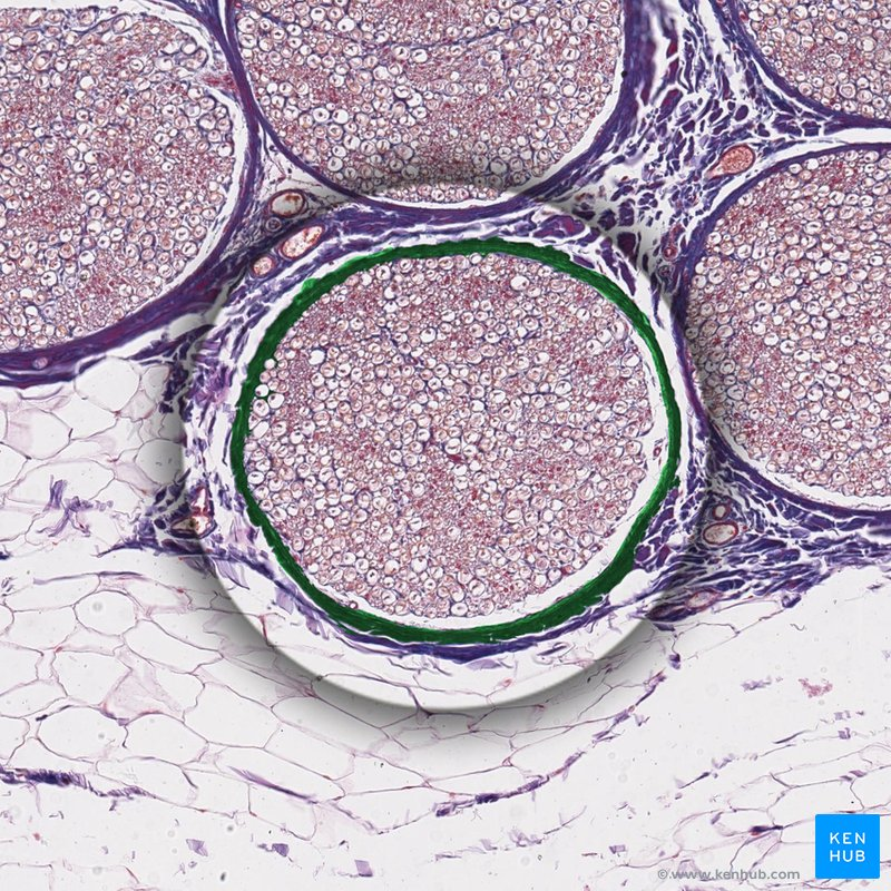
sympathetic (autonomic) ganglion
cluster of nerve cells in sympathetic nervous system acting as relay station between central NS and target organs (controls fight or flight)

neuron
excitable neural cell in nervous system that transfer nerve impulses through electrical and chemical signals

dendrites
a neuron's bushy, branching extensions that receive messages and conduct impulses toward the cell body (soma)

cell body (soma)
bulbous, central part of a neuron that coordinates information-processing tasks and keeps the cell alive; contains nucleus and other cell organelles

axon hillock
cone shaped region of a neuron cell body where it joins the base of the axon

axon
the long threadlike part of a nerve cell along which impulses are conducted away from the cell body to other neurons, muscles, or glands

schwann cells
primary glia in the PNS, Supporting cells of the peripheral nervous system responsible for the formation of myelin; insulate, support, and help regenerate nerve cells

myelin sheath
a fatty, insulating tissue layer segmentally encasing the axons of some neurons; enables vastly greater transmission speed as neural impulses hop from one node to the next

nodes of ranvier
a gap in the myelin sheath of a nerve, between adjacent Schwann cells; where ion exchange occurs to regenerate nerve cells

axon terminals
branches at the end of the axon that contain tiny pouches, or sacs, called synaptic vesicles; release neurotransmitters across synapse to communicate with target cells

cerebrum
largest and uppermost part of the brain; responsible for voluntary muscular activity, vision, speech, taste, hearing, thought, and memory

cerebral cortex
outer region of the cerebrum, containing sheets of nerve cells; has heavily folded ridges (gyri) and grooves (sulci) increase surface area for higher level processing; gray matter of the brain

white matter
deep brain tissue made of myelinated axons that connect different regions of cerebral cortex and lines the cerebrum to rest of nervous system

frontal lobe
associated with reasoning, planning, parts of speech, movement, emotions, and problem solving

parietal lobe
receives sensory input for touch, pressure, temperature, pain, and body position
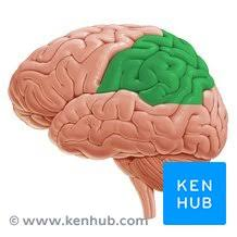
temporal lobe
memory, hearing, language comprehension, facial recognition

occipital lobe
interpreting visual information from eyes (color, shape, motion)

corpus callosum
the large band of neural fibers (200-300 million) connecting the two brain hemispheres and carrying messages between them

gyri
ridges of the brain on cerebral cortex

sulci
shallow grooves that separate gyri in cerebral cortex

central sulcus
separates frontal and parietal lobes; sits between motor and sensory areas

lateral sulcus
separates the parietal and frontal lobes from temporal lobes

parieto-occipital sulcus
separates the parietal lobe from the occipital lobe on medial surface of brain

fissures
deep grooves in the brain; divide brain into distinct hemispheres and functional lobes

longitudinal fissure
separates left and right hemispheres; contains falx cerebri

transverse fissure
separates cerebrum from cerebellum and brainstem

diencephalon
core structure of brain located deep inside the head; sits just above brainstem and between cerebral hemispheres

thalamus
largest section of diencephalon; acts as mater sorting and relay station for sensory and motor signals to and from the cerebral cortex

hypothalamus
below thalamus; regulated body's internal balance, autonomic function, hunger, thirst, sleep wake cycles, and endocrine system via pituitary gland

pineal gland
small pine cone shaped endocrine gland located deep in the center if brain; produces secretes melatonin through pinealocytes

pituitary gland
pea sized endocrine organ at base of brain, behind bridge of nose; master gland

anterior pituitary
growth hormone, thyroid-stimulating hormone, adrenocorticotropic hormone, prolactin, luteinizing hormone, and follicle-stimulating hormone

posterior pituitary
antidiuretic hormone and oxytocin

brain stem
stalk-like lower part of the brain; connects the cerebellum and cerebrum to spinal cord

midbrain
top section of brainstem handling vision, hearing, motor control, and eye movements

pons
middle section of brain stem, bridging cerebrum and cerebellum while managing facial movements, balance, and sleep cycles

medulla oblongata
bottom section of brainstem; connects directly to spinal cord and manages life-sustaining reflexes

reticular formation & reticular activating system (RAS)
complex net-like network of interconnected nuclei and nerve fibers running through core of brainstem; RAS runs within it and is dedicated to controlling arousal, wakefulness, sleep-wake transitions, and conscious awareness

cerebellum
major structure of the hindbrain located at back of skull, beneath cerebral cortex and brainstem that controls fine motor skills; has 3 functional regions

arbor vitae
branched, tree-like network of white matter in cerebellum at lower back of brain; made of axons covered in myelin sheath

ventricles
4 interconnected, fluid filled cavities that produce, store, and circulate cerebrospinal fluid (CSF) to cushion and protect brain and spinal cord

lateral ventricles (1st and 2nd)
largest ventricles; one in each cerebral hemisphere featuring distinct horns (frontal, temporal, occipital)

3rd ventricle
narrow, slit-like ventricle along midline of brain between the two halves of the thalamus

4th ventricle
lower hindbrain ventricle; between brainstem (pons and medulla) and cerebellum

cerebrospinal fluid (CSF)
plasma-like clear fluid circulating in and around the brain and spinal cord; cushions and protects within CNS

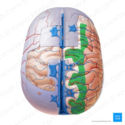
meninges
three protective membranes that surround the brain and spinal cord

dura mater
tough, thick, outermost layer of the meninges; lines inside of skull featuring 2 fused layers (periosteal and meningeal) and forms dural folds like the flax cerebri and tentorium cerebri (control stability)

arachnoid mater
weblike middle layer of the three meninges; features filament called trabeculae and houses finger-like projections (arachnoid granulations) that absorb CSF into nervous system
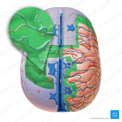
pia mater
thin, delicate inner membrane of the meninges; tightly shrink-wraps and contours directly to the surface of the brain, following every fold and carrying blood vessels into the tissue