PSYC 3220: Mod 2
 Call Kai
 Learn
Learn Practice Test
 Spaced Repetition
 Match
Match Flashcards
 Knowt Play
1/125

There's no tags or description
Looks like no tags are added yet.
Name | Mastery | Learn | Test | Matching | Spaced | Call with Kai | Chat |
|---|
No analytics yet
Send a link to your students to track their progress
126 Terms

how is the visual pathway of humans different compared to animals (prey)?
both our eyes are in the front, so each eye sees the left and right side of the world, with only a fraction being exclusive to each eye

how does the visual pathway of humans work?
parts of the left visual fields from both eyes cross onto the right hemisphere of the brain, and parts of the right visual field of both eyes cross onto the left hemisphere of the brain
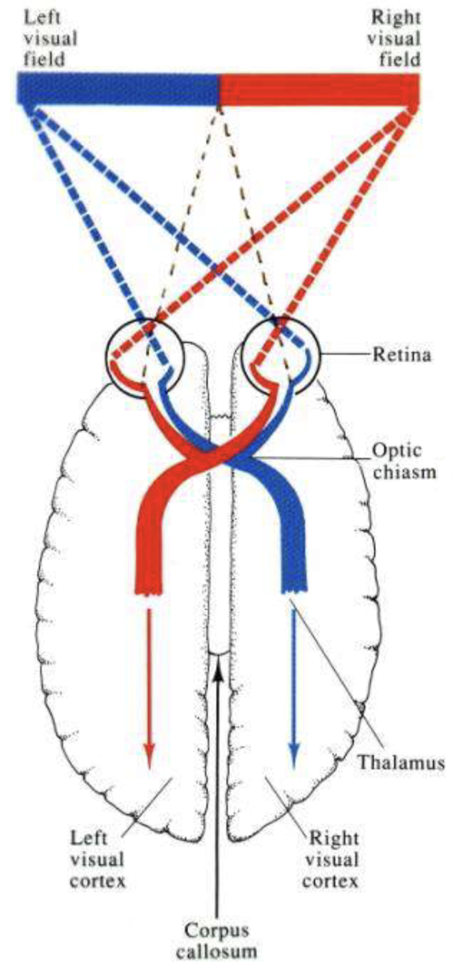
![<p>What if you had an injury that cut off one of your optic nerves [e.g left optic nerve while left eye works fine still]?</p>](https://assets.knowt.com/user-attachments/67afcff1-a05b-4cb0-baaa-94996f88fc2d.png)
What if you had an injury that cut off one of your optic nerves [e.g left optic nerve while left eye works fine still]?
Regardless of eye function, the information goes no where so you have no response from the eye. Many people can have this and not notice because they have a working optic nerve in the other eye.


What if you had a tumor in your chaism?
You lose all the fibers that cross, losing the inside vision of your two eyes. But because of the way it criss crosses, the result will be not being able to see outer part of eyes. The left world ends up on right side of left eye and vice versa.


What if you had an aneurysm on the left side of the chaism (internal carotid)?
You knock out only one of the four pathways, you lose the outside of the left eye which from from the right world. Many people could easily not realize they have this.


What would happen if you lost one of your optic tracts?
You would lose either the entire left world in both eyes or vice versa (losing both the uncrossed from the right eye and what is crossed from the left eye), becoming truly half blind.

Where do the optic nerve fibers lead to?
Around 90% of the retinal ganglion cells in the eye end up at the 2 LGNs, rest goes to superior colliculus (primitive side of brain)
What does the superior colliculus do?
Where a minority of the retinal ganglion cells send information, handles blinking, pupil size, very fast and primitive perception to help you survive
blindsight
eyes send information just fine, but the brain isn’t getting the message due to brain damage (side note: possible for superior colliculus to allow blind people to perceive moving sitmuli unconsciously)
Each LGN recieves information from ____ and eventually sends the information to ____
both eyes, due to optic crossing (chiasm), visual cortex
where do we have conscious perceptions?
visual cortex

Layers 1-2 in LGN
produce dorsal (top) pathway and go to a different part of the brain later on than the other layers, has larger cells (mostly from rods since rods have more neural convergence so you need physically larger cells to grab all the connections you need), useful for information that gives sensitivity, motion, “where”

We know that the world is organized spatially and that the image on the retina is also organized spatially, how is the information organized on the LGNs?
Spatially, if I stimulate the image on the retina, e.g A, it will stimulate cell A in the LGN. We get another copy of the image in the brain now. However the LGN has 6 layers so it is 6 copies per layer, per LGN.
Each layer in an LGN contains information from
only one eye, the information is segregated at this point and has not been put together
Layer 3-6 in LGN
smaller cells, mostly from cones, pick up on details (e.g to read small letters or pick out a car in a parking lot), color perception comes largely from temporal lobes so it makes sense that it sends information downwards towards that area, resolution, color, what
why are the layers in the LGN different?
they specialize in different things, multiple copies because there are multiple purposes for information
what are the two chromatic channels for color opponency in the LGN?
blue/yellow and red/green, these exact combinations are what we pit against each other to help us find things in the world
in the retina we had 3 cone pigments, what changes in the LGN?
we add a new one, yellow, which the brain wires itself to add during development
color contrast
the same wavelength of light can appear to be a different color depending on what color its next to, e.g blue looks bluer next to yellow, and red looks reddish next to green, determines what colors are complementary

what determines which colors are complementary to one another?
the way the brain is wired determines what complementary colors are (color contrast), blue is across from yellow on the color wheel, green is across from red, etc. but there is no blueish-yellow or reddish green (we can have blueish-red = purple), because these are opponent processes

Explain how this opponent cell in LGN works
A single cell can be excited OR inhibited, depending on both the color of light and where that light falls. The center has two possible responses: red light hits the center → cell is excited (+). Green light hits the center → cell is inhibited (−). The surround also has two possible responses: green light hits the surround → cell is excited (+). Red light hits the surround → cell is inhibited (−).

The red apple falls in the center of this receptive field, while the green leaves fall in the surround
Both regions provide excitatory input to this particular cell, showing that makes the neuron especially responsive to certain color boundaries, and higher visual areas can use this to distinguish objects
How are cone inputs combined to produce the two main color-opponent channels?
L versus M cones: the brain compares long-wavelength and medium-wavelength cone signals. Example: R+G− means red-ON, green-OFF.
S versus combined L and M cones: short-wavelength input is compared against a combination of long- and medium-wavelength inputs.
Example: B+Y− means blue-ON, yellow-OFF.
Combined L and M cones because yellow is found in between them

How do single-cell recordings reveal color opponency in LGN neurons?
Researchers record a single LGN neuron's firing rate while presenting different wavelengths of light (color) to its receptive field. For a red-ON/green-OFF center, red increases firing and green decreases firing, demonstrating that different wavelengths can excite or inhibit the same neuron.


How do single-cell recordings produce the blue-yellow opponent-response graph?
Researchers shine different wavelengths of light one at a time while recording the same LGN neuron's firing rate. For a B+Y− neuron, blue wavelengths increase firing (excitation), while yellow wavelengths decrease firing (inhibition). They plot the responses across wavelengths to show color opponency.
Color opponency results in ___ and ____
color adaptation, afterimages, for example, if you stare at something blue for too long and look away, you will see a yellow version or moving in the opposite direction if it was motion based
how does color opponency result in after images
the opponent process is like a scale, if a shirt is 50 units of blue and you have stared at it, you adapted to that color, causing you to perceive less of that color, tipping the scale up by 10. You look away to the white wall, removing the 50 units of blue, however on the other end of the scale, you have tipped up by 10 units. You get the yellow afterimage on the white wall.
visual cortex
found in the back of the head, processes vision but is not the final place, it sends that to other parts of the brain
cortical magnification
parts of your vision magnified in your brain, we have a much larger area of the brain processing center (fovea), this doesn’t mean you see the world as distorted, but that you have more brain power dedicated to processing that information
80% of the cortex is devoted to the ___ 10% of the visual field
central

spatial organization and cortical magnification on the visual cortex
world is still laid out spatially, but magnification is occurring
why were single cell recordings in visual cortex groundbreaking
first time scientists were measuring activity at the level of individual neurons
preliminary cells to get information in the visual cortex
circular receptive fields similar to LGN cells, require having light in the right location to fire, pass information to other cells in cortex

second layer of cells to get visual cortex information
simple cells, light has to be in the right location and a type of bar or edge (vertical, horizontal, oblique)

why was it so surprising to discover simple cells?
They expected to only find circular receptive fields. But they saw responses for all types of lines (bright lines, dark lines, edges, diff orientations).
third layer of cells to get visual cortex information
complex cells, require light to be in certain location, certain orientation, and has to be moving in a particular direction as well.

fourth layer of cells to get visual cortex information
end-stopped cells, require light to be in the right location, right orientation, going in a specific direction, and a particular length, width or corners of particular angles

How did Hubel & Wiesel perform single cell recordings on the visual cortex?
perpendicular penetrations of the cortex, sticking probe into cortex and latching onto first neuron, seeing what orientation that neuron is looking for, then probing further (column of neurons)
oblique penetration of the cortex, going sideways across the cortex to get a row of columns with progression of preferred orientations

what did Hubel & Wiesel’s perpendicular penetration of the cortex reveal?
we get a column of cells with the same prefered orientation and overlapping receptive fields


What did Hubel and Wiesel find using oblique penetrations of the visual cortex?
Moving the electrode sideways across the cortex revealed columns of neurons with changing orientation preferences.
In one direction, neurons preferred different angles but had overlapping receptive fields (same general location).
In another direction, we get a row of columns with a progression of receptive fields, this is where we get the full image by mapping out the world side to side


ocular dominance columns
Alternating columns of neurons that respond more strongly to input from either the left or right eye, helping the visual cortex organize information from both eyes.
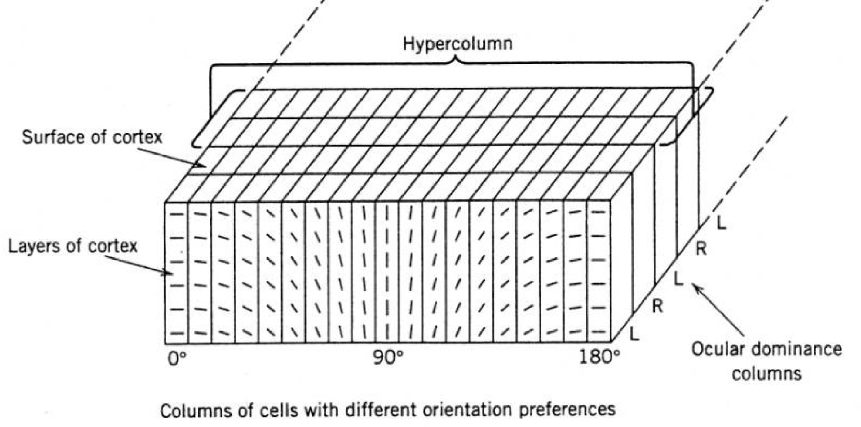
hypercolumn
a slice of the cortex, in one direction its the left and right eye region, and in another direction its the full circle of orientations
what are binocular cells, where are they found?
cells that look at both eyes simultaneously (complex cells and beyond), found in visual cortex in the ocular dominance columns, sums inputs from the two eyes and achieves depth perception, giving you 3-d vision

There were some cells in single cell recording on the visual cortex, that did not respond no matter what light or where we shined it. We only found a small bit of activity in one tiny spot. What does this mean?
This cell is binocular, it has a receptive field in each eye and is looking for the light to fall on both receptive fields at the same time. Its an individual neuron looking for a particular point in the 3-d space.
When are binocular cells developed
During childhood, no one is born with them
When are cortical cells developed
during childhood, in experiments where monkeys or cats were raised in cages, they never developed ability to make sense of different angles, or with cats only exposed to vertical lines, there was no ability to detect horizontal lines
ventral pathway
the what pathway, where you identify what something is, includes color and shape perception, naming
as we go through pathways in the brain we look at things
more and more particularly
single cell recordings in temporal cortex
we see individual cells that are extremely specific, such as only looking for a Y in a certain direction or stripes of specific brightnesses, they identify what something is


What does this image show
Cells responding to individual colors with spatial organization, we see the rainbow laid out on the ventral pathway
A monkey loses their temporal lobes, how does this affect their ability to perceive what things are and where things are? Assume the monkey’s tasks are to pick a triangle block instead of a square block to get a treat
They struggled to learn which object contained food because their object recognition was impaired (picked randomly every time), but they could still perform the location-based task.
Shows ventral damage mainly affects WHAT an object is.
double disassociation
damaging one pathway impaired one type of visual task while leaving the other relatively intact
the ventral pathway is ___ identification, the dorsal pathway is ___ action
conscious, unconscious, using vision to make motor control work
Researchers damaged the parietal lobes (dorsal pathway) of monkeys. The monkeys were tested on remembering which location contained food. What happened?
They struggled with the location-based task but could still learn which object contained food.
Shows dorsal damage mainly affects WHERE an object is.
A woman with ventral pathway damage was asked to rotate a card to match the angle of a slot. What happened?
She struggled to match the slot's orientation because her conscious perception of its angle was impaired.
Shows the ventral pathway helps with consciously judging what we see.
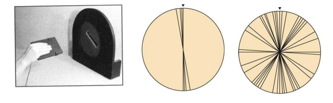
Woman with ventral pathway damage was asked to quickly insert a card into the slot. What happened?
She could correctly rotate and insert the card, even though she couldn't accurately report the slot's orientation.
Shows the dorsal pathway can guide actions using visual information without accurate conscious perception.

What can happen when a person has damage affecting the dorsal pathway but relatively preserved ventral processing?
They may recognize an object and its orientation but struggle to accurately reach toward or grasp it. Conscious recognition and visually guided action can be impaired separately
Healthy participants briefly viewed a target, then closed their eyes and both reported its distance and reached toward it. If both responses came from one shared perception, what would researchers expect?
Their errors should be correlated. If they thought the target was too close, both their distance report and reach should tend to be too short.
In the healthy human experiment comparing distance reports and reaching movements, were the errors correlated?
No. Errors in conscious distance reports did not reliably predict errors in reaching. Visual information used for conscious judgments and actions may be processed separately.
Researchers told healthy participants to expect targets in a closer distance range. What happened to their distance judgments and reaches?
Participants tended to report targets as closer, but their reaches changed very little.
Shows expectations can bias conscious perception without similarly affecting visually guided action.
Researchers told healthy participants to expect targets in a farther distance range. What happened to their distance judgments and reaches?
Participants tended to report targets as farther away, but their reaches remained relatively unchanged. Shows conscious distance judgments were more affected by expectations than reaching movements.
What did the follow-up experiment manipulating expected target distance demonstrate?
Expectations changed conscious distance judgments much more than reaching.
This supports the idea that conscious perception and visually guided actions use partly separate visual processing systems.
How does the magnocellular pathway connect to the dorsal visual pathway?
The magnocellular layers (1–2) of the LGN contain large neurons specialized for rapid changes and motion. This information contributes strongly to the dorsal pathway, which processes motion and helps guide actions.
What does the existence of specialized cortical areas within the dorsal and ventral pathways tell us about visual processing?
Each pathway contains smaller areas specialized for different tasks (motion, color, faces, object recognition, guiding actions, etc.).
Damage to one area can impair a specific ability while leaving others intact, showing that visual processing is divided into specialized systems.
a seperate cortical area includes face detection, why?
we evolved a separate piece of our brain to recognize faces as opposed to objects because we don’t want to miss it, and we will sacrifice false positives for this (e.g illusions that appear to be faces), shows how the brain hates randomness and tries to make sense of it in any way possible
how does the brain apply a mental razor to perceiving objects from a retinal mosaic?
through perceptual segregation, groups parts into objects, and segregates parts from other objects
goals of structualism
discover the basic elements of mental experience, discover laws for their combination into complex mental experiences
atomisitc
basic, tiny building blocks of perception and trying to understand how they go together
introspection in structuralism
analysis of one’s own conscious experiences, not really a viable scientific technique, failed and was replaced by Behaviorism in America and Gestalt psychology in Europe
gestalt psychology
perception is holistic, following configurations, its the relationship between the parts that determines the perception, not the parts themselves (e.g in music, we don’t hear exact notes or instrument, but can identify the song/harmony)
parts are perceived differently depending on the whole through
lightness constancy, lightness contrast, color contrast, etc.

Law of Pragnanz
gestalt organization principle responsbile for perceptual segregation, meaning good figure, simplicity, the brain follows laws of nature and laws of nature follow pragnanz, your brain will naturally organize things to have a good pragnanz
what does the law of Pragnanz mean for the ghost in the machine?
A soul/ghost doesnt follow the laws of nature, humans started to think the mind and brain could be some form of machinery that has to follow laws of Newton
What are the main Gestalt organizing principles?
Prägnanz (simplicity), proximity, similarity, good continuation, closure, and common fate
What is the Gestalt principle of proximity?
We group things that are close together as belonging together.
What is the Gestalt principle of similarity?
We group things that look alike, such as objects with the same color or shape.
What is the Gestalt principle of good continuation?
We tend to see lines and patterns as continuing smoothly, even when interrupted.
What is closure, and how does it explain illusory contours?
The brain fills in missing edges to see a complete shape, like an illusory triangle.
What is the Gestalt principle of common fate?
We group things that move together as part of the same object, like dots moving in one direction.
figure ground problem
you have to perceive a figure as distinct and seperate from its background

contrast sensitivity
key element to perceiving objects, helps you perceive the contrast between objects, associated more with dorsal stream

acuity
ability to resolve small details, associated more with ventral stream
what is crucial for nighttime visibility, especially for pedestrians
contrast, drivers use ambient light to percieve different objects, and if your clothes do not reflect off enough of that contrast, the driver wont see you in time or at all
what is the medium for hearing perception
mechanical energy in the form of sound waves (traveling through air, water and solids)
you hear in terms of
events
amplitutde
loudness, height of wave (more ampl —> louder sound)
frequency
pitch or tone (higher frequency —> higher pitch)
complex wave/sound
several sound waves added together, with a fundamental frequency and overtones (simple waves)

Fourier analysis
mathematical procedure for taking complex wave and breaking it down into parts, and determining which individual sound waves make up the wave
fundamental frequency
the lowest frequency of a complex tone (NOT LOUDEST or HIGHEST AMPLITUDE)

harmonics
waves that accompany the fundamental frequency

timbre
quality given to a complex tone by its harmonics (almost like different color/flavor that we sense with instruments)
what are the main steps involved in hearnig
sound waves -→ outer ear collects → middle ear amplifies → inner ear transduces → brain processes

What is the role of the outer ear?
Collects sound waves from the environment and funnels them toward the eardrum.
What is the role of the middle ear?
Uses the eardrum and three small bones to transfer and amplify vibrations into the inner ear.
What are the three middle-ear bones?
Hammer (malleus), anvil (incus), and stirrup (stapes). They act as levers.
Why does the middle ear need to amplify sound?
Air vibrations must move fluid in the cochlea, which requires efficient energy transfer.
Why do middle-ear muscles contract during loud sounds?
They reduce vibration transmission, helping protect the inner ear from sustained loud sounds.
What is the function of the Eustachian tube?
Equalizes air pressure across the eardrum and helps drain fluid from the middle ear.
What is the role of the inner ear (cochlea)?
Converts mechanical vibrations into neural signals and separates sound into frequencies.