approach to abd imaging + parenchyma
 Call Kai
 Learn
 Practice Test
 Spaced Repetition
 Match
 Flashcards
 Knowt Play
1/39

There's no tags or description
Looks like no tags are added yet.
40 Terms
Radiology vs US
R- better for size + shape
US- better for internal architecture + lumens (unless gas)
Purpsoe of Low kV high mA technique in Rx
Avoid scatter, improve contrast resolution
How to improve abd rx - when to take pic
Exp pause
How to improve abd rx - View
VD, R lat
Positive vs negative contrast media
P- white (I, Barium)
N - black (air, CO2, N2O)
Contrast medium - issue with barium
GIT only (oral ROA)
Granulomatous infl in body cavities (dont use if rupture)
Contrast medium - When to use iodinated
GIT, urinary, IV, myelography
Contrast medium - Safest type
Non ionic low osmolarity iodinated
Contrast medium - iodinated side effects
Anaphylaxis, shock in body cavity (better than barium), local irritant, kidney failure
Contrast medium - Side effect of gas
Air embolism
What state of matter does not work with US
Gas
Which to do first - US or rx + why
Rx - no gel artefact
Which cases need CT as blind spots
Pelvic canal
Surgical access assessment
Vascular malformations
Ectopic ureters
Röntgen signs
Number
Size
Shape + margins
Opacity
Location

Opacity types 5
Gas
Fat (dark)
Soft tissue/ fluid (efface fat)
Bone/ mineral
Metal

Mass effect shows what
Swelling, mass, space occupying lesions → displacement

Types of mineralisation + causes
Dystrophic (focal) - tissue damage
Metastasic (diffuse) - hypercalcaemia
Other - urinary calculi, ingesta, osseous neoplasia

Which vit toxicity can lead to metastatic mineralisation
Vit D
Common mineralisation in old cats
Dystrophic from adrenal damage

Abd rx - peritoneum appearance
Some fluid
Fat in omentum + retroperitoneum

Abd rx - Causes of loss of serosal detail
Lack of fat
Peritoneal fluid
Carcinomatosis/ sarcomatosis (many metastatic micro nodules)
Brown fat (higher water content)
Peritonitis
Visceral crowding
Age brown fat disappears
4-6mon

Abd rx - Causes of pneumoperitoneum
Post sx (<4w)
Ruptured GIT/ septic abd
Penetrating trauma
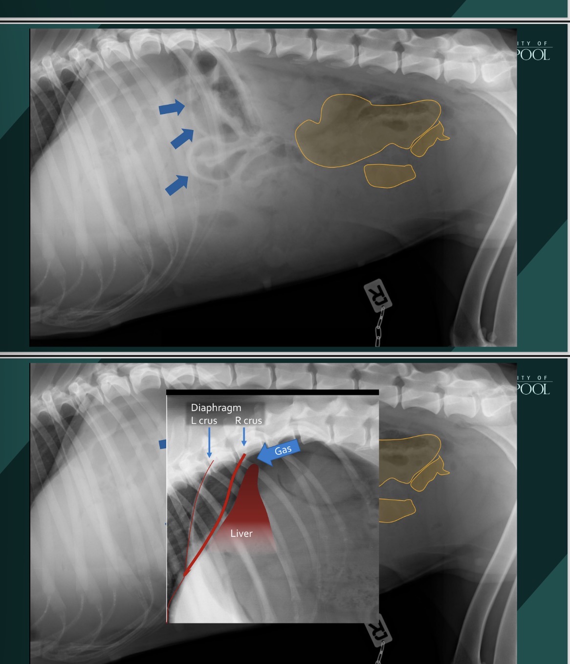
Abd rx - Pneumoperitoeum appearance
Inc contrast between gas+ soft tissue → inc serosal surface brightness
Gas bubbles dont conform to GIT
Abd rx - Common location of pneumoperitoenum
Diaphragm + dor liver margin
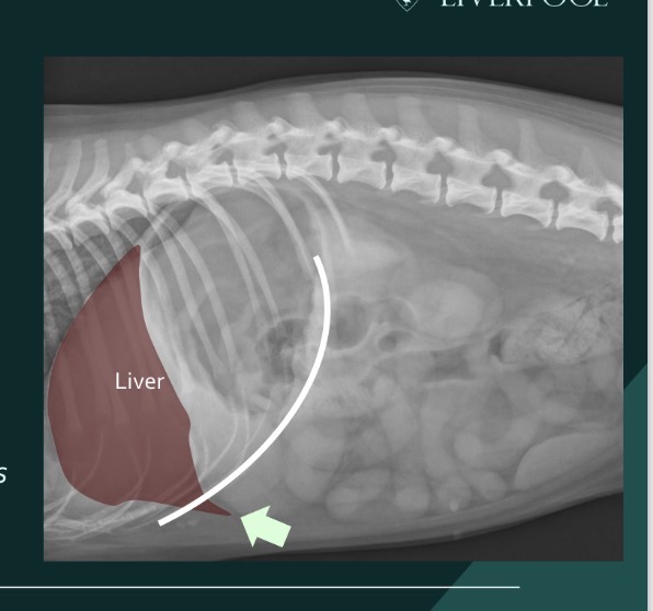
Abd rx - Normal liver appearance
Cr
Acute Vencd angle
Pokes past costal arch

Abd rx - Gastric axis
Point in middle of fundus → middle of pylorus
Shows size of liver (displaced axis)

Abd rx - Hepatomegaly appearance
Rounded margins
Extend further cd to costal arch
Cd displaced gastric axis
Mass effect

Abd rx - Causes of hepatomegaly
General - nodular hyperplasia, lymphoma, mets, acute hepatitis, metabolic hepatopathy
Focal - neoplasia, abscess

Abd rx - Microhepatia appearance
Contained within costal arch
Little soft tissue cr to stomach
Cr displaced gastric axis

Abd rx - Microhepatia causes
Portosystemic shunt, primary portal v hypoplasia
Chronic hepatitis, fibrosis, cirrhosis

Abd rx - Normal spleen (tail not visible in cat)
Tail - triangle cd to liver (mobile)
Head - triangle on L superimposing R kidney (fixed)
Body - L


Abd rx - Splenomegaly appearance
Rounded margins
Tail cd displaced (visible in cats)

Abd rx - Splenic mass vs general splenomegaly causes
M- neoplasia (haemangiosarcoma, MCT), haematoma, nodular hyperplasia
G- sed, GA, infiltration (lymphoma), infl (hyperplasia, splenitis)

Retroperitoneum - normal
Dor to peritoneal cavity
Contain kidney, ureter, bladder neck, great vessels, adrenal glands, LN, prostate
Continues with mediastinum + aorta
Fat opacity + streaky

Retroperitoneal mass causes
Any organ (kidney, LN)
Fluid (effusion, H)

Adrenal glands - normal
Not visible
Mineralise in old cats


Adrenal glands - When does mineralisation happen
Cat - old
Dog - neoplasia (adenoma, carcinoma, phaeochromocytoma)

Abd LN - opacity (normally not visible)
Soft tissue

Abd LN - List of them
Retroperitoneal - para aortic, renal, med iliac, int iliac, sacral
Peritoneal - gastric, pancreaticoduodenal, splenic, jejunal, colic