L56 - kVP, mAS, Image Storage
 Call Kai
 Learn
Learn Practice Test
 Spaced Repetition
 Match
Match Flashcards
 Knowt Play
1/74

There's no tags or description
Looks like no tags are added yet.
Name | Mastery | Learn | Test | Matching | Spaced | Call with Kai | Chat |
|---|
No analytics yet
Send a link to your students to track their progress
75 Terms
What are the Three Major Factors of a High-Quality Radiographic Image?
Appropriate exposure
Appropriate contrast/latitude
Low scatter radiation
What is Radiographic Exposure?
The amount of ionization in air produced by radiation
Associated with overall image blackness and opacity

What is the Difference Between an Overexposed and Underexposed Radiograph?
Overexposed: Too dark/black
Underexposed: Too light/white

What Three Factors determine Overall Radiographic Exposure?
mAs (milliampere-seconds)
kVp (peak kilovoltage)
Focal spot-film distance (FFD)
What does mAs stand for, and How is It Calculated?
mAs = strength of electric current within given time
mAs = milliamperes (mA) × exposure time (seconds)

What is the Primary Function of mAs in Radiography?
Controls the quantity (number) of X-ray photons produced
How does Increasing mAs affect X-ray Production and Image Exposure?
Increasing mAs produces more X-ray photons → increasing detector exposure and image blackness

What is the Relationship Between mAs and Radiation Exposure?
A linear relationship: doubling mAs doubles X-ray photon quantity and radiation exposure
How does Increasing mAs affect Motion Artifacts?
If exposure time increases, motion artifacts increase because the patient has more time to move during the exposure
Why does Increasing mA Without Changing Exposure Time NOT Increase Motion Artifacts?
Because exposure duration remains unchanged, even though more X-ray photons are produced
How should mAs be Adjusted for Underexposed vs. Overexposed Images?
Underexposed → Increase mAs
Overexposed → Decrease mAs
Does Changing mAs Directly affect Radiographic Contrast?
No
mAs primarily controls X-ray quantity and overall exposure, not contrast
What does kVp stand for, and What does It represent?
Peak kilovoltage
The voltage difference between the cathode and anode of the X-ray tube
What is the Primary Function of kVp in Radiography?
Controls X-ray photon energy, penetration, and radiographic contrast
How does Increasing kVp affect X-ray Energy and Penetration?
Higher kVp → Faster electrons → Higher-energy X-rays → Greater tissue penetration
How does Increasing kVp affect Overall Image Exposure?
Greater penetration allows more X-rays to reach the detector → increasing image blackness

How should kVp be Adjusted for Underexposed vs. Overexposed Images?
Underexposed → Increase kVp
Overexposed → Decrease kVp
What is the Major Difference Between kVp and mAs?
kVp: Controls X-ray energy and contrast
mAs: Controls X-ray quantity and exposure time
What is Focal Spot-Film Distance (FFD)?
The distance between the X-ray focal spot (anode) and the film/detector
What does the Inverse Square Law state?
Radiation intensity decreases with the square of the distance from the source.
I ∝ 1/d².

What happens to X-ray Intensity When FFD is Doubled or Tripled?
Double distance → ¼ of original intensity
Triple distance → ⅑ of original intensity
Why is the Inverse Square Law Important for Radiation Safety?
Increasing distance reduces radiation exposure, supporting ALARA (As Low As Reasonably Achievable)
What is Radiographic Contrast?
The difference in radiographic opacity between adjacent structures
Black and White

What is Radiographic Latitude?
The range of tissue densities displayed as useful shades of gray
Shades of Grey

What is the Difference Between High Contrast and High Latitude?
High contrast: Strong black-and-white differences with fewer shades of gray
High latitude: More shades of gray, allowing subtle tissue differences to be visualized

Which Machine Setting Primarily Controls Contrast and Latitude?
kVp
How does Low kVp affect Contrast and Latitude?
Low kVp → High contrast → Low latitude
How does High kVp affect Contrast and Latitude?
High kVp → Low contrast → High latitude
Why does Low kVp produce High Contrast?
Lower-energy X-rays favor photoelectric absorption, which depends strongly on tissue atomic number and produces greater differences in tissue absorption
Why does High kVp produce High Latitude?
Higher-energy X-rays penetrate tissues more readily, reducing differences in absorption and producing more shades of gray.
Why can Lowering kVp Alone cause an Underexposed Image?
Lower kVp reduces X-ray energy and penetration → so fewer photons reach the detector
How can Contrast be Increased While Maintaining Overall Exposure?
Decrease kVp and increase mAs to compensate for reduced penetration
How can Latitude be Increased While Maintaining Overall Exposure?
Increase kVp and decrease mAs to compensate for increased penetration
What is the 15% Rule in Radiography?
A 15% change in kVp produces approximately the same change in detector exposure as doubling or halving mAs
What happens When kVp is Increased by 15%?
Detector exposure approximately doubles
What happens When kVp is Decreased by 15%?
Detector exposure approximately halves
How can kVp and mAs be Adjusted to Increase Contrast Without Changing Exposure?
Decrease kVp by approximately 15% and double mAs

How can kVp and mAs be Adjusted to Increase Latitude Without Changing Exposure?
Increase kVp by approximately 15% and halve mAs
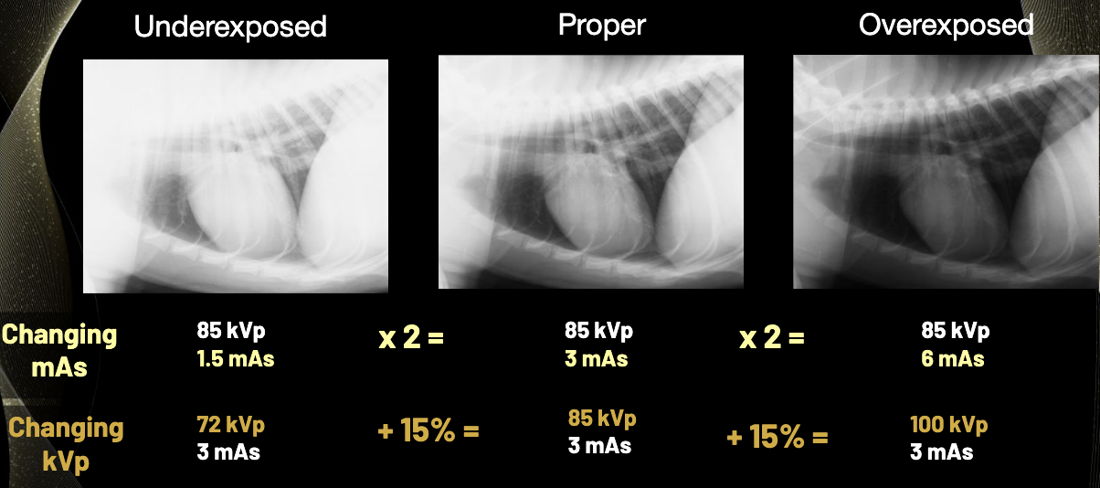
A Radiograph is Underexposed at 85 kVp and 1.5 mAs. How can Exposure be Corrected Using mAs?
Double mAs from 1.5 to 3 mAs while maintaining 85 kVp
A Radiograph is Overexposed at 85 kVp and 6 mAs. How can Exposure be Corrected Using mAs?
Halve mAs from 6 to 3 mAs while maintaining 85 kVp
What kVp and mAs Technique is Preferred for Thoracic Radiographs?
High kVp + Low mAs
Why is High kVp Preferred for Thoracic Radiographs?
The thorax already has natural contrast between air, soft tissue, and bone
Higher kVp increases latitude and improves visualization of subtle pulmonary structures
Why is Low mAs Preferred for Thoracic Radiographs?
Lower mAs allows shorter exposure times, reducing motion artifacts from respiration
At What Respiratory Phase should Thoracic Radiographs be Taken?
End inspiration → when the lungs are maximally inflated
Why can Excessively Low kVp be Problematic for Thoracic Radiographs?
Excessively high contrast can obscure peripheral pulmonary vessels and small nodules
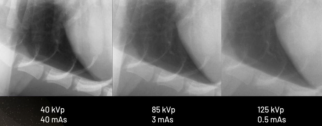
Why can Excessively High kVp be Problematic for Thoracic Radiographs?
Excessively low contrast can reduce bone detail and visibility of rib margins

What kVp and mAs Technique is Preferred for Abdominal Radiographs?
Low kVp + Higher mAs

Why is Low kVp Preferred for Abdominal Radiographs?
It increases contrast between abdominal organs and surrounding fat, improving visualization of organ margins
Why is Higher mAs Often Needed for Abdominal Radiographs?
To maintain adequate detector exposure when using lower kVp
At What Respiratory Phase should Abdominal Radiographs be Taken?
Near end expiration → when the diaphragm moves cranially and provides more space for abdominal organs
What is Scattered Radiation, and How does It affect Image Quality?

What Three Patient or Imaging Factors Increase Scattered Radiation?
Greater tissue density
Greater tissue thickness
Larger X-ray field size
Higher X-ray energy also increases scatter contribution
What is the Most Important Controllable Factor for Reducing Scatter Radiation?
Field size, controlled through collimation

What is Collimation, and Why is It Important?
Restricting the X-ray beam to the anatomical region of interest
It reduces scatter, improves contrast, and decreases patient radiation dose.

How does a Bladder Paddle Shot Improve Radiographic Image Quality?
Compression displaces overlapping intestinal loops
Decreases tissue thickness
Reduces scatter
Improves visualization of urinary bladder stones
What is the Function of an Anti-Scatter Grid?
Absorbs scattered X-rays traveling at different angles while allowing more primary radiation to pass toward the detector, improving image contrast

Why must mAs be Increased When Using an Anti-Scatter Grid?
The grid absorbs some useful primary X-rays, so mAs must be increased to maintain adequate detector exposure without unnecessarily changing contrast.

Why may Anti-Scatter Grids be Unnecessary for Very Small Patients?
Small patients, such as kittens, puppies, and birds, produce less scatter because of their smaller tissue thickness
What are the Three Major Radiographic Image Recording Systems?
Screen-film radiography
Computed radiography (CR)
Digital radiography (DR)

How does Screen-Film Radiography Record and Display Images?
Uses photosensitive film inside a cassette
Intensifying screens convert X-rays into light, exposing silver halide film that is chemically developed and viewed on a light box.

What are the Major Advantages and Disadvantages of Screen-Film Radiography?
Advantages:
Inexpensive equipment
Permanent physical images
High resolution
Disadvantages:
Requires chemical development
Darkroom/storage space
Has low tolerance for exposure errors
How does Computed Radiography (CR) Record Images?
X-rays
Storage phosphor plate
Stored X-ray information
Laser scanning in CR reader
Visible light
Digital image

What is the Major Advantage of Computed Radiography (CR)?
Reusable storage phosphor cassettes that can be erased with intense light after image processing.
What is the Difference Between Direct and Indirect Digital Radiography (DR)?
Indirect DR: X-rays → Visible light → Electronic signal → Digital image
Direct DR: X-rays → Electronic signal → Digital image

What are the Major Advantages and Limitations of Digital Radiography (DR)?
Advantages:
Rapid image acquisition
Post-processing
Higher tolerance for minor exposure errors
Easy image sharing
Limitations:
Expensive equipment
Severely incorrect exposures cannot always be corrected through post-processing
kVp vs. mAs SUMMARY
Feature | kVp | mAs |
Primary function | X-ray energy | X-ray quantity |
↑ Setting | ↑ Energy and penetration | ↑ Number of photons |
↓ Setting | ↓ Energy and penetration | ↓ Number of photons |
↑ Image exposure | Yes | Yes |
↑ Contrast | Decrease kVp | No direct effect |
↑ Latitude | Increase kVp | No direct effect |
Motion artifacts | No direct effect | Increase if exposure time increases |
Radiation exposure | Affects patient dose | Directly proportional to mAs |
Remember:
kVp = ENERGY + CONTRAST
mAs = QUANTITY + EXPOSURE TIME
High vs. Low kVp — Contrast and Latitude SUMMARY
Feature | Low kVp | High kVp |
X-ray energy | Low | High |
Penetration | Low | High |
Contrast | HIGH | LOW |
Latitude | LOW | HIGH |
Image appearance | More black and white | More shades of gray |
Interaction emphasized | Photoelectric absorption | Greater penetration |
Best clinical application | Abdomen | Thorax |
Reason | Organs have similar opacities | Already has natural contrast |
Remember:
Low kVp → High contrast → Low latitude
High kVp → Low contrast → High latitude
The 15% Rule — How to Adjust kVp and mAs SUMMARY
Goal | kVp Adjustment | mAs Adjustment |
Double exposure using kVp | ↑ 15% | No change |
Halve exposure using kVp | ↓ 15% | No change |
Double exposure using mAs | No change | Double |
Halve exposure using mAs | No change | Halve |
Increase contrast, maintain exposure | ↓ 15% | Double |
Increase latitude, maintain exposure | ↑ 15% | Halve |
Important: When changing contrast without changing overall exposure, adjust BOTH kVp and mAs.
Increasing Contrast Without Changing Exposure SUMMARY
Start with a properly exposed image.
Decrease kVp by approximately 15%.
This increases contrast but decreases exposure
Double mAs to compensate
Result: Higher contrast with approximately the same exposure
Increasing Latitude Without Changing Exposure SUMMARY
Start with a properly exposed image.
Increase kVp by approximately 15%.
This increases latitude but also increases exposure
Halve mAs to compensate.
Result: Higher latitude with approximately the same exposure
Thorax vs. Abdomen — Clinical Application SUMMARY
Feature | Thorax | Abdomen |
Preferred kVp | HIGH | LOW |
Preferred mAs | LOW | HIGHER |
Primary priority | Latitude + minimize motion | Contrast |
Natural contrast | High (air, soft tissue, bone) | Low (similar soft-tissue opacities) |
Reason for kVp | Visualize subtle lung structures | Differentiate organs from fat |
Exposure time | Short | Longer tolerated |
Respiratory phase | End inspiration | Near end expiration |
If kVp is too high | Loss of bone detail | Loss of organ contrast |
If kVp is too low | Peripheral lung detail may be obscured | Inadequate penetration if too low |
Remember:
Thorax = HIGH kVp + LOW mAs + INSPIRATION
Abdomen = LOW kVp + HIGHER mAs + EXPIRATION
Scatter Radiation — Factors and Methods of Reduction SUMMARY
Factor/Method | Effect on Scatter | Clinical Importance |
↑ Tissue density | ↑ Scatter | Patient-dependent |
↑ Tissue thickness | ↑ Scatter | More interactions |
↑ Field size | ↑ Scatter | Most important controllable factor |
Collimation | ↓ Scatter | Improves contrast and reduces patient dose |
Compression | ↓ Scatter | Bladder paddle shot |
Anti-scatter grid | ↓ Scatter reaching detector | Improves contrast but requires higher mAs |
Remember: More scatter = lower image contrast
How an Anti-Scatter Grid Works SUMMARY
X-rays pass through the patient.
Some X-rays undergo Compton scattering.
Scattered X-rays travel in different directions.
Metal strips in the grid absorb many scattered photons.
More primary radiation relative to scatter reaches the detector.
Image contrast improves.
Increase mAs as needed to compensate for radiation absorbed by the grid.
Screen-Film vs. CR vs. DR — Image Recording SUMMARY
Feature | Screen-Film | Computed Radiography (CR) | Digital Radiography (DR) |
Recording material | Photosensitive film | Storage phosphor plate | Electronic detector |
Cassette | Yes | Yes | No separate cassette-reading step |
Image viewing | Light box | Computer | Computer |
Processing | Chemical | Laser reader + digitization | Digital |
Reusable | Film: No | Plate: Yes | Detector: Yes |
Image availability | After development | After reader processing | Rapid |
Main advantage | High resolution, inexpensive | Reusable cassettes | Fast, post-processing |
Main disadvantage | Darkroom, storage, artifacts | Separate reader required |
Image Recording Pathways SUMMARY
1. Screen-Film Radiography
X-rays → Intensifying screen → Visible light → Silver halide film → Chemical development → Image viewed on light box
2. Computed Radiography (CR)
X-rays → Storage phosphor plate → Stored information → Laser scanning → Visible light → Digital image
3. Digital Radiography (DR)
Indirect DR: X-rays → Visible light → Electronic signal → Digital image
Direct DR: X-rays → Electronic signal → Digital image