622 11/9 L Autoimmunity and Type 1 Hypersensitivity
 Call Kai
 Learn
 Practice Test
 Spaced Repetition
 Match
 Flashcards
 Knowt Play
1/88

There's no tags or description
Looks like no tags are added yet.
89 Terms
Hypersensitivity
an excessive, harmful response by the adaptive immune system
usually requires exposure to an antigen (sensitization)
4 Main Types of Hypersensitivities
type i
type ii
type iii
type iv
there can be overlap (ex- penecillin allergy)
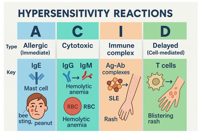
Type I Hypersensitivity (Immediate)
occurs within seconds to minutes, most common, mediated by IgE antibodies and mast cells
Atopy
a predisposition to produce IgE antibodies in response to common environmental antigens and to develop type i hypersensitivity responses
Triggers of Type I Hypersensitivity
allergens (pollen, foods), animal dander (skin cells shed from animals with fur or feathers), insects (cockroaches), insect venom (bee stings), arachnids (dust mites), drugs, fungi
Examples of Type I Hypersensitivity
allergici rhinitis (hay fever), food allergies, drug allergies, allergic asthma (acute or chronic), uricaria, eczema, anaphylaxis

Type II Hypersensitivity (Antibody-Mediated/Cytotoxic)
usually occurs within 2-4 hours, mediated mainly by IgG and IgM anitbodies and complement activation
sometimes cytocoxic
Examples of Type II Hypersensitivity
blood transfusion reactions, autoimmune hemolytic anemia, erythroblastosis fetalis (hemolytic disease of newborns), goodpasture’s syndrome
Type III Hypersensitivity (Immune-Complex Mediated)
usually occurs within hours or days, mediated mainly by deposition of antigen/antibody complexes (IgG/IgM) and complement activation
Examples of Type III Hypersensitivity
systemic lupus erythematosus (SLE), serum sickness, post-infectious glomerulonephritis, arthus reaction
Type IV Hypersensitivity (Delayed-Type/Cell-Mediated)
usually occurs withtin 24-72 hours, mediated mainly by T cells
antibodies NOT involved
Examples of Type IV Hypersensitivity
contact dermatitis (poison ivy reaction), tuberculin skin test, acute cellular organ transplant rejection
Hypersensitivity Reactions Image
2 Phases of Type I Hypersensitivity
sensitization phase
effector phase (early and late)

Sensitization Phase of Type I Hypersensitivity (Simple)
first phase, initial exposure to antigen, asymptomatic, triggers immune memory
Effector Phase of Type I Hypersensitivity (Simple)
second phase, occurs upon subsequent exposure to the same antigen, causes inflammation and/or tissue damage (early effector phase and late effector phase)
9 Steps of Sensitization Phase
initial contact with allergen (skin, lungs, GI tract)
apc (usually dendritic cell) internalizes and processes antigen then migrates to secondary lymphoid site
apc presents peptide fragments with MHC II to naive CD4+ T cells
the naive T cells then differentiate to type 2 helper T cells (Th2 cells)
Th2 cells secrete IL-4, IL-5, and IL-13
IL-4 and IL-13 trigger B cell class switching to IgE
B cells differentiate into long-lived plasma cells and secrete IgE
IgE binds to FceRs on mast cells in tissues and basophils in blood
these mast cells/basophils are now sensitized to the original antigen
What type of cell do CD4+ T cells differentiate into during sensitization of type I hypersensitivity?
Th2 cells
What cytokines do the Th2 cells of the sensitization reaction of type I hypersensitivity secrete?
IL-4, IL-5, IL-13
What does the secretion of IL-4 and IL-13 in the sensitization reaction of type I hypersensitivty lead to?
class swithcing to IgE
What do B cells differentiate into during sensitization of type I hypersensitivity?
long-lived plasma cells that secrete IgE
What is the importance of IgE in the sensitization phase of type I hypersensitivity?
IgE binds to FceR receptors on mast cells in tissues and basophils in blood
this allows the mast cells and basophils to be sensitized to the original antigen
6 Steps of Early Effector Phase of Type I Hypersensitivity
when the same allergen as before is encountered subsequently, it binds and crosslinks the IgE on mast cells and basophils
corsslinking of two or more IgE molecules triggers degranulation of mast cells and basophils
degranulation releases pre-formed mediators of inflammation: histamines, heparin, and proteases
this occurs within seconds to minutes of re-exposure
within 1-6 hours, mast cells begin secreting newly-synthesized mediators (seperate from degranulation)
these mediators, together with the products of degranulation, are responsible for the physiological changes associated with immediate hypersensitivity reactions
What pre-formed mediators of inflammation are released during degranulation of mast cells and basophils?
histamine, heparin, proteases
What is the role of mast cells and basophils in the early effector phase of type I hypersensitivity?
when the allergen is present it immediately crosslinks the IgE on the mast cells and basophils, triggering them to degranulate and release pre-formed mediators of inflammation (histamine, heparin, proteases)
a few hours later, the mast cells secrete other mediators seperate from degranulation that work with the degranulation products to create the physiological reactions of type i hypersensitivity
Newly-Synthesized Mediators of Inflammation Secreted by Mast Cells During Early Effector Phase of Type I Hypersensitivity (seperate from degranulation)
lipid-derived mediators: platelet-activating factors (PAF), prostaglandins, leukotrines
cytokines: IL-4, IL-5, IL-13, TNF-a
chemokines: CCL3 and IL-8
2 Categories of Chemical Mediators of Type I Hypersensitivity
preformed
newly synthesized
Preformed Mediators
released upon mast cell degranulation, primary mediators
Preformed Mediators of Type I Hypersensitivity
histamine
heparin
proteases
Histamine
preformed mediator of type i hypersensitivity, vasodilation, increases vascular permeability
increase smooth muscle contraction (in bronchi and intestines)
increase mucus, increase itching
Heparin
preformed mediator of type i hypersensitivity, anticoagulant that decreases the formation of blood clots
Proteases
preformed mediator of type i hypersensitivity, may damage local rissues and contribute to tissue remodeling
activate PARs (protease activated receptors) resulting in inflammation, pain, and itching
3 Categories of Newly Synthesized Mediators of Type I Hypersensitivity
lipid-derived mediators (secreted by mast cells)
chemokines (secreted by mast cells and Th2 cells)
cytokines (secreted by mast cells and Th2 cells)
3 Lipid Derived Mediators of Type I Hypersensitivity (Newly Synthesized)
platelet-activating factor
prostaglandins
leukotrines
2 Chemokines of Type I Hypersensitivity (Newly Synthesized)
CCL3
IL-8
4 Cytokines of Type I Hypersensitivity (Newly Synthesized)
IL-4, IL-13
IL-5
TNF-a
Platelet-Activating Factor (PAF)
newly synthesized lipid-derived mediator of type i hypersensitivity, increases vasodilation, increases vascular permeability
increases smooth muscle contraction, recruits leukocytes, enhances arachidonic acid catabolism
Prostaglandins
newly synthesized lipid-derived mediator of type i hypersensitivity, derived from arachidonic acid, increase vasodilation
increase vascular permeability, increase smooth muscle contraction, recruits leukocytes
Leukotrines
newly synthesized lipid-derived mediator of type i hypersensitivity, derived from arachidonic acid, increase vasculr permeability
increase smooth muscle contraction, recruits leukocytes
CCL3 and IL-8
newly synthesized chemokine mediators of type i hypersensitivity
recruit monocytes, macrophages, neutrophils, T cells, eosinophils, basphils, and NK cells
IL-4 and IL-13
newly synthesized cytokine mediator of type i hypersensitivity, trigger B cell class switching to IgE, induce differentiation of Th2 cells, increase mucus, recruit eosinophils
IL-5
newly synthesizedcytokine mediator of type i hypersensitivity, activation and proliferation of eosinophils
TNF-a
newly synthesized cytokine mediator of type i hypersensitivity, only from mast cells, increases adhesion molecule expression
recruits and activates neutrophils, eosinophils, and mast cells
Late Effector Phase of Type I Hypersensitivity
within 2-6 hours after initial exposure to antigen, a late effector phase reaction may begin
often occurs after immediate reaction has subsided
does not require exposure to allergen
Mediators of Late Effector Phase of Type I Hypersensitivity
eosiophils, neutrophils, and Th2 cells recruited by earlier signals
Late Effector Phase of Type I Hypersensitivity Damage and Resolution
may cause prolonged inflammation and additional tissue damage
usually resolves within 1-2 days
Rhinitis and Sinusitis
clinical manifestation of type i hypersensitivity, occurs at nasal mucosal
increased mucus, sneezing, coughing, watery eyes, sinus pressure
Food Allergies
clinical manifestation of type i hypersensitivity, occurs in intestines
increases peristalsis leads to vomiting and diarrhea, may also cause itching and swelling in mouth and lips, hives, asthma
Acute Asthma
clinical manifestation of type i hypersensitivity, occurs in bronchi
excess mucus and bronchial contstriction → difficulty breathing, coughing, wheezing
Chronic Astham
clinical manifestation of type i hypersensitivity, occurs in bronchi
accumulation of eosinophils → airway remodeling → excess mucus, fibrosis, smooth muscle hypertrophy
Atopic Dermatitis/Eczema
clinical manifestation of type i hypersensitivity, occurs in epidermis, may last days or weeks
dry, red, itchy, scaly patches (mutations in filaggrin gene are a risk factor)
Urticaria/Hives
clinical manifestation of type i hypersensitivity, occur in dermis, usually last <25 hrs
smooth, red or skin-toned, itchy welts
Anaphylaxis
clinical manifestation of type i hypersensitivity, systemic, may cayse anaphylactic shock
vasodilation, adema, and smooth muscle contraction in multiple tissues
Anaphylactic Shock
sudden drop in blood pressure, dizziness, fainting, difficulty breathing, nausea, vomiting, hives, and flushing
Anaphylaxis Common Triggers and Treatment
insect stings, food allergies, drug allergies
epinephrine, hospitalization
Top 9 Food Allergies in USA
shellfish, eggs, fish, milk, peanuts, sesame, soy, tree nuts, wheat
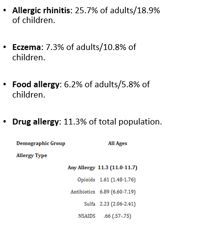
Percentages of Physican Diagnosed Allergies in USA
2 Ways to Detect Type I Hypersensitvity
intradermal skin test
skin prick test
Intradermal Skin Test
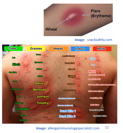
used to detect type i hypersensitivity, 0.5 mL of potential allergens are injected intradermally into back or forearm
looks for wheal and flare reaction within 30 mins (central raised wheal surrounded by red flare)
environmental allergens only (NOT for food)
Downsides of Intradermal Skin Test
may give false positives, may sensitze patient to new allergens
pt must be monitored for late phase reaction and anaphylaxis (rare)
Skin Prick Test (SPT)
used to detect type i hypersensitivity, skin of back or forearm lightly scratched with lancet and a drop of diluted antigen is placed on the scratch
look for wheal and flare reaction within 30 mins
used for environmental AND food allergies

Downside of Skin Prick Test
less sensitive than intradermal injection, but it is usually done first and may be followed by intradermal injection
Allergen-Specific Immunotherapy (ASIT)
long-term treament for allergies, involves repeated subcutaneous injections of slightly increasing allergen dose
regular injections give for 3-5 years to maintain tolerance, works best for allergic rhinitis
How does ASIT gradually increase tolerance?
formation of inhibitor Tregs
supression of Th2 cells
shift from IgE to IgG4 (“blocking” antibodies)
IgG4 Formation as a Result of ASIT
<5% of total IgG in blood, low affinity Fc receptors, do not activate complement
do not form large antigen/antibody complexes
bind allergen and blocks IgE → prevents mast cell degranulation
Autoimmunity
an abnormal adaptive immune response against self antigens, often resulting in chronic inflammation and damage
may be organ specific or systemic
What causes autoimmunity and which hypersensitivity reactions are involved?
caused by a failure of immune clearance
self-reactive antibodies and/or self reactive T cells are involved in some type ii, iii, and iv hypersensitivity reactions
4 Type II Hypersensitivities Involving Autoimmunity
autoimmune hemolytic anemia
goodpasture’s syndrome
graves’ disease
myasthenia gravis
Autoimmune Hemolytic Anemia
type ii hypersensivity involving autoimmunity, antibodies against rbc antigens
opsonization, phagocytosis, and complement-mediated killing of rbcs
Autoimmune Hemolytic Anemia Clincal Presentation and Treatment
anemia, hemoglobinuria, jaundice
corticosteroids, plasmapheresis, splenectomy
Goodpasture’s Syndrome
type ii hypersensivity involving autoimmunity, antibodies against type IV collagen in basement membrane of kidney golmeruli and lung alveoli
complement activation, inflammation, tissue damage
Goodpasture’s Syndrome Clinical Presentation and Treatment
bloody cough, kidney failure
corticosteroids, plasmapheresis, dialysis, kidney transplant
Graves’ Disease
type ii hypersensivity involving autoimmunity, antibodies against thyroid-stimulating hormone receptor (TSH-R)
overstimualtion of thyroid
Graves’ Disease Clinical Presentation and Treatment
hyperthyroidism (weight loss, fatigue, goiter, bulging eyes)
antithyroid druges, thyroid ablation (radioactive iodine), thyroidectomy
Myasthenia Gravis
type ii hypersensivity involving autoimmunity, antibodies against ACh receptors at NMJ
block muscle activation, cause complement activation, inflammation and tissue damage
Myasthenia Gravis Clinical Presentation and Treatment
progressive muscle weakness, paralysis, drooping eyelinds, double vision
acetylcholinesterase inhibitors, corticosteroids, plasmapheresis
Type III Hypersensitibity Involving Autoimmunity
systemic lupus erythematosis
Systemic Lupus Erythematosus (SLE)
type iii hypersensivity involving autoimmunity, antibodies against DNA, nuclear proteins, and other self-antigens
systemic deposition of antigen/antibody complexes, complement activation, inflammation, tissue damage
SLE Clinical Presentation and Treatment
arthritis, kidney damage, vasculitis, “butterly rash” on face, extreme fatigue, fever
corticosteroids, NSAIDs, immunosuppresants, monoclonal antibodies against cytokine BAFF
affects 1/130 in USA (90% female)

Type IV Hypersensitivity Involving Autoimmunity
rheumatoid arthritis (RA)
multiple sclerosis (MS)
Rheumatoid Arthritis (RA)
type iv hypersensivity involving autoimmunity, has features of type ii/iii/iv reactions
complement activation, inflammation, tissue damage, antigen/antibody complex deposition, T cell mediated tissue damage
Rheumatoid Arthritis Clinical Presentation and Treatment
joint pain and swelling (inflammed synovium, cartilage and bone erosion)
immunosuppressants, coricosteroids, NSAIDs, monoclonal antibodies against TNF-a

2 Types of Rheumatoid Arthritis
seropositive
seronegative
Seropositive RA
IgM antibodies against Fc region of IgG (rheumatoid factor) and/or antibodies against various chemically modified (citrullinated) proteins in joints
Seronegative RA
no autoantibodies or other autoantibodies present
Multiple Sclerosis (MS)
type iv hypersensivity involving autoimmunity, autoreactive CD4+ T cells, inflammatory cytokines, macrophage-mediated destruction of myelin sheath of neurons
may involve features of type ii reactions (IgG autoantibodies)
Multiple Sclerosis Clinical Presentation
ranges in severity but can include motor weakness, fatigue, numbness and tingling in limbs, blurry vision paralysis
Detection of Multiple Sclerosis
MRI scans used to detect neural lesions in CNS
multiple genetic risk factors, strongest is HLA-DRB1 (75% females)
Multiple Sclerosis Treatment
monoclonal antobodies against a4b1 (VLA-4) block leukocyte migration to CNS
monolonal antibodies against B cell marker CD20 depletes B cells
fingolimod blocks T cell migration
corticosteroids, plasmapheresis