IPHY 3415 (Cadaver Lab) Exam 3
 Call Kai
 Learn
 Practice Test
 Spaced Repetition
 Match
 Flashcards
 Knowt Play
1/277

There's no tags or description
Looks like no tags are added yet.
278 Terms
conducting zone
portions of respiratory system whose function is to conduct air to the alveoli
respiratory zone
where gas exchange occurs, consists of respiratory bronchioles and alveoli in the lung
nasal cavity
extends from the external nares (nostrils) in front, to the choanae (posterior nares) behind
boundaries of nasal cavity
hard palate and roof
paranasal sinuses
mucus lined, are filled cavities found in some of the bones of the skull; each sinus drains into the nasal cavity
maxillary sinus

maxillary sinus site of drainage
middle meatus
frontal sinus

frontal sinus site of drainage
middle meatus
sphenoid sinus

sphenoid sinus site of drainage
sphenoethmoidal recess
ethmoid sinus

ethmoid sinus site of drainage
superior and middle meatus
uvula
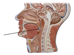
palatine tonsil

pharyngeal tonsil

soft palate
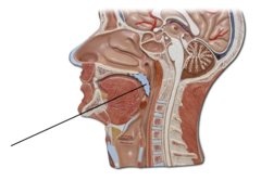
lingual tonsil
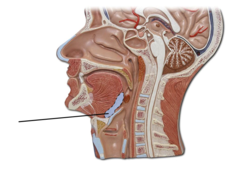
regions of the pharynx
nasopharynx, oropharynx, laryngopharynx
boundaries of nasopharynx
posterior nares to tip of soft palate
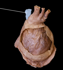
superior vena cava (anterior view)
structures of nasopharynx
pharyngotympanic opening, pharyngeal tonsil
chordae tendineae

boundaries of oropharynx
from soft palate to epiglottis
papillary muscles

structures of oropharynx
palatine tonsils, lingual tonsils
pectinate muscles

boundaries of laryngopharynx
epiglottis to esophagus
left ventricle (superanterior view)
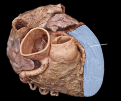
structures of laryngopharynx
none
inferior vena cava (inferior view)

epiglottis (medial view)

superior vena cava (superolateral view)

arytenoid cartilage (posterior view)

right atrium (anterosuperior view)

epiglottis (posterior view)

right ventricle (anteroinferior view)

cricoid cartilage (medial view)

apex of heart

larynx

left auricle (anterior view)

cricoid cartilage (posterior view)

pectinate muscles (anterosuperior view)

vocal fold
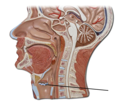
interatrial septum (anterosuperior view)

main bronchus

epiglottis (anterior view)

thyroid cartilage
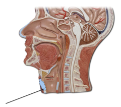
trachea

vocal fold (medial view)

segmental bronchus

tracheal cartilage (posterior view)

corniculate cartilage (posterior view)

vocal fold (posterior view)

tracheal cartilage (anterior view)

cricoid cartilage (anterior view)

cricothyroid ligament (anterior view)

thyrohyoid ligament

thyrohyoid ligament (medial view)
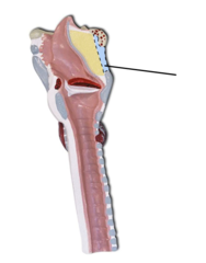
epiglottis (head model view)

cricotracheal ligament

vestibular fold (medial view)
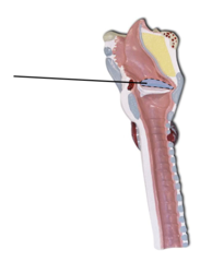
thyroid cartilage (medial view)

thyroid cartilage (anterior view)
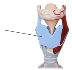
thyroid cartilage (full model view)

rima glottidis (posterior view)

tracheal cartilage (medial view)
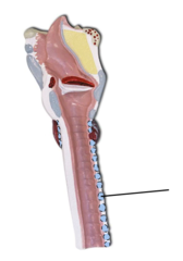
cricothyroid ligament (medial view)

cricothyroid ligament (full model view)

cricotracheal ligament (posterior view)

oropharynx

posterior vein of left ventricle (inferior view)

laryngopharynx
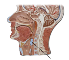
ascending aorta (anterior view)

nasooharynx

left coronary artery (anterior view)

pharyngotympanic (auditory) tube
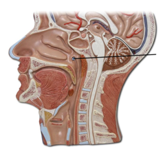
left subclavian artery (posterior view)

carina

brachiocephalic trunk (anterosuperior view)

tracheal cartilage (anterior cadaver view)

pulmonary trunk (anterosuperior view)

trachea (anterior cadaver view)

left common carotid artery (anterosuperior view)
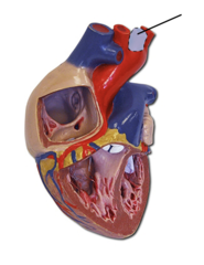
lobar bronchus

great cardiac vein (posterior view)

right bronchus is
wider, shorter, and more vertically oriented; foreign objects are more likely become lodged here
ligamentum arteriosum (anterior view)

base of lung

middle cardiac vein (posterior view)

costal surface of lung

right coronary artery (anterior view)

mediastinal surface of lung

pulmonary trunk (anterior view)

diaphragmatic surface of lung
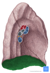
interventricular septum (anterior view)

root of the lung

left subclavian artery (anterior view)

hilum of lung
area through which the root of the lung passes through
right coronary artery (posterior view)
