311 Session 4 Brain Organization and Neural Pathways (Lobes - limbic system - basal ganglia - cerebellum)
 Call Kai
 Learn
Learn Practice Test
 Spaced Repetition
 Match
Match Flashcards
 Knowt Play
1/38

There's no tags or description
Looks like no tags are added yet.
Name | Mastery | Learn | Test | Matching | Spaced | Call with Kai | Chat |
|---|
No analytics yet
Send a link to your students to track their progress
39 Terms
What are the four lobes of the brain
Frontal
Parietal
Temporal
Occipital


Identify this structure
Insula

Identify this sulcus
Longitudinal fissure: divides cerebrum into left and right hemispher

Identify orange and yello
Orange: parieto-occipital fissure
Yellow: Calcarine sulcus
Why does the corpus callosum contain white matter and what does it connect
Connectes left and right hemispheres of brain
Contains white matter because it is made primarily of myelinated axons travelling between the two hemisphere
Where does the primary visual cortex lie
along the banks of the calcarine sulcus
Where does the primary auditory cortex lie
Superior temporal surface of the lateral sulcus
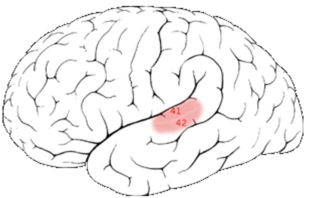
What are the limbic structures
Hippocampus: new memory formation + learning
Amygdala: Emotion + emotional memory
Hypothalamus: Homeostasis + motivaiton
Mammillary bodies: memory/ recall
Cingulate gyrus: Emotion + Motivaiton
What does this image show? Identify the structures


Limbic system
How are the mamillary bodies related to the hypothalmus?
Mammillary bodies are part of the hypothalamus, they’re small rounded structures on the inferior surface of the hypothalamus
Where is the hypothalamus relative to the third ventricle
The hypothalamus forms the inferior (lower) floor and the side walls of the third ventricle
What exactly is the basal ganglia
A group of subcortical nuclei (a collection of interconnected clusters of gray matter neurons) located deep within the brains white matter
What is the main function of the basal ganglia
Control and fine-tune voluntary movements by promoting desired motor actions while suppressing unwanted ones
What are the structures of the basal ganglia
Caudate nucleus
Putamen
Globus pallidus

Where can you see the caudate nucleus in a transverse vs a coronal section


Identify this structure

Internal Capsule
What structures form the Lentiform nucleus
Putamen and Globus Pallidus

What is the most medial vs lateral part of the lentiform nucleus
Medial: globus palligus
Lateral: Putamen

What structures make up the dorsal Striatum (aka striatum)
Caudate Nucleus
Putamen


What does this image show and identify the structures
Basal ganglia


Identify this sturcture
Substantia Nigra

Identify this structure
Subthalamus
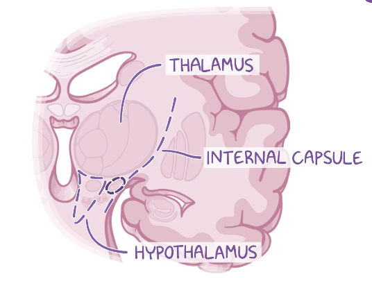
What are the surrounding structures relative to the subthalamus
Anterior: Thalamus
Lateral and posterior: hypothalamus
How does the basal ganglia influence movement
Influence movement INDIRECTLY by influencing the motor cortex via the thalamus
information enters the B.G → corpus striatum → leaves through GPi → thalamus
How does the caudate nucleus relate to the lateral ventricle?
The caudate nucleus follows the C-shaped curve of the lateral ventricle. Its head lies beside the anterior horn, its body runs along the lateral wall/floor of the body of the ventricle, and its tail curves inferiorly with the temporal horn toward the amygdala.

Which basal ganglia components lie in the cerebrum, diencephalon, and midbrain
Cerebrum: caudate nucleus, putament, GP
Diencephalon: Subthalamic nucleus
Midbrain: substantia nigra
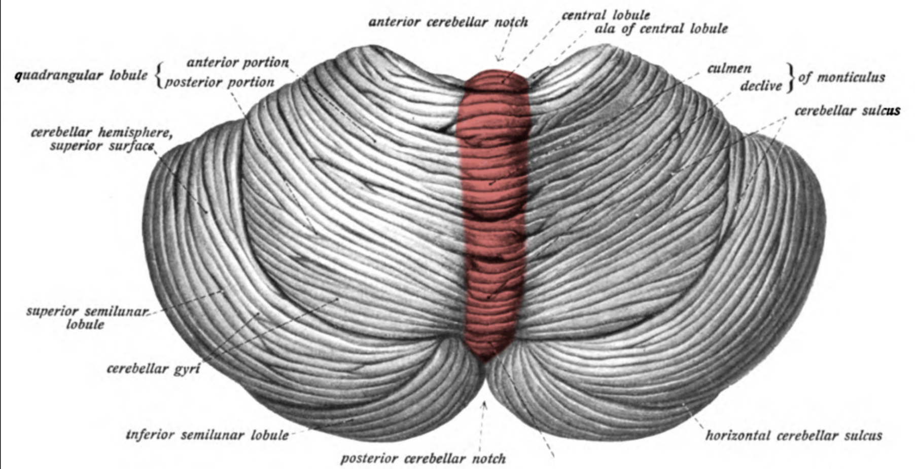
Identify this structure
Vermis

Name this structure
Tonsils

Name the following structures

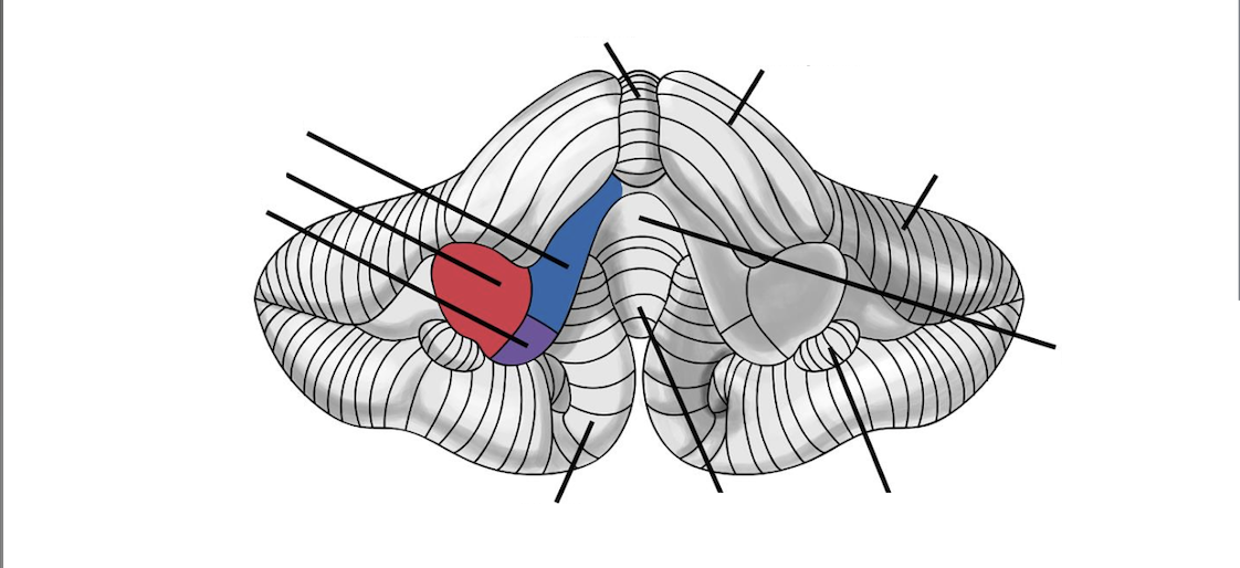
Name these structures

What is the cerebellums main role
Plays a role in coordinating voluntary muscle movements, maintain balance, and regulate posture
How does the cerebellum play a role in movement coordination
Coordinates the timing, force, and sequence of muscle activity so movements are smooth and precise. Without proper cerebellar funciton, muscles can still contract, but movements may be poorly timed or inaccurate
How does the cerebellum play a role in balance / posture
Uses information about body position and head movement to help maintain balance, posture, and stability
How does the cerebellum play a role adjusting movements
Recieves feedback about how a movement is actually happening and helps correct errors while the movement is occuring
Which features of the cerebellum identify the midline, inferior surface, and branching internal white matter? What does each help explain
Midline: Vermis = midline structure connecting the two cerebellar hemispheres
Inferior surface: Tonsils = rounded structure on the inferior, near the foramen magnum
Arbor Vitae: Internal white matter = inside the cerebellum, carries information between the cerebellar cortex and other regions
What does the primary fissure of the cerebellum separate?
Separates the anterior lobe from the posterior lobe
What are the main function of the cerebellar peduncles
The cerebellar peduncles are white matter tracts that connect the cerebellum to the brainstem, carrying information into and out of the cerebellum for the coordination and adjustment of movement
Where are the fourth ventricle and brainstem relative to the cerebellum? Why do the tentorium, posterior cranial fossa, and tonsils matter?

Cerebellum lies posterior to the brainstem, with the fourht ventricle between the pons and the cerebellum
Tentorium cerebelli → dural fold that separates the cerebellum from the occipital lobes.
Posterior cranial fossa → bony compartment containing the cerebellum and brainstem.
Cerebellar tonsils → inferior portions of the cerebellum near the foramen magnum. Increased intracranial pressure can push them downward through the foramen magnum (tonsillar herniation), potentially compressing the brainstem.
Why can movement be poorly coordinated even when the muscles can still contract
Because the cerebellum doesnt initiate muscle contraction, it coordinates and fine tunes movement. It compares intended movement with what is actually occuring and hleps adjust timing,force, accuracy, balance, and posture