Wrist/Forearm
 Call Kai
 Learn
 Practice Test
 Spaced Repetition
 Match
 Flashcards
 Knowt Play
1/75

There's no tags or description
Looks like no tags are added yet.
76 Terms
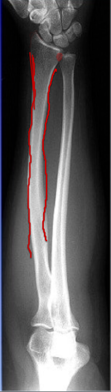
Body/Shaft of Radius
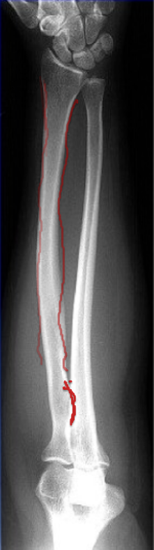
Radial Tuberosity
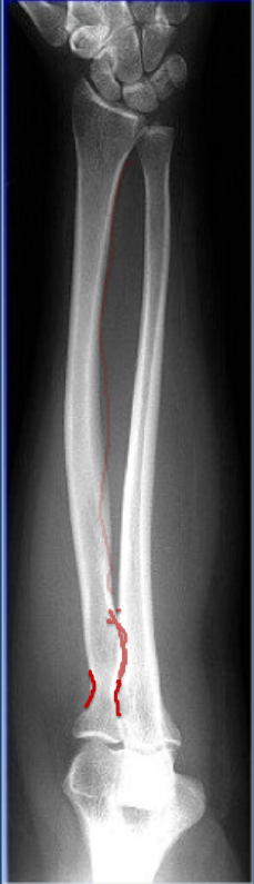
Narrowing
Radial Neck
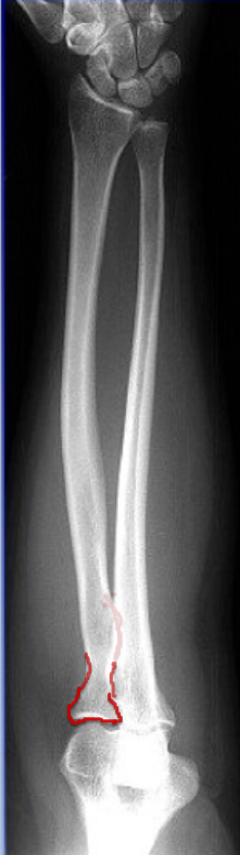
Radial head
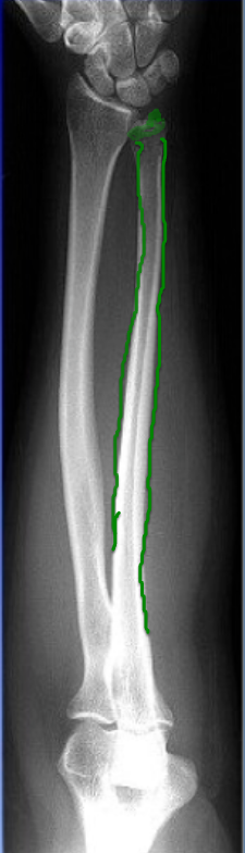
Body of Ulna
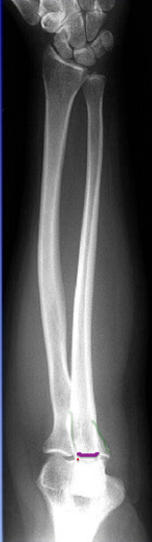
Ridge
Coronoid process
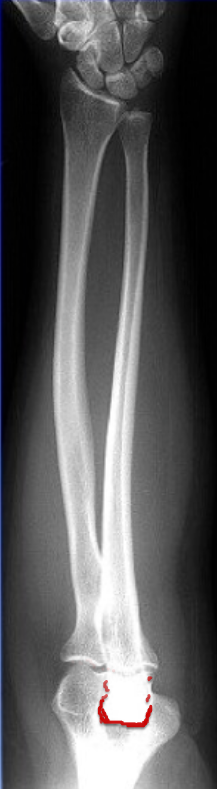
The End
Olecranon process
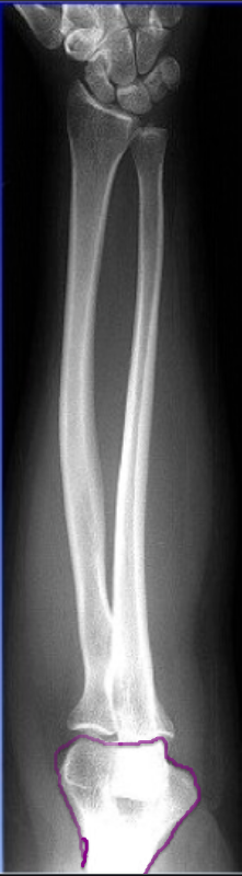
Distal Humerus
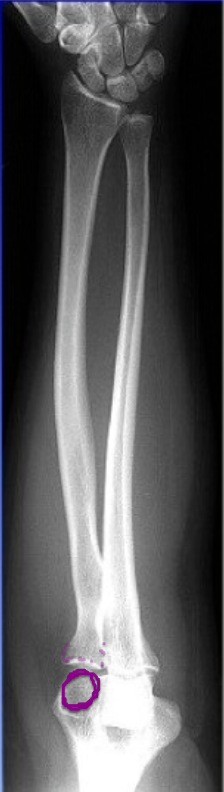
Radius articulates with humerus on lateral aspect with
Capitulum
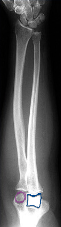
Ulna articulates with
Trochlea
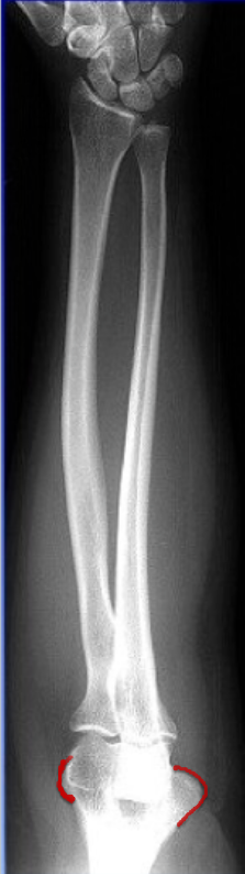
What is on lateral aspect
Lateral epicondyle
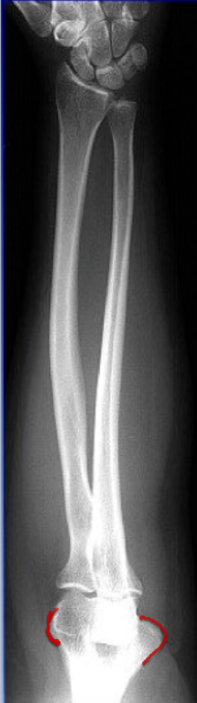
What is on medial aspect
Medial epicondyle
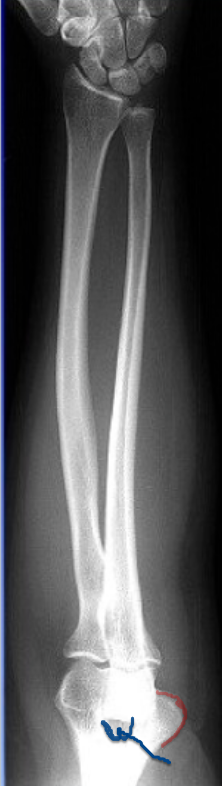
Dark spot ( indent so less bone) ( more radiolucent) (posterior aspect of humerus)
Olecranon fossa
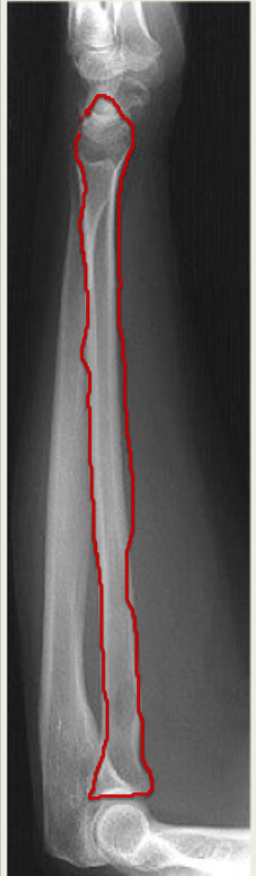
Radius
Bigger on distal aspect on lateral
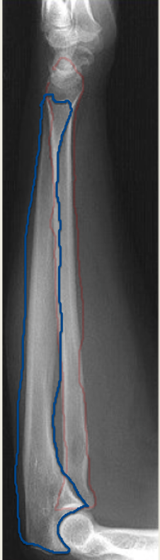
Ulna
Bigger on proximal aspect on lateral
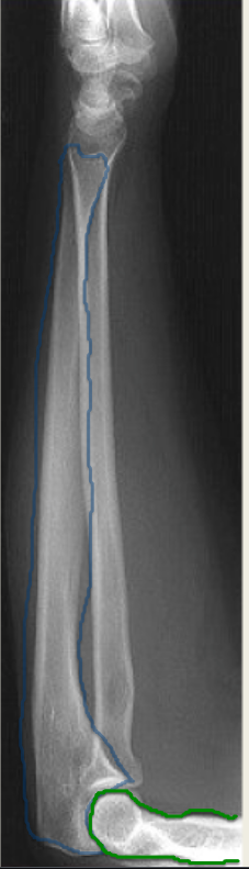
Humerus
On lateral we cannot see
Radial Tuberosity
lives on medial aspect
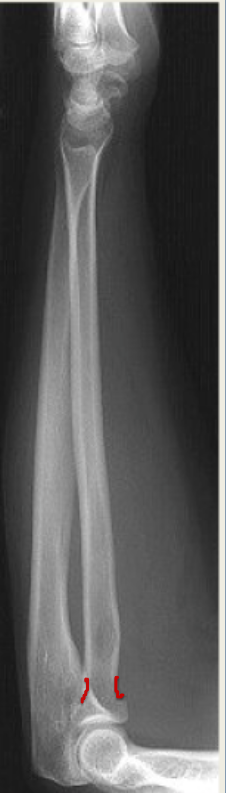
Radial neck
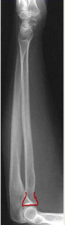
Radial head
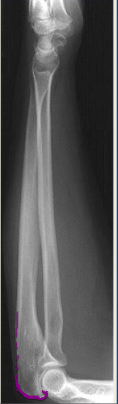
Olecranon process
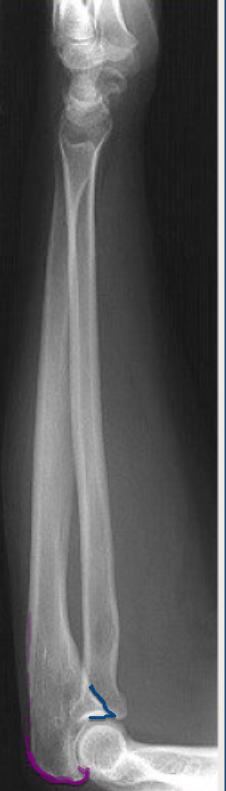
(blue)
Coronoid process
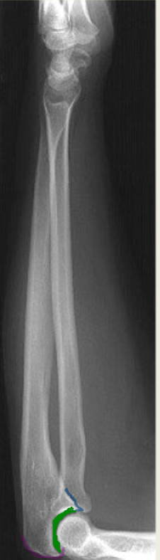
Where Coronoid process and Olecranon process meet and also indentation
Trochlear notch / Semilunar notch
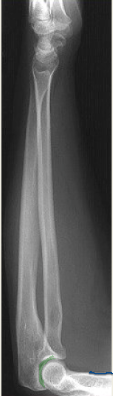
Shaft of Humerus
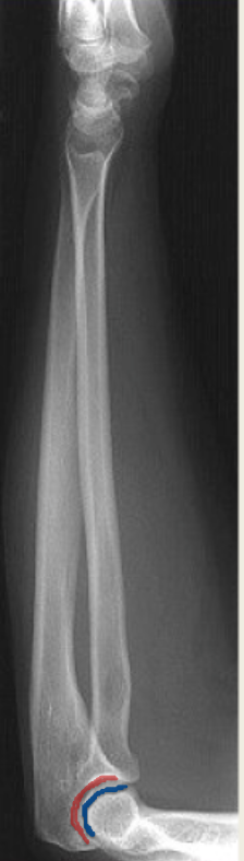
Red- Trochlear notch Blue is what you are looking at
Capitulum , Trochlea , & Trochlear sulcus
Superimposing in three circles
Good forearm
Wrist and an elbow included with soft tissue
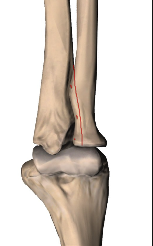
Ulna on left side & Radius on right side → No rotation
slight overlap on AP
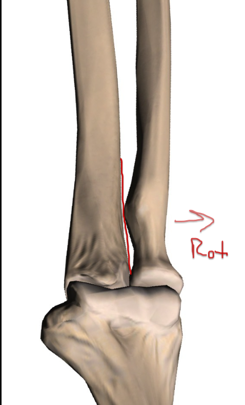
Ulna on left side & Radius on right side → Lateral rotation on AP
Right AP forearm
Separation not touching/no overlap
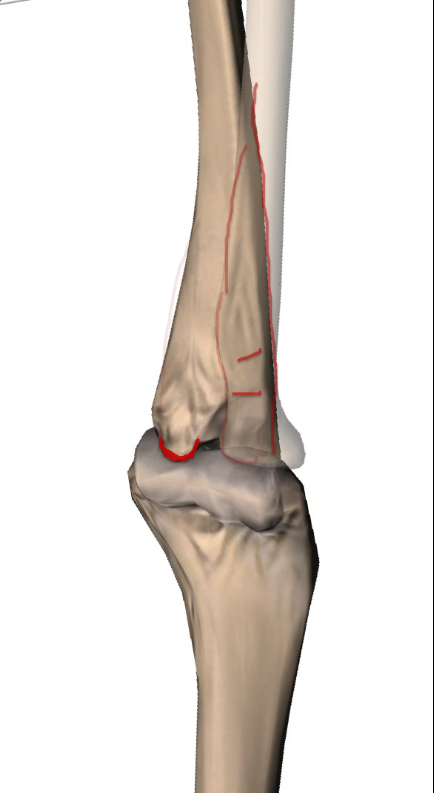
Ulna on left side & Radius on right side → Medially rotated on AP
This is right forearm AP
Lots of overlap
Coronoid process moved medially
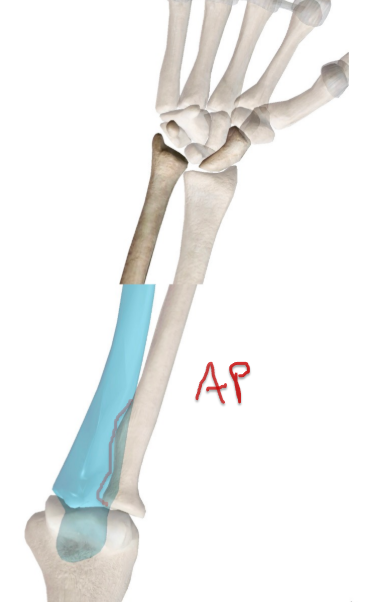
How to tell it’s a good AP → No rotation
Distal Portion
Nice separation on metacarpals
evaluation of carpal bones → they are all laid out
Distal Radioulnar joint → No overlap
Proximal Portion
Slight overlap of Radial head, neck and tuberosity with the Ulna
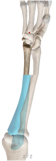
Medial/internal Rotation in AP
Distal Portion:
Overlap of carpal bones they are coming together → See medial aspect better than lateral
Pisiform separating from other carpal bones
4th and 5th metacarpal no overlap of the heads , space b/w shafts are closer and slight overlap of the bases
Distal radioulnar joint starts to overlap
Proximal Portion:
Lots of overlap of Radial head, neck and tuberosity with the Ulna
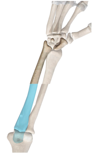
External/Lateral rotation in AP
Distal Portion:
3rd-5th metacarpals start to overlap
Trapezium and scaphoid you can see better
Overlap of the Distal Radioulnar joint
Proximal portion:
Very little superimposition of the radius and ulna
4th and 5th big metacarpal separation
Medially rotated cause it’s an AP
If you have lateral rotation in the wrist you will see
Radial tuberosity on the anterior aspect
radius and ulna CAN appear superimposed
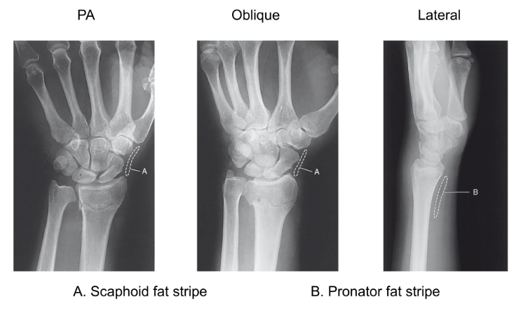
Fat pad sits adjacent to the
Joint capsule
Why do we want to see the fat stripe?
So if the joint is swelling due to Joint effusion (joint fluid)
It’s gonna press on that fat stripe and make it noticeable to the doctor
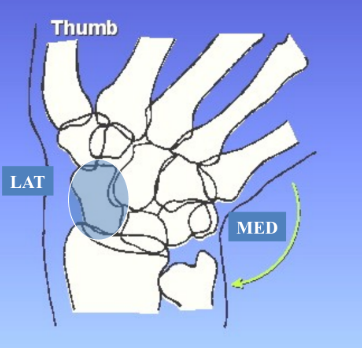
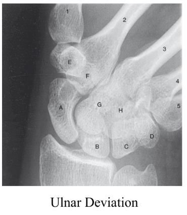
What deviation is this
Ulnar deviation
We see the scaphoid better
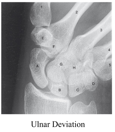
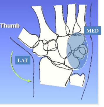
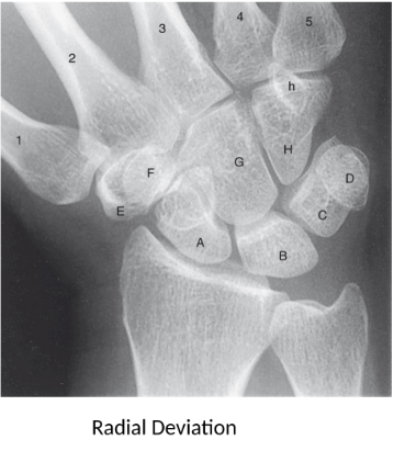
What deviation is this?
Radial deviation
We see the Lunate , Triquetrum, Pisiform and Hamate better
Wrist Exam Criteria
Multiple:
PA & 45 degree oblique ( 8×10 CW)
Lateral 8×10 LW
Single:
1 on 1 8×10 LW
PA or PA Axial Scaphoid
8×10 LW
kV: 60-70
SID: minimum 40 inches
Another reason for Ulnar Deviation
Flattens the scaphoid as it is naturally tilted
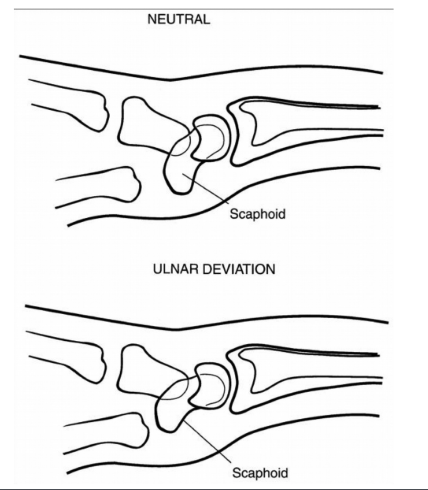
Why use angle or sponge for navicular view?
Foreshortened if no angle
Allows to see scaphoid ( ← angled and tilted in wrist ) properly
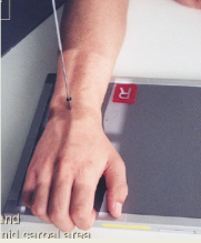
Why do we curl our fingers during PA wrist
wrist and carpal area gets closer to IR
CR perpendicular to midcarpals
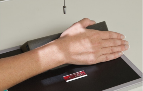
45 degree lateral oblique wrist criteria
CR to midcarpals
Anatomy: Distal radius, ulna , carpals and metacarpals.
Support wrist to eliminate motion
Wrist special projections ?
Scaphoid projections:
PA axial w/ ulnar deviation
Modified stretcher method
Tangential-carpal canal
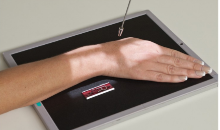
PA axial w/ ulnar deviation
Angle tube 10-15 degrees proximally towards elbow
ulnar deviation
CR to scaphoid ( ¾ inch distal and medial to radial styloid process)
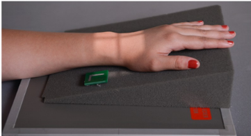
If you can’t angle or patient can’t deviate → Modified stretcher method ( Hand elevated 20 degrees no deviation)
CR perpendicular to scaphoid
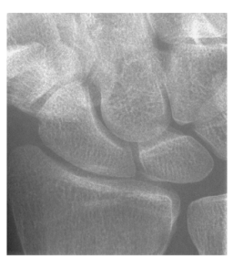
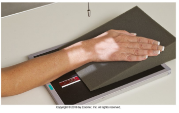
If you can’t angle → Modified stretcher method ( Hand elevated 20 degrees with deviation)
CR perpendicular to scaphoid
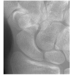
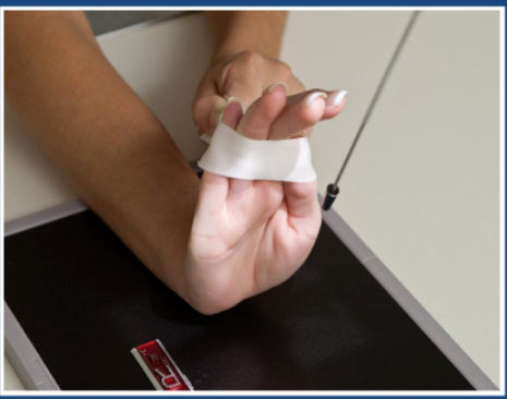
Tangential-carpal canal method ( Tangential , Inferosuperior projection)
CR 25-30 degrees to hand
Rotate hand & wrist 10 degrees towards radius → so Pisiform and Hamate aren’t superimposed
Pull back with tape so fingers aren’t in the way of central ray
Center proximal to base of 3rd metacarpal
8×10 IR
Olecranon Fossa is part of ?
Humerus
Olecranon Process is part of ?
Ulna
In AP position the Trochlear notch is in b/w
Coronoid process and Olecranon process
Ulna indent is what
Trochlear notch
Humerus indent
Trochlear Sulcus
Trochlear Sulcus ( groove) is part of the
Humerus
Trochlear notch is holding part of
the humerus that is sitting in the notch
Trochlear notch/Semilunar notch is part of the
Ulna
Radial head is the most
proximal part of the radius
Radial Tuberosity lives
Medially and slightly anterior on Radius
Radius articulates w/ distal Humerus on the
Lateral aspect
Positioning of Forearm exam criteria
2 exposures on one IR or 1 on 1
11×14 lw for smaller patients ( This depends on length of bones )
14×17 lw for large patients
Collimation: include min 1 inch beyond wrist and elbow joints
SID : 40 inches
Non-grid
kV: 65-75
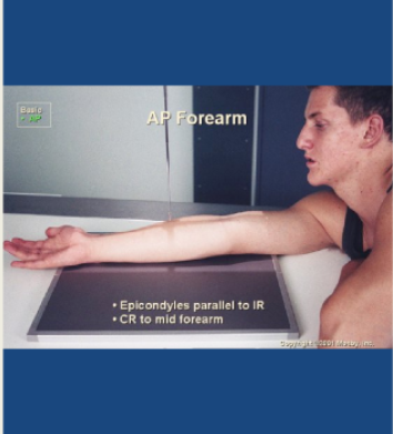
AP forearm
shoulder, elbow and wrist same plane
Hand supinated
Humeral epicondyles parallel to IR ( DON’T FOCUS WE WILL LEARN LATER)
elbow and wrist joints included
CR to mid forearm
Don’t forget we naturally medially rotate our arms when reach out so lean on side of interest
AP forearm image analysis Criteria : Position
Position:
Long axis of forearm aligned with long axis of IR
No rotation → look at Radial head , neck and tuberosity → Should be slightly superimposed by the Ulna ( no more than quarter of an inch)
Olecranon process situated w/ olecranon fossa ← Just telling us if arm is extended
AP forearm image analysis Criteria : Collimation & CR
Collimation:
Collimation to skin margins along length of forearm
Include both wrist and elbow joints (1-1.5 inches)
CR to midpoint of forearm (radius & ulna)
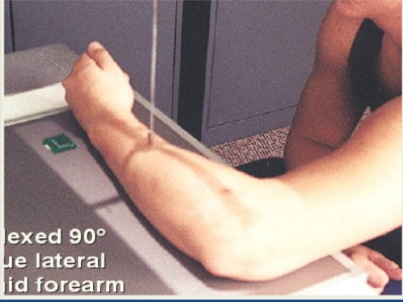
Lateral Forearm
Shoulder , wrist and elbow on same plane
Epicondyles perpendicular to IR
Elbow 90 degrees
Wrist true lateral
CR to mid forearm
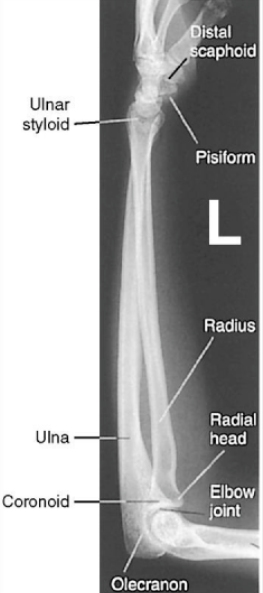
On Lateral Position → Radius is ___________ on distal aspect
Ulna is ___________ on proximal aspect
Bigger
Ap forearm we mark the
Lateral side and position that first
Lateral forearm image analysis Criteria : Position
90 degrees
everything on same plane
No rotation: head of ulna superimposed over the radius ( that’s the wrist )
Radial head superimposed with coronoid process
Lateral forearm image analysis Criteria : Collimation & CR
Collimation to skin margins along length of forearm
Include both wrist and elbow joints (1-1.5 inches)
CR to midpoint of forearm (radius & ulna)
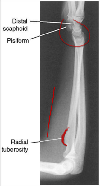
What rotation is this?
Lateral/external rotation
if you have external rotation in wrist you will most likely see the radial tuberosity
pisiform is popping out and anterior to scaphoid
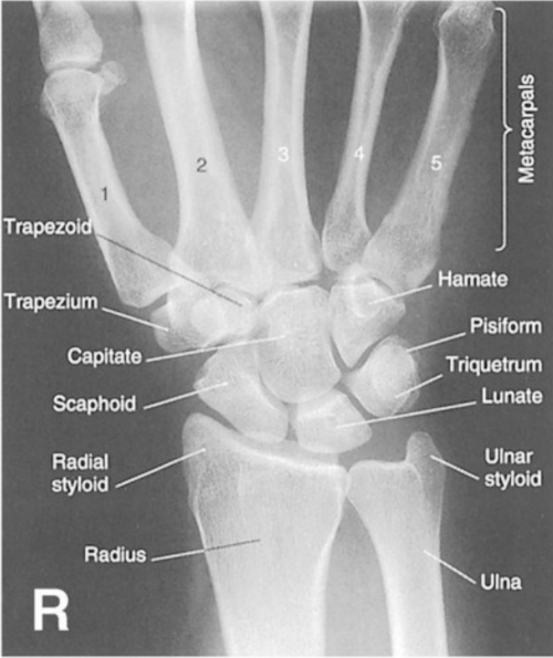
What projection/position is this?
PA wrist (right)
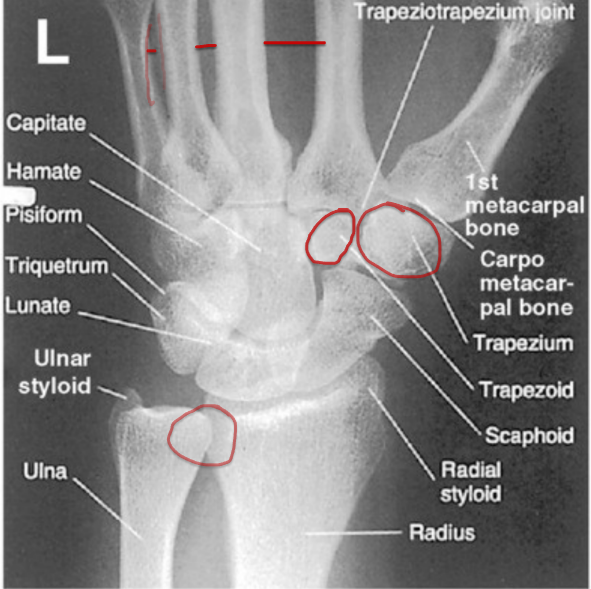
What projection/position is this?
PA external oblique wrist (left)
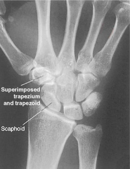
This is an oblique wrist what rotation does it have ? Not a good image
Medially rotated like PA
corrective action: externally rotate wrist to 45 degrees
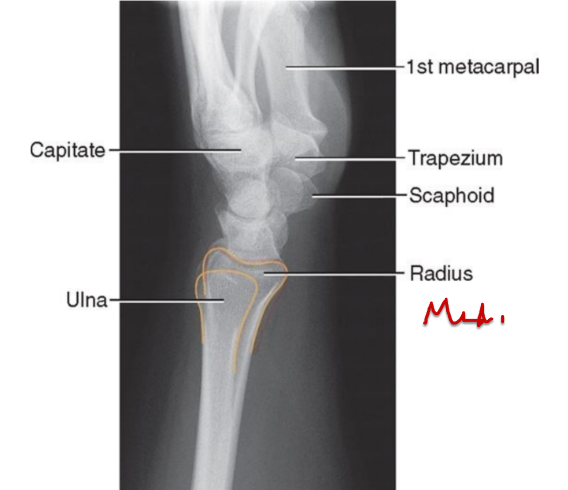
What rotation does it have? ( Lateral wrist)
Medially rotated
relationship of ulna and radius
scaphoid anterior to pisiform
corrective action: Externally rotate to 90 degree angle
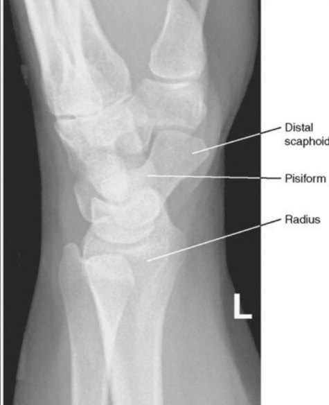
What rotation does it have? ( Lateral wrist)
Medially rotated
relationship of ulna and radius
scaphoid anterior to pisiform
corrective action: Externally rotate to 90 degree angle
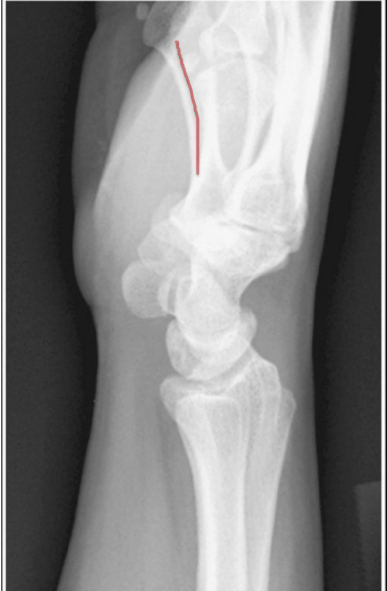
What rotation does it have? ( Lateral wrist)
Externally rotated
pisiform anterior to scaphoid
relationship of ulna and radius
5th metacarpal anterior to 2nd metacarpal
Corrective action: medially rotate to 45 degree angle
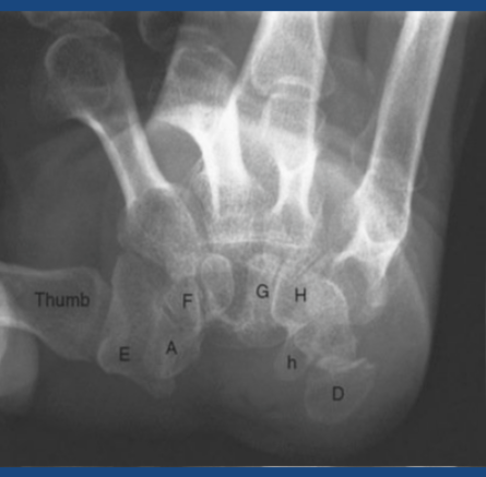
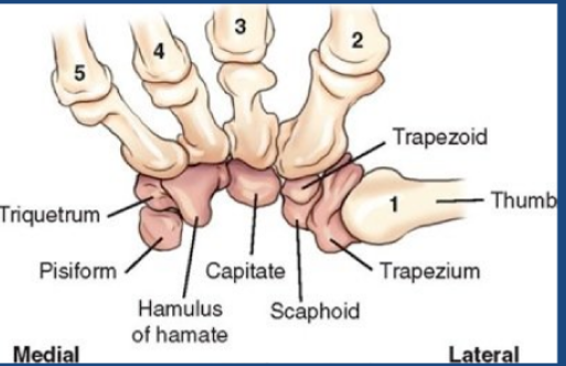
Carpal canal radiograph what are the bones
a.
d.
e.
f.
h.
A. scaphoid
d. Pisiform
e. Trapezium
F. trapezoid
h. hamulus