CLIN MED: Cardiology
 Call Kai
 Learn
 Practice Test
 Spaced Repetition
 Match
 Flashcards
 Knowt Play
1/184

There's no tags or description
Looks like no tags are added yet.
185 Terms
DILATED CARDIOMYOPATHY (DCM)
-characterized
-systolic or diastolic, impairs??
-describe dilation
-age/race
stretching/thinning of the cardiac chambers
impaired contraction/systolic function) not in slides
Dilation
-starts in left ventricle
-the inside of the chamber enlarges
-then the dilation spreads to the right ventricle and atria
-any age, but 20-60 years
-men, black

PRIMARY CAUSES OF DCM
Genetic/hereditary conditions - Inherited autosomal dominant
Inflammatory/infectious (myocarditis)
--> viral agents are Coxsackie B, adenovirus, parvovirus, human herpesvirus 6, COVID-19, and Chagas disease.
Toxic (drugs, alcohol)
-alcohol (reversible)
-Chemo dug doxorubicin
- cocaine
-Dobutamine, diuretics, ampicillin, tetracycline, and tricyclic antidepressants.
-vitamin B1 deficiency (NIS)
Idiopathic- cause unknown
ADDITIONAL CAUSES OF DCM
Peripartum Cardiomyopathy
-late pregnancy or up to 5 months after delivery.
Iron Overload
-hemochromatosis or repeated blood transfusions.
Thiamine Deficiency
-wet beriberi when associated with DCM.
-Uncommon in developed countries, except in malnourished people.
DCM
-leads to...
-sxs
L and R systolic heart failure due to the left ventricle inability to pump blood effectively.
SXS
-Fatigue and dizziness
-Pulmonary congestion crackles, orthopnea and PND
-Lower extremity edema
-S3 heart sound (gallop NIS)
-Jugular vein distension (JVD)
DCM - DIAGNOSIS
-test choice
-gold standard
-imaging
-labs
Echocardiogram- test of choice
-low ejection fraction
-thin, nonfunctional myocardium
Myocardial biopsy: gold standard
-Not often required because most cases of DCM have causes (heavy alcohol use).
Chest x-ray- cardiomegaly, often taking up more than 1/2 of the width of the chest cavity (pleural effusion NIS)
BNP level- (will be elevated)
DCM- TREATMENT/PROGNOSIS
-general treatments (no meds)
-systolic HF meds
-peripartum period with cardiomyopathy
-refractory patients
First line: treat underlying cause
-Stop Causative drugs
-Thyrotoxicosis and beriberi also require specific treatment.
-Systolic HF:
-ACE + β-blockers (if stable) + diuretic (if edema)
-ACE doesn't work = ARB --> hydralazine + nitrate
Peripartum period with cardiomyopathy: require anticoagulants.
Refractory patients = heart transplantation
Left Ventricular Hypertrophy (LVH)
(under hypertrophic cardiomyopathy)
-patho
-causes
-demographics
LVH is an increased thickness of the left ventricle.
CAUSES
-MC: LV pressure
-HTN and aortic stenosis.
-African Americans
-elderly
-obesity
-HTN
-obesity

HYPERTROPHIC CARDIOMYPATHY (HCM)
-most common ________ ______
-demographics
-occurs in absence of
HCM is the MC inherited cardiomyopathy-- autosomal dominant mutations.
DEMOGRAPHIC:
-young and fit patients (often athletes) with sudden cardiac arrest.
Occurs in the absence of chronic hypertension, valvular disease, ischemic heart disease, or congenital heart disease.
HCM- PATHOLOGY
Wall thickening - as a result of LVH resulting in
-decreased ventricular chamber size
-reduced end-diastolic volume (EDV)
-cardiac output.
-Diastolic dysfunction results in ineffective filling during diastole and reduced cardiac output.

HCM- 3 main pathology
Hypertrophy
-typically asymmetric, not concentric as in other forms of LVH.
-The most typical pattern is asymmetric septal hypertrophy with dominant thickening of the interventricular septum closest to the aortic valve.
Ventricular Outflow Obstruction.
-from asymmetrical hypertrophy of papillary muscles or interventricular septum.
Mitral valve abnormality
-blood backs up through the valve, leading to mitral regurgitation = dilation of the left atrium.
HCM- SIGNS AND SYMPTOMS
-sounds (2)
-increases or decreases sounds
S4 sound
Crescendo-decrescendo systolic murmur due to the outflow obstruction
A systolic mitral regurgitation murmur due to the abnormal anterior motion of the mitral valve.
-Squatting decreases the murmur intensity.
-Valsalva maneuver and standing increases murmur intensity

HCM -diagnosis
-test of choice (describe findings)
-other imaging
Echocardiography - test of choice/first
-information on the ejection fraction and valve regurgitation
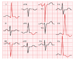
ECG:
-LVH --high voltage in the lateral ECG leads (I, V5-V6).
-Criterion for LVH is to add the S amplitude in V1 to the R amplitude in V5 or 6.
-If more than 35 mm, there is LVH.
Chest X-Ray - show cardiomegaly
Imaging.
-Cardiac MRI and cardiac CT - Used as confirmatory tests, or when the echocardiogram is uncertain.
-Genetic testing - confirm that a particular genetic variant is present in multiple family members
HCM-TREATMENT
-treatment overview
-drugs
-what should be avoided
Lifestyle changes, medications, and/or surgery.
-BP control (LVH is secondary to hypertension) (
HCM - TREATMENT: management
-most patients
-Patients with dyspnea or chest pain
-diuretics
Most patients with HCM are asymptomatic and do not need treatment.
Patients with dyspnea or chest pain:
-should be started on verapamil (CCB).
Diuretics should be used cautiously, and only in cases of severe volume overload.
--> Dehydration can potentially worsen the outflow obstruction.
HCM-TREATMENT
-with extreme septal hypertrophy and outflow track obstruction
Septal myomectomy (cutting out the extra muscle)
Alcohol septal ablation
-A minimally invasive procedure that reduces the proportion of tissue blocking the blood flow, due to thickening of the heart muscles.
-ASA involves injection of pure alcohol into the target septal branch of the left anterior descending coronary artery.
RESTRICTIVE CARDIOMYOPATHY (RCM)
-results in
-impairs
-most common cause
-treatment
Loss of compliance = ventricles stiff due to protein infiltration or fibrosis and scarring.
-impairs ventricular filling and decreased cardiac output.
Most common cause is idiopathic.
TREAT UNDERLYING CAUSE

RESTRICTIVE CARDIOMYOPATHY (RCM)
-age
-characteristic findings
-results in
diagnosed in children 5 to 6 years of age, but can occur in any age.
characteristic findings
-diastolic heart failure
-As a result, pulmonary congestion along with signs of exertional dyspnea, orthopnea, and PND.
--> right heart failure sxs such as lower extremity edema and hepatic congestion.
PATHOLOGY - RESTRICTIVE CARDIOMYOPATHY
-lack of
-results in
Lack of compliance or stretch results in failure of the ventricle to fully expand to fill normally with blood from the atria during diastole.
Results in
-reduced end-diastolic volume, stroke volume and cardiac output.
•Commonly, fibrosis of the tissue or infiltration of the myocardium by various deposits prevent normal chamber relaxation.
CAUSES: RESTRICTIVE CARDIOMYOPATHY
-primary
-secondary
PRIMARY
Idiopathic: MCC United States.
-Endomyocardial fibrosis: Nutritional deficiencies and inflammation from parasitic infections cause myocardial fibrosis.
-Endocardial fibroelastosis: Overactivation of collagen deposition in the endocardium causes fibrotic stiffening.
SECONDARY
-Post-radiation fibrosis: Radiation leads to scarring and fibrosis that causes increased stiffness.
-Progressive systemic sclerosis: Autoimmune disease that leads to extensive fibrosis in skin and multiple organs.
-Sarcoidosis: (granulomas).
-Hemochromatosis: Iron overload leads to hemosiderin deposition in cardiac myocytes, causing stiffness.
-Amyloidosis: Causes deposition of abnormal amyloid in the myocardium.
AMY HAS amyloidosis, hemo, amyloid, sarcoid
S&S: RESTRICTIVE CARDIOMYOPATHY
-buzz words
-S4 heart sound
RHF signs- Increased jugular venous pressure (JVP), edema, hepatomegaly.
(R) Kussmaul sign, ---a jugular venous pressure that either increase or does not change with inspiration. (normally the pressure should decrease with inspiration).
DIAGNOSIS: RESTRICTIVE CARDIOMYOPATHY
-imaging (2)
-labs
Echocardiography best diagnosis
-normal systolic function
-significant enlargement of the atria (dilation)
-diastolic dysfunction --> Elevated left ventricle filling pressures with normal ejection fraction.
Endomyocardial biopsy - gold standard
-but it is rarely needed to confirm diagnosis.
-apple green birefrinegence (amyloidosis) NIS
Serum studies- determine underlying secondary causes.
-Patients with amyloidosis may have reduced renal function or proteinuria
MRI is helpful in patients with diagnosing endocardial fibrosis, "V"-like shape
Chest x-ray --may show sarcoidosis
Heart catheterization measure pressures in the heart.
CLASSIFICATION OF HEART FAILURE
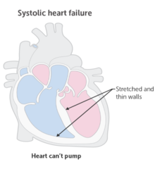
Heart Failure with reduced Ejection Fraction (HF rEF),
-Systolic Heart Failure.
-weakened, thin, overly compliant ventricles
-S3 (gallop) commonly heard on auscultation
-Dilated cardiomyopathy
Heart Failure with preserved/normal Ejection Fraction (HF pEF),
-Diastolic Heart Failure.
-stiffened, thick or noncompliant ventricles
-S4 heard on auscultation
-HCM, restrictive

HF rEF (SYSTOLIC HF)
-reduced EF value
-characterized
HF rEF
-reduced ejection fraction =
HF rEF (Systolic HF)
-affect on stroke volume and EF = EDV
-because heart ____________--> leads to
-leads
1. reduced stroke volume and EF
2. increase EDV
Because the heart cannot pump blood adequately, there are increases in EDV.
-This leads to fluid backing up into the alveoli of the lungs, and resultant pulmonary congestion and dyspnea.
Causes of systolic HF
-Ischemic heart disease
-Dilated cardiomyopathy
-Viruses, COVID-19
-Toxins (alcohol, cocaine)
-Chemotherapy drugs such as doxorubicin
HF pEF (Diastolic HF)
-describe
-what is maintained/characterized by
Preserved ejection fraction
-inability of the ventricle to adequately relax and fill with blood.
The contractility of the ventricle is maintained (unlike systolic HF):
-Decreased ventricular compliance (increased stiffness)
-Reduced EDV
-Reduced stroke volume
-Preserved EF

DIASTOLIC HF
-compliance
-decreased compliance
-reduced EDV leads to
Compliance is the ability of the heart muscle fibers to lengthen in response to pressure of the ventricle.
-Decreased compliance impairs ventricular filling and decreases the EDV.
-Reduced EDV reduces preload. The reduced preload reduces the stroke volume.
DIASTOLIC HF - causes
Causes of diastolic failure include:
-Hypertension
-Aortic stenosis
-Hypertrophic obstructive cardiomyopathy
HIGH-OUTPUT HEART FAILURE
-occurs when
-patient type
Occurs when the demand for blood from the body exceeds the capacity of a normal heart.
-severe anemia
-pregnancy
-AV malformations with shunting of blood
-hyperthyroidism.
LEFT SIDED HEART FAILURE
-occurs when
-lung sounds
-as left sided HF progresses....
L = lungs, blood backs up in lung
Left ventricle does not pump properly, depriving the organs of blood (and oxygen).
-heart cannot empty properly = blood builds in lungs and/or the venous system.
detected
-lung crackles on exam
As left sided HF progresses... reduced renal perfusion causing increased sodium and water retention (edema).

LEFT SIDED HEART FAILURE - SIGNS AND SYMPTOMS
-Cough- dry, hacking cough (mc)
-dyspnea (mc)
-Productive frothy or blood-tinged sputum
-Orthopnea (shortness of breath when lying down)
-PND (waking up breathless from sleep)
RIGHT SIDED HEART FAILURE
-describe
-most common cause
-occurs in isolation?
R = rest of body (pulmonary conditions, COPD NIS)
Blood from right ventricle backs up into the systemic circulation --> hepatomegaly, jugular venous distention, and peripheral edema.
MCC of right-sided HF is left-sided HF.
-Right heart failure uncommonly occurs in isolation, except cor-pulmonale and pulmonary HTN.

SIGNS AND SYMPTOMS OF RIGHT SIDED HEART FAILURE
-Weight gain
-jugular venous distention (JVD)
-Liver congestion
-Ascites
-Reduced GFR
-Peripheral edema
CONDITIONS THAT CONTRIBUTE TO HF
#1 Coronary heart disease
Cigarette smoking - Increases BP, decreases exercise tolerance and increases the tendency for blood to clot.
Obesity
Diabetes -from microvascular dysfunction and increased fatty acid utilization and decreased NO bioavailability.
Valvular heart disease- an increasingly common cause of HF at older ages, with calcific aortic stenosis being the most common disorder requiring surgery.
Hypertension increases the risk of HF at all ages.
-high bp
Heart failure - diagnosis
-test of choice
Echocardiogram - test of choice
-Differentiates HF with/without preserved LV function
-Estimate the ejection fraction (EF).
-Diagnose abnormalities of diastolic filling, ventricular wall thickness, valve disease, and pericardial fluid
-Detects the presence of masses
Heart failure
-EKG findings
-CXR buzz word fidings
EKG - not definitive
-Systolic HF and diastolic
- T wave inversions and ST abnormalities.
Chest Xray useful to compare baseline (initial test?)
-Cardiomegaly
-Alveolar infiltrates ("bat-wing" pattern)
-Pulmonary edema
-Kerley B lines (horizontal lines at the periphery of the lungs)
-Pleural effusions
LEFT VENTRICULAR EJECTION FRACTION
-Hyperdyanmic
-Normal
-Mild LV dysfunction
-Moderate LV dysfunction
Hyperdyanmic: >65% EF
Normal: 55-65%
Mild LV dysfunction: 45-54%
Moderate LV dysfunction: 30-44
Heart failure - lab testing
BNP (Brain Natriuretic Peptide)
Heart failure -adjunctive labs
-CBC- anemia can exacerbate HF
-Serum electrolytes, blood urea nitrogen, and creatinine may indicate associated conditions.
-Hyponatremia generally indicates severe HF, though other causes should be considered
-Renal impairment can be caused by or contribute to HF; baseline renal function is important when starting or adjusting diuretic regimens.
-Liver function tests may be affected by hepatic congestion
-A1c/Fasting blood glucose to detect underlying diabetes mellitus
NYHA HF FUNCTIONAL CLASSIFICATION SYSTEM
I - no symptoms or limitation in ordinary physical activity,
II -Mild symptoms
III - Comfortable only at rest.
IV - symptoms even while at rest.
FINDINGS SPECIFIC FOR HF
-symptoms
-signs
-imaging
-bnp levels
echo
Symptoms: orthopnea, PND
Signs: JVD, S3 heart sounds, laterally displaced point of PMI
Chest x-ray: pulmonary edema
BNP levels: elevated
Echo: showing EF
Heart failure treatment goals
Decrease preload: (amount of stretch on the heart's muscle fibers at the end of diastole, before the heart contracts)
-preload reduction is a mainstay of HF therapy since it reduces the patient's symptoms.
Decrease afterload/pressure the heart has to pump against
-increases the cardiac output for any given level of contractility, increasing tissue perfusion. It reduces the work of contraction.
Increase contractility: Inotropic drugs (do not increase mortality, not used routinely.) increase the force of contraction directly, increasing the cardiac output.
Decrease myocardial remodeling
First line treatment of HF
ACE Inhibitors and ARBS
REDUCE AFTERLOAD
-Reducing the effects of angiotensin II (AT-II), reduce systemic vascular resistance (SVR) resulting in reduced afterload.
REDUCE PRELOAD
Decreasing renal sodium and water reabsorption, intravascular volume, leading to reduced preload.
REDUCE CARDIAC REMODELING
-ACE inhibitors and ARBs decrease cardiac remodeling, improving prognosis and mortality for the long term.
First line treatment of HF
-names
-used with
-also indicated for management of patients with _______
ACE inhibitors (1st line!)
"pril"
-lisinopril
-enalapril
-captopril
-quinapril
-benazepril
-Usually in combination with a diuretic.
First line treatment of HF - ACE inhibitors
-Major adverse effects are ACE-I
-organs
Major adverse effects are ACE-I
-cough
-hyperkalemia
-creatine levels increase
kidney injury in those with bilateral renal artery stenosis and angioedema.
Kidney dysfunction is more frequent in patients with DM, older patients of those with low systolic dysfunction (monitor these groups closely).
Angiotensin receptor blocker ARBS (first line) - HF
-names
-reduces
-ARB vs ACE
Used if contraindicated to ACE
"sartan"
-losartan, valsartan, candesartan, eprosartan, olmesartan
reduces: preload, afterload, cardiac remodeling
ARB vs ACE
-ARB does not have vasodilator effects for heart, vessels and other tissues.
First line treatment HF- beta blockers
-decrease ____ load
-names
-used how
Decreases
-preload
-afterload
-reduce cardiac remodeling
-reduces mortality
Primary B-blockers:
-Carvedilol, Metoprolol succinate, Bisoprolol
Patients with stable HF along with ACE inhibitors.
First line treatment HF- beta blockers
-works how
-avoid
-can cause ________ in men
Block RAAS
AVOIDED:
-asthma or bronchospasm
-acute decompensated HF
-bradycardia
-heart block
-cardiogenic shock.
-Can cause erectile dysfunction in men
Heart failure - Diuretics
-effective for
-combination with ______ for first line
-loop diuretics (specific patients, adverse reactions)
Effective for symptomatic relief
ACEI + diuretic should be initial treatment along with BB (at discharge).
Loop diuretics (Furosemide, bumetanide, torsemide)
-patient with pulmonary or severe peripheral edema.
-common adverse reactions: hypo-kalemia/natremia/calcemia
POTASSIUM SPARING DIURETICS
-works how
-compared to loop diurectics
-combo with
-reduces mortality in patients with
-side effects
Spironolactone- Aldosterone antagonist
-Less potent than loop diuretics.
-in combination with Loop diuretics
Reduces mortality in patients with severe heart failure (NYHA Class III-IV).
Side effects
-GI
-Kidney dysfunction
-Hyperkalemia
-Gynecomastia
Heart failure - additional treatment
-Neprilysin inhibition medication
--> combo
--> works how
--> used when
--> Cons
ARB combination drug - sacubitril/valsartan
Kidney to excrete sodium (and water) = reduce preload
Used replacement for ACEI - reduced EF who remain symptomatic on an ACEI, BB and mineralocorticoid inhibitor (spironolactone).
CONS
-expensive
-significant hypotension
Heart failure - additional treatment - NITRATES
-works how
-drug name
-affect on preload
-combo (what line)
-side effects
Dilates vein (isosorbide dinitrate or nitroglycerin)
Reducing preload.
-isosorbide dinitrate or nitroglycerin.
Combo + hydralazine second line (X ACE/β-blockers)
Side effects
-headache, dizziness, hypotension, and tachycardia.
Heart failure - additional treatment - hydralazine
-describe/reduces
-combo (form of med)
-side effects
Arterial dilator - Reduces Afterload
Combination:
-nitrate + hydralazine reduces preload and afterload
-nitrates + oral hydralazine produces greater hemodynamic effects.
Side effects
-reflex tachycardia and dizziness and, occasionally, drug-induced lupus.
Heart failure - additional treatment - calcium channel blockers
-works how/decreases _______
-useful for
-contraindicated
Dilating arterioles = decreasing afterload
-amlodipine and felodipine ONLY (other CCB are bad)
**Useful in the treatment of diastolic HF.
Contraindicated in systolic HF.
Heart failure - additional treatment - digital glycosides
-works how/lessens ______
-Used when?
-adverse side effects
Digoxin increases cardiac contractility.
-only symptomatic relief
Digoxin is only used in chronic HF when other drugs fail.
-Cardiac arrhythmias: heart block, bradycardia, ventricular/atrial tachycardia
-Hyperkalemia, nausea, vomiting
-yellow vision
TREATMENT OF SYSTOLIC HF
1. Start with ACE inhibitor (or ARB).
2. Add a β-blocker when patient is stable
--> ACE/ARBs, and B-blockers reduce both morbidity and mortality.
3. Use a loop diuretic if there is edema
4. If these drugs fail, or are not tolerated, initiate spironolactone, hydralazine/nitroglycerin, or digoxin
TREATMENT OF DIASTOLIC HF
no drugs shown to reduce remodeling or mortality.
Treatment includes:
-hypertension management and fluid overload (blood pressure reduced to
Treatment of acute decompensated HF with pulmonary edema
ABCs (Airway, breathing, and circulation)
Reducing preload or filling pressures
L - Lasix
M - Morphine
N - Nitro
O - Oxygen
***mechanical ventilation if respiratory compromised
-Ionotropic medications (Dobutamine) if hypotensive
Implantable cardioverter defibrillators (ICD) (HF devices)
-indications include
Indications include:
-Patients with systolic HF who survive a cardiac arrest
- less than 30 ejection fraction
-Ventricular tachycardia
**decrease risk of ventricular arrhythmias + lowers mortality

Biventricular Pacing (RESYNCHRONIZATION) (use of heart failure devices)
-describe issue
-leads
-best responders have
-indicated
Abnormal intraventricular conduction resulting in dys-synchronous and inefficient contractions.
-Multisite pacing with leads stimulate the RV from the apex and LV from the lateral wall via coronary sinus.
-increase in EF, improvement of sxs and exercise tolerance.
Indicated
-Class II, III
-ambulatory Class IV HF
-EF of 35% or less
-LBBB pattern with QRS duration >120ms.

Systemic Hypertension
-occurs how
-demographic
-risk factors
Force of blood exerts too much pressure on blood vessels
-males, African Americans, older adults
Risk Factors
-Age
-Family history
-Tobacco/alcohol
-Overweight/obesity
-Lack of physical activity
-Stress
-diabetes, CKD, OSA
-Excessive sodium intake
Hypertension diagnosis
-normal BP
-elevated BP
-stage 1 HTN
-stage 2 HTN
at least two elevated blood pressure readings taken at least 2 weeks apart
Normal BP: < 120/80
Elevated BP: 120-129/
Primary (Essential) Hypertension (most common cause)
-describe
-BP (stage 1)
Hypertension without an identifiable cause
Accounts for 95% of hypertension
130-139 systolic and or 80-89 diastolic = hypertensive (need 2 separate reading and 2 separate visits)
Secondary Hypertension
-accounts for
-occurs due to
-causes
Accounts for 5% of hypertension cases
-Hypertension occurring due to another medical condition.
Causes
-Renal parenchymal disease (most common cause)
-renal artery stenosis
-hyperaldosteronism
-Cushing's syndrome
-pheochromocytoma
-certain medications

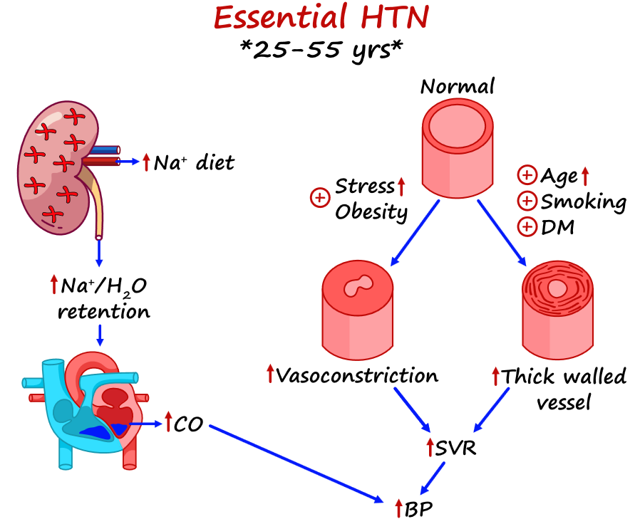
PRIMARY HTN- PATHOLOGY Increased Systemic Vascular Resistance (SVR)
-common
-describe
-catecholamines
-renin angiotensin aldosterone system
-decreased vascular compliance
most common mechanism for primary HTN
-vessel walls thicken, the lumen becomes narrower = increase BP
Catecholamines
-norepinephrine and adrenaline bind to vascular smooth muscles = vasoconstriction
Renin-angiotensin-aldosterone system
-kidney to release renin --> increases angiotensin II (AT-II) = increases aldosterone--renal sodium and water retention
Decreased vascular compliance
-increasing age, small arteries harden and thicken = raise BP t
PRIMARY HTN- PATHOLOGY
Increased Cardiac Output due to.. (describe)
-salt causes
-increased blood volume increases (2)
-peripheral vessels __________ due to
Due to increased stroke volume (common)
-expanded blood volume and occurs in patients who are "salt sensitive."
Increased blood volume increases stroke volume and cardiac output
Peripheral vessels constrict due to sodium retention, raising SVR and contributing to elevated blood pressure
Primary Essential Hypertension
-exacerbating factors
Obesity
Sleep apnea-CPAP
Increased Salt Intake
Alcohol -Raises BP perhaps by ^plasma catecholamines
Cigarette Smoking - Increases BP by ^norepinephrine
Exercise -Aerobic exercise lowers BP in previously sedentary individuals
Polycythemia -increases blood viscosity and may raise blood pressure
NSAIDS
Primary Essential Hypertension
-exacerbating factors part 2
Metabolic Syndrome (upper body obesity, insulin resistance, hypertriglyceridemia)
-Associated with both the development of HTN and an increased risk of adverse cardiovascular outcomes
Affected patient usually also have
Low HDL
Elevated catecholamines
Elevated inflammatory markers such as C-reactive protein
Secondary HTN
-age
-signs
After age of 50
-HTN resistant to three medications is another clue
(Multiple medications are usually required to control HTN in persons with DM)

Secondary Causes - Renal Disease (Chronic Kidney Disease)
-which disease
-patho
-treatment
glomerular diseases
-increased tubular sodium reabsorption = ↑ cardiac output.
Low renal perfusion, leading to activation of the RAAS --> ↑ SNS activity.
ACE/ARB

Secondary Causes - RENAL ARTERY STENOSIS
-patho
-demographic
DO NOT GIVE ACE
Reduced renal blood flow --> RAAS activation --> reabsorb sodium/water --> increased SVR --> HTN
Older patients - hx of atherosclerosis
Younger female - fibromuscular dysplasia
-overgrowth of the smooth muscles of the arteries and is the cause in young women without risk for atherosclerosis.

Secondary Causes - RENAL ARTERY STENOSIS
-labs
-diagnostic imaging
-treatment
Elevated creatinine (prerenal acute kidney injury)
Renal arteriogram - gold standard
-alternating narrowing and dilation-like beads on a string with fibromuscular dysplasia.
Give Antihypertensive agents (control blood pressure)
DO NOT GIVE ACE (↑ creatinine)
SECONDARY CAUSE - Primary HYPERALDOSTERONISM
-occurs
-caused by
-correction
unexplained hypokalemia + new hypertension
Aldosterone-producing adrenal adenoma or Idiopathic bilateral adrenal hyperplasia
-sodium and water reabsorption --> elevated blood volume (increases CO and BP)
Correction of the high aldosterone levels by medications or surgery corrects the hypertension
SECONDARY CAUSE - CUSHING SYNDROME
-patho
-sxs
-treatment
Cortisol secreting tumor on the adrenal gland
-High cortisol = ↑ sodium and water retention
-Increased expression of angiotensin-II receptors, increased effects of catecholamines and SVR
SXS
Fatigue, central obesity, muscle wasting, and osteoporosis
TREATMENT
medications or surgery corrects the hypertension

SECONDARY CAUSE - PHEOCHROMOCYTOMA
-patho
-sxs
-treatment
rare catecholamine-secreting tumor (epinephrine and norepinephrine).
SXS: headaches, sweating, tremors, flushing, agitation, and tachycardia.
Catecholamines ↑ blood pressure by ↑ cardiac output and SVR.
TREATMENT medications or surgery corrects the hypertension.

SECONDARY CAUSE - OBSTRUCTIVE SLEEP APNEA
-sxs
-describe
-diagnosed by
-treatment
SXS - obesity, loud snoring at night, daytime fatigue, depression, and excessive daytime sleepiness.
airway compression = cessation of breathing.
-activates SNS --> ↑ cardiac output and SVR = hypertension
Polysomnography.
Treatment - CPAP + medications
Medication cause of HTN
Oral contraceptive pills- estrogen and estrogen replacement
-estrogen induces hepatic production of angiotensinogen (aldosterone)
NSAIDs - COX-2 inhibition of prostaglandins (pgs normally decrease constriction)
Pseudoephedrine α-agonist used as a decongestant
-dose-related increases in BP (α-receptor mediated vasoconstriction)
Cocaine and amphetamines - α-receptor-induced vasoconstriction
Chronic systemic glucocorticoid use - ↑ sodium and water retention and potentiating the vasoconstrictive effect
Hypertensive Cardiovascular Disease - Heart complications
-Persistent high BP causes (4)
Persistent high BP
1. ↑ cardiac demand
2. ↑ afterload --> ↑ cardiac work and LV pressure.
3. LV hypertrophy (diastolic dysfunction)
4. Poor filling and reduced stroke volume --> diastolic heart failure.

Hypertensive Cerebrovascular Disease and Dementia
-predisposing cause
-systolic or diastolic
-incidence reduced/increased by
Major predisposing cause of hemorrhagic and ischemic stroke
-Correlate more with systolic than diastolic blood pressure
Incidence
-reduced: antihypertensive therapy
-increased: vascular and Alzheimer types of dementia
Hypertensive Kidney Disease
-associated with
-demographic
Nephrosclerosis
--> may result from other factors such as smoking and DM contributing to HTN
Prevalent in persons of sub-Saharan African ancestry
HYPERTENSIVE RETINOPATHY
-describe
-leads to
Narrow retinal artery lumen --> atherosclerotic = ↓ flow
-arteriovenous nicking
-microaneurysms
-small retinal hemorrhages
--> eventually cause vision loss
HTN
-sxs
Largely asymptomatic for many years
-MC = headache which is also very nonspecific
Other S/S
-Chest pain
-Confusion/altered mental status
-Blurred vision
-SOB/Signs CHF
-LVH - Displaced PMI
-Decreased urine output
TWO CATEGORIES FOR URGENT PRESENTATIONS
Uncontrolled hypertension (urgency)
-Usually asymptomatic
-Incidental finding
Hypertensive Emergency
-Uncontrolled HTN → end-organ damage
-Hypertensive encephalopathy
--> HA, somnolence, vomiting which usually improve with prompt control of HTN
-Cerebral infarct or hemorrhage
-Aortic Dissection/Rupture
HTN - laboratory findings
-recommended tests
-rule out
Recommended tests
-Hemoglobin
-Urinalysis and serum --> Creatinine
-Fasting blood glucose (FBS)
-Plasma lipids
-Serum uric acid (hyperuricemia is relative contraindication to diuretic therapy)
-Serum electrolytes
Rule out secondary causes of hypertension
-Hypokalemia- Renal artery stenosis and Hyperaldosteronism
-Urine cortisol level- Cushing's syndrome
-Urine or plasma metanephrines- Pheochromocytoma
-Plasma aldosterone and renin level- Hyperaldosteronism
HTN - imaging
ECG and Radiographs
-ECG criteria are specific but not sensitive for Left Ventricular Hypertrophy
-"strain pattern" ST-T wave changes
CXR not necessary
Treatment Therapy: Nonpharmacologic
Lifestyle modification: initially or in addition to meds
-DASH diet (high fruits, vegetables, low-fat dairy, low in saturated/total fats)
-Weight reduction
-Reduced ETOH consumption
-Reduced salt intake
-Gradually increase activity (exercise)
-Smoking cessation
Potassium supplementation NIS
Stepwise approach to hypertension management.
1. lifestyle changes (exercise, weight loss, dietary modifications)
2. one medication (unless BP is markedly elevated)
3. additional medications
First line treatment
-black/elderly and non black
1st line black/elderly
-Thiazide diuretics
-CCBs
1st line for NON-BLACK patients
-Thiazide diuretics
-ACE
-ARBs
-CCBs
THIAZIDE DIURETICS
-names
-pros
-especially helpful in specific patients
Hydrochlorothiazide + chlorthialiadone
First-line: decreases Na/H2O reabsorption, vasodilate
-dual effect on kidney and blood vessels
-prevents renal calcium stones
Patients
HTN + osteoporosis! (hypercalcemia)
THIAZIDE DIURETICS
-hyper/hypo
-contraindications
-interactions
Hypokalemia + hyponatremia
Hyper
-glycemia
-lipidemia
-uricemia --> increased risk of gout
-Ca+
Contraindications
-sulfa allergy
-pregnancy
Interactions
-lithium toxicity
LOOP Diuretics
-names
-electrolyte issues
-duration
-specific patients
Lasix/Furosemide, Bumex, torsemide
Hypokalemia (volume depletion)
Shorter duration of action
-patient who need them for fluid mgt (CHF) and those patients with renal disease
Angiotensin-Converting Enzyme Inhibitors (ACE Inhibitors)
-patho
-decrease and protect
-best for ______ patients
-Reduce mortality when
↓ aldosterone
-decrease vascular resistance + Na retention by the kidney.
-protect kidney from progressive htn damage
Initial drug of choice --> diabetes + CKD
Reduce mortality
-after MI a
-history of CHF with reduced EF.
****ACE inhibitors should be initiated first-line in these patients unless contraindicated.

ACE Inhibitor
-side effects (4)
-more effective in
-less effective in
-Dry cough (related to increased bradykinin effect)
-Hyperkalemia
-Azotemia
-Angioedema
-More effective in younger whites
-Less effective in blacks & elderly
-Less effective in predominately systolic hypertension
Angiotensin II Receptor Blocking Agents (ARBs)
-patho
-comparing to ACE side effects
-reduce
Vasodilatation and ↓ aldosterone
-cough is not a common side effect.
Reduce cardiovascular event rates (including myocardial infarction and CHF).
•Eprosartan (Teveten)
•Olmesartan (Benicar)
•Medoxomil (Edarbi)
•Valsartan (Diovan)
•Telmisartan (Micardis)
•Losartan (Cozaar)
•Candesartan (Atacand)
•Irbestartan (Avapro)

Angiotensin II Receptor Blocking Agents (ARBs)
-risk
-side effect
-patients who
Skin rashes
Risk of hypotension and renal failure (pts with bilateral renal stenosis and hyperkalemia)
Use for pts who develop cough with ACEIs
Calcium-Channel Blockers
-types
-reduce
-preferable in what patients
Nondihydropyridine - dilitiazem and verapamil
Dihydropyridine - dipine
Reduce the influx of Ca++
Preferable
-blacks
-elderly patients

Calcium-Channel Blockers -Nondihydropyridine CCBs and Dihydropyridine CCBs
-effectiveness
-reduce
-sometimes used for
-examples
Nondihydropyridine CCBs (verapamil and diltiazem)
-Less effect on vascular smooth muscle
-block calcium influx into sinoatrial (SA) nodal cells = reduce SA nodal conduction
-used to manage stable angina and coronary artery spasm as alternatives to β-blockers.
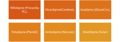
Dihydropyridine CCBs (nifedipine and amlodipine)
-potent peripheral vasodilator of vascular smooth muscle cells
-arteriolar dilation
Calcium-Channel Blockers
-side effects
-contraindications
Side effects
-Peripheral edema
•Lightheadedness
•Headache
•Bradycardia
•Constipation
•Flushing due to vasodilation
•Reflex tachycardia (dihydropyridines only)
Contraindications
-acute coronary syndromes ≠ dihydropyridine CCBs such as nifedipine because of the reflex tachycardia they cause
Second/third line drugs (HTN) -beta blockers
-used for patients who
-works by
-work better in
No longer used unless patients have ischemic heart disease or CHF
-second-line agents = inferior protection against stroke
Reducing heart rate and contractility
younger white patients
BETA BLOCKERS
-preferred in
-adverse effects
-contraindications
Preferred in patients -MI (cardiovascular issues!)
Adverse Effects
-Bronchospasm --> asthma or COPD
-Bradycardia --> dizziness and falls
-Masking hypoglycemic symptoms, such as tachycardia, tremor, sweating, and anxiety in patients prone to hypoglycemic episodes.
-Erectile dysfunction
-Nasal Congestion
Contraindications.
-asthma and bronchospasm
-Bradycardia and heart block
-Cardiogenic shock with low cardiac output and hypotension (decompensated HF)