Week 7: neurological assessment
 Call Kai
 Learn
 Practice Test
 Spaced Repetition
 Match
 Flashcards
 Knowt Play
1/109

There's no tags or description
Looks like no tags are added yet.
110 Terms
Structure and function of the nervous system
Central nervous system
peripheral nervous system
information flow of NS
Afferent (Sensory)
Efferent (Motor)
central nervous system
(CNS) consists of…
brain: Controls thinking, memory, emotions, movement, and many body functions.
spinal cord: Carries messages between the brain and the rest of the body and coordinates certain reflexes
integration and control center: is the body's control center. It receives information, processes it, and decides how the body should respond
fight or flight
peripheral nervous system
(PNS) consists of…
12 cranial nerves: Nerves that originate from the brain.
31 spinal nerves: Nerves that branch from the spinal cord
connects body to CNS: is the body's communication network. It connects the CNS to the rest of the body.
rest and digest
afferent
(sensory) = receptors to CNS
goes from body to CNS to body
efferent
(motor) = muscle and glands
body to body
two functional divisions (of PNS)
somatic system
autonomic system
somatic system
controls skeletal muscles (voluntary)
Controls voluntary movements, such as walking, and carries sensory information.
autonomic system
controls smooth muscle, cardiac muscles and glands
works unconsciously and maintains homeostasis
Controls involuntary functions, such as heart rate, digestion, and sweating.
developmental competence (infants)
Neurologic system is not completely developed at birth
Movement is directed by reflexes (should disappear by 1 y.o)
Persistence of primitive reflexes is an indication of CNS dysfunction
Sensation is also limited at birth
Sensory and motor development proceed with gradual acquisition of myelin (more accurate responses with myelination)
primitive reflexes of infant
stepping: 0-2 months
rooting: 0-4 months
moro: 0-6 months
grasp: 0-6 months
tonic neck: 0-7 months
babinski: 0-12 months
neurologic changes in older adults
Neuron loss leads to loss of muscle tone, impaired fine coordination and agility, loss of vibratory sense at the ankle, decreased reflexes.
Velocity of nerve conduction decreases causing slower reaction times.
Delayed impulse conduction at synapses can cause diminished sensation to touch and pain, taste, and smell.
subjective neurologic assessment
headache history
head injury details
dizziness and vertigo
seizure characteristics
tremors and weakness
sensory and speech issues
headache history
Frequency, onset timing, pattern recognition, presence of auras or premonitory feelings, associated symptoms like light/sound sensitivity, nausea/vomiting, and red flags 'worst headache of my life' (WHOL).
head injury details
Mechanism of injury, eyewitness or observer descriptions, and, if repetitive, the number of head injuries sustained.
dizziness and vertigo
Dizziness includes lightheadedness, wooziness, and spinning sensations; vertigo specifically refers to a rotational or spinning sensation affecting either the patient or the environment. Syncope involves fainting or near-fainting episodes.
seizure characteristics
Course and duration, warning signs, motor activity onset and progression, associated symptoms such as color changes, loss of consciousness, automatisms, incontinence, postictal phase details, precipitating factors, medications, and coping strategies.
tremors and weakness
Tremors: involuntary shaking, vibrating, or trembling caused by anxiety, intention, or resting.
Weakness: paresis (partial paralysis) or paralysis (total motor loss).
sensory and speech issues
Incoordination, numbness or tingling (paresthesia), difficulty swallowing (dysphagia), saliva management, difficulty speaking (dysarthria, dysphasia), vision changes, and relevant past medical history.
objective assessment
mental status
cranial nerves
muscle strength
mnemonic for nerve names
Oh Once One Takes The Anatomy Final Very Good Vacations Are Happening
cranial nerves
Olfactory (I)
Optic (II)
Oculomotor (III)
Trochlear (IV)
Trigeminal (V)
Abducens (VI)
Facial (VII)
Vestibulocochlear (Acoustic) (VIII)
Glossopharyngeal (IX)
Vagus (X)
Spinal Accessory (XI)
Hypoglossal (XII)
cranial nerve I
olfactory
main function: smell
type: sensory
simple test: identify familiar scents
cranial nerve II
optic
main function: vision
type: sensory
simple test: Suellen chart, visual fields
cranial nerve III
oculomotor
main function: eye movement, pupil constriction
type: motor
simple test: PERRLA, extra ocular movements
cranial nerve IV
trochlear
main function: downward and inward eye movement
type: motor
simple test: follow finger down toward nose
cranial nerve V
trigeminal
main function: facial sensation, chewing
type: both
simple test: light touch on face; clench teeth
cranial nerve VI
abducens
main function: lateral eye movement
type: motor
simple test: move eyes side to side
cranial nerve VII
facial
main function: facial expression, taste (ant. 2/3 tongue)
type: both
simple test: smile, frown, puff cheeks, taste test
cranial nerve VIII
vestibulocochlear
main function: hearing and balance
type: sensory
simple test: whisper test, tuning fork, balance
cranial nerve IX
Glossopharyngeal
main function: taste (post 1/3), swallowing, gag
type: both
simple test: test gag reflex, assess swallowing
cranial nerve X
vagus
main function: speech, swallowing, autonomic control
type: both
simple test: say ahhh observe uvula rise midline
cranial nerve XI
spinal accessory
main function: shoulder and neck movement
type: motor
simple test: shrug shoulders, turn head
cranial nerve XII
hypoglossal
main function: tongue movement and articulation
type: motor
simple test: stick out tongue, note deviation
olfactory (I)
function: smell
sensory
Have the client close their eyes, block one nostril, and identify a familiar scent (like coffee or vanilla)
optic (II)
function: vision and visual fields
sensory
Test visual acuity using a Snellen eye chart, test peripheral visual fields by confrontation, and inspect the ocular fundus with an ophthalmoscope
oculomotor (III)
Extraocular eye movements, pupil constriction, and eyelid elevation
motor
Check pupils for size, shape, direct/consensual reaction to light, and accommodation (PERRLA), and inspect for ptosis (drooping eyelid)
trochlear (IV)
downward and inward eye movement
motor
Ask the client to follow your finger as you move it down toward their nose
trigeminal (V)
Facial sensation; motor control of chewing muscles
both
Motor: Have the client clench their teeth while you palpate the masseter and temporalis muscles for strength and symmetry.
Sensory: Lightly touch a cotton wisp to the client's forehead, cheeks, and chin while their eyes are closed
abducens (VI)
Lateral (outward) eye movement
motor
Ask the client to move their eyes side-to-side.
(Note: CN III, IV, and VI are commonly assessed together during extraocular movement testing.)
Facial (VII)
Facial expressions; taste on the anterior 2/3 of the tongue
both
Motor: Observe facial symmetry while asking the client to smile, frown, close eyes tightly, raise eyebrows, and puff out cheeks.
Sensory: Taste test on the anterior portion of the tongue
Vestibulocochlear (VIII)
Hearing and balance
sensory
Assess hearing using normal conversation, the whispered voice test, or a tuning fork
glossopharyngeal (IX)
Swallowing, gag reflex, and taste on the posterior 1/3 of the tongue
both
Test swallowing and taste on the back of the tongue. Works closely with CN X
vagus (X)
Speech, swallowing, gag reflex, and parasympathetic autonomic control
both
Ask the client to say "ahh" and observe that the uvula and soft palate rise symmetrically in the midline; touch the posterior pharynx to test the gag reflex
spinal accessory (XI)
Movement of sternocleidomastoid and trapezius muscles (neck and shoulder)
motor
Ask the client to shrug shoulders against your downward resistance and turn their head side-to-side against your hand's resistance
hypoglossal (XII)
Tongue movement and speech articulation
motor
Ask the client to stick out their tongue. Look for midline alignment and note any tremor or deviation to one side
examine pupils
check for PERRLA
PERRLA
Pupils
Equal
Round
Reactive to Light (direct and consensual response)
and
Accommodation
normal pupil size
Generally 3–5 mm in ambient light
anisocoria
Unequal pupil sizes (can be a normal anatomical variation in ~20% of people or a sign of increased intracranial pressure)
ptosis
Drooping of the upper eyelid (CN III defect or neuromuscular disease)
nystagmus
Back-and-forth oscillation of the eyes (mild end-point nystagmus at extreme lateral gaze can be normal, but persistent nystagmus is abnormal)
motor system evaluation
muscle size
muscle strength
muscle tone
involuntary movements and pronator drift
muscle size
Evaluate the size of muscles to identify any atrophy or hypertrophy which may indicate underlying neurological or muscular conditions.
muscle strength
Assess muscle strength by testing resistance against force in various muscle groups to detect weakness or asymmetry.
using the 0-5 muscle strength scale
using the 0-5 muscle strength scale
5: Normal strength
4: Slight weakness
3: Movement against gravity only
2: Movement with gravity eliminated
1: Flicker of contraction: trace
0: No movement: no muscle contraction at all
muscle tone
Check for muscle tone abnormalities including limited range of motion, flaccidity (decreased resistance, hypotonia), spasticity, and rigidity which indicate central nervous system dysfunction.
involuntary movements and pronator drift
Observe for involuntary movements such as tics, tremors, fasciculations, myoclonus, and chorea.
Tics - sudden, brief and repetitive movements (e.g. blinking)
Tremors - rhythmic shaking, common in Parkinsons
Fasciculations -Small muscle twitches seen under the skin (ALS)
Myoclonus - sudden quick jerks
Chorea-irregular jerky, dance-like movements (Huntington’s)
Test for pronator drift in upper and lower extremities as a sign of subtle motor weakness.
Tics
sudden, brief and repetitive movements (e.g. blinking)
tremors
rhythmic shaking, common in Parkinsons
fasciculations
small muscle twitches seen under the skin (ALS)
myoclonus
sudden quick jerks
cerebellar function tests
cerebellum: controls motor coordination, smooth movements, and balance.
balance tests
balance tests
gait
normal
tandem walking
the romberg test
gait
observe as the person walks 10 to 20 feet, turns, and returns to starting point
normal gait
gait is smooth, rhythmic, and effortless; opposing arm swing is coordinated; the person turns smooth; step length about 15 inches from heel to heel
tandem walking
Ask the person to walk straight line in heel-to-toe fashion
romberg test
Ask the person to stand up with feet together and arms at sides; when in stable position, ask the person to close eyes and to hold position for about 20 seconds
pronator drift
The client stands with arms extended forward, palms up, and eyes closed for 20 seconds. A positive drift occurs when one arm downwardly drifts and pronates (palms turn down), indicating an upper motor neuron lesion (e.g., stroke)
negative romberg test
A negative Romberg (expected) means the client maintains posture with minimal swaying. A positive Romberg means the client loses balance or steps out when eyes are closed (indicates sensory ataxia or vestibular impairment)
sensory system assessment
Tests sensory pathways conveying pain, temperature, touch, position, and vibration from peripheral nerves to the brain.
sharp vs dull sensation
light touch
vibration sense
stereognosis
graphesthesia
sharp vs dull sensation
Test patients by distinguishing sharp sensations from dull sensations to evaluate pain perception and sensory nerve function.
light touch
Use a wisp of cotton to lightly touch the skin in multiple dermatomes to assess the patient's ability to perceive gentle tactile stimuli.
vibration sense
Apply a tuning fork over bony prominences using lower pitch frequencies (128Hz or 256Hz) to assess vibration sense as these have a slower decay.
stereognosis
Evaluate the patient's ability to identify familiar objects by touch without looking, testing finely localized touch perception
graphesthesia
Test the patient's ability to recognize numbers or letters traced on the palm, assessing cortical sensory processing.
deep tendon reflexes
Automatic body responses that happen when a tendon is tapped with a reflex hammer.
Help nurses to check functions of nerves and spinal cord.
Limb should be relaxed on assessment, and muscle partially stretched
Bilateral comparison: responses should be equal
DTR 4-Point Grading Scale
reflex response graded on 4 point scale
4+: Very brisk, hyperactive with clonus (indicates CNS disease).
3+: Brisker than average (may be normal or indicative of disease).
2+: Average / Normal.
1+: Diminished / low normal.
0: Absent (no response)
biceps reflex
C5 to C6
Support the person’s forearm on yours; place your thumb on biceps tendon and strike a blow on your thumb
Normal response is contraction of biceps muscle and flexion of forearm

triceps reflex
C7 to C8
Tell the person to let arm “just go dead” as you strike triceps tendon directly just above the elbow
Normal response is extension of forearm

brachioradialis reflex
C5 to C6
Hold the person’s thumbs to suspend forearms in relaxation and strike forearm directly, about 2 to 3 cm above radial styloid process
Normal response is flexion and supination of forearm

quadriceps reflex
L2 to L4 (“Knee Jerk”)
Let lower legs dangle freely to flex knee and stretch tendons; strike tendon directly just below patella
Normal response is extension of lower leg

achilles reflex
(“Ankle Jerk”)
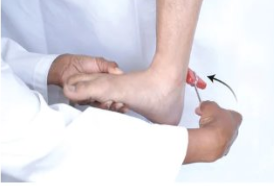
Position the person with knee flexed; hold foot in dorsiflexion and strike Achilles tendon directly
Normal response is foot plantar flexes against your hand
glasgow coma scale (GCS)
is a neurological assessment tool nurses use to determine a patient's level of consciousness and how well they respond to stimulation and it measures 3 responses: Eye Opening, Motor Response, and Verbal Response.
It is commonly used for patients with:
Head injuries or brain trauma
Stroke
Altered mental status
Unconsciousness or coma
eye opening
This measures how easily the patient opens their eyes, (maximum 4 points)
4 = Spontaneous, Opens eyes without being asked
3 = To speech, Opens eyes when spoken to
2 = To pain, Opens eyes only with painful stimulation
1 = No response, Does not open eyes
motor response
This measures how well the patient can move their body in response to commands or stimulation (max 6 points)
6 = Obeys commands, Follows instructions, such as squeezing your hand
5 = Localizes pain, Purposefully reaches toward the painful stimulus to remove it
4 = Withdraws from pain, Pulls the affected body part away from pain
3 = Abnormal flexion, Arms bend inward abnormally (decorticate posturing)
2 = Abnormal extension, Arms straighten and extend abnormally (decerebrate posturing)
1 = No response, No movement
verbal response
This measures how well the patient can speak and whether their responses make sense (max 5 points)
5 = Oriented, Knows who they are, where they are, and the time or situation
4 = Confused conversation, Can speak but is confused or disoriented
3 = Inappropriate words, Says random words that do not make sense in the conversation
2 = Incomprehensible sounds, Makes sounds such as moaning but no understandable words
1 = No response, Makes no verbal sounds
EVM
eyes, verbal, motor
scoring maximum is 15
decreasing GCS score is a warning sign that the patient's neurological condition may be worsening and needs prompt reassessment and escalation
neuro check
mental status
orientation
cranial nerves tested
strength and movement
mental status
Evaluate the patient's level of consciousness, alertness, and cognitive function to establish baseline neurologic status.
orientation
Check patient's awareness of person, place, and time to evaluate cognitive orientation and neurological status
cranial nerves tested
Examine cranial nerves 2, 3/4/6, 7, and 8 through visual acuity, eye movements, facial muscle function, and hearing assessments.
strength and movement
Assess muscle strength and voluntary movements to identify any weakness or abnormal motor function.
diabetic neuropathy screening
Use a monofilament to touch the skin at each numbered site for 1-2 seconds.
Ask the patient to identify when and where they feel the touch.
This test helps assess the presence of neuropathy by detecting sensory loss.
Perform at multiple standardized sites on the feet to ensure comprehensive screening.
Early detection through this screening can prevent complications such as ulcers or infections
stroke symptoms
Weakness or numbness in face, arms, legs; especially on one side of body
Confusion, trouble speaking or understanding
Changes in vision, such as blurry vision or partial/complete vision loss in one or both eyes
Trouble with walking, dizziness, loss of balance or coordination
Severe headache with no explanation
stroke
occurs due to a sudden interruption of blood flow to the brain.
FAST tool
is a quick assessment used by nurses and other healthcare professionals to recognize the warning signs of a stroke
Face, Arms, Speech, Time
Face
face drooping
ask the patient to smile.
Does one side of their face droop?
Is their smile uneven?
Abnormal: One side of the face does not move normally.
Arms
arm weakness
Ask the patient to raise both arms.
Does one arm drift downward?
Is one arm weaker than the other?
Abnormal: The patient cannot keep both arms raised equally
Speech
speech difficulty
Ask the patient to repeat a simple sentence, such as "The sky is blue."
Is their speech slurred?
Are they having trouble speaking?
Are they using incorrect words?
Abnormal: Speech is slurred, unusual, or difficult to understand