HEENT Lecture 2
 Call Kai
 Learn
Learn Practice Test
 Spaced Repetition
 Match
Match Flashcards
 Knowt Play
1/160

There's no tags or description
Looks like no tags are added yet.
Name | Mastery | Learn | Test | Matching | Spaced | Call with Kai | Chat |
|---|
No analytics yet
Send a link to your students to track their progress
161 Terms
inflammation of the nasal mucosa is what?
Rhinitis
______ Rhinitis:IgE-mediated inflammatory response to allergens (e.g., pollen, mold, dust mites, animal dander). Often associated with a history of atopic disease.
Allergic
Infectious:Usually viral; _______ is the most common cause of the common cold.
rhinovirus
_______ (Nonallergic): Nonallergic, noninfectious rhinitis triggered by factors such as temperature changes, strong odors, smoke, or humidity.
Vasomotor Rhinitis
Samter Triad / Aspirin-Exacerbated Respiratory Disease (AERD):
Asthma
Chronic rhinosinusitis with nasal polyps
Aspirin/NSAID sensitivity
ClinicalManifestations RHINITIS
______
Nasal congestion. Clear, watery rhinorrhea
Nasal ______ - especially allergic rhinitis
Postnasal ______
Cough
May have associated eye, ear, or throat symptoms
sneezing
itching
drainage
RHINITIS
PhysicalExamination
Allergic
Pale or violaceous, boggy turbinates
Clear rhinorrhea
Conjunctival findings may occur
Allergic ____:dark blue-gray/purple discoloration beneath the eyes
Allergic ____:repeated upward rubbing of the nose
____ nasal creasemay develop from chronic rubbing
Viral
Erythematous, edematous nasal mucosa/turbinates
____ (Nonallergic)
Boggy nasal mucosa
Nasal congestion and watery rhinorrhea following irritant exposure
Symptoms are often transient
shiners
salute
transverse
vasomotor
MANAGEMENT OF ALLERGIC RHINITIS
Avoidance of allergen and environmental control; exposure _________.
Intranasal ________: Mometasone, ________, Triamcinolone.
Indications: most effective medication for Allergic rhinitis (moderate to severe or persistent) especially with ______ _______
reduction
glucocorticoids; fluticasone
nasal polyps
MANAGEMENT OF ALLERGIC RHINITIS
Decongestants:
-Intranasal: Oxymetazoline, _________, Naphazoline.
_____: Pseudoephedrine.
MOA: may improve congestion but have ____ effect on rhinorrhea, sneezing, or pruritus.
Should not exceed >3-5 days of intranasal decongestant treatment in order to prevent nasal mucosal dependency and ______ __________ (rebound congestion).
phenylephrine
oral
little
Rhinitis medicamentosa
MANAGEMENT OF _______ RHINITIS
2nd-generation oral ________ (eg, _______, Cetirizine, Fexofenadine), antihistamine nasal spray (eg, Azelastine,Olopatadine)
ALLERGIC
antihistamine; loratidine
MANAGEMENT OF ALLERGIC RHINITIS
Mast cell stabilizers (Cromolyn nasal spray).**************
Acute viral Rhinosinusitis (common cold)
Symptomatic inflammation of the _____ ______ and ______ sinuses. Acute if ___ weeks of symptoms.
The vast majority of cases of acute rhinosinusitis (ARS) are due to ____ infection (common cold) -Rhinovirus, influenza, parainfluenza, & other viruses.
nasal cavity
paranasal
<4
viral
Acute rhinosinusitis
CLINICAL MANIFESTATIONS
_____ ______ & obstruction, clear rhinorrhea, hyposmia (decreased smell).
Viral symptoms: cough, __, malaise.
Symptoms of Eustachian tube dysfunction: ear pain, fullness or pressure, hearing loss, or tinnitus.
facial pain, pressure or fullness that is worse or localized with ___ ____ & leaning forward;headache, malaise, purulent nasal discharge, fever, nasal congestion, maxillary teeth discomfort, ear pressure or fullness, halitosis.
nasal congestion
HA
bending down
Physical examination Acute viral Rhinosinusitis (common cold):
• ______, _____ nasal mucosa without significant intranasal purulence.
erythematous
engorged
MANAGEMENT - Acute viral Rhinosinusitis (common cold):
_______ management as symptoms are self-limited, with symptoms resolution usually within ___ days. Although symptoms may persist >10 days, there is usually some improvement by day 10. If fever is present, it is usually present early in the illness & disappears within the first 24-48 hours.
supportive
7-10
MANAGEMENT - Acute viral _______ (common cold):
Sinusitis: symptomatic management - _____ are considered the mainstay of therapy for Acute sinusitis (eg, _______, NSAIDs), nasal lavage (saline irrigation), & intranasal Glucocorticoids. Decongestants (promote sinus drainage), antihistamines, and mucolytics.
Rhinosinusitis
analgesic
Acetaminophen
_____
Bacteria secondarily infect and inflame the sinus cavity, with <4 weeks duration.
Acute bacterial infection occurs in only 0.5-2% of episodes of Acute Rhinosinusitis (vast majority of sinusitis is viral).
ARBS
TERMINOLOGY - Acute Bacterial Rhinosinusitis (ARBS
Acute rhinosinusitis-symptoms< ___ weeks.Symptoms usually resolveof improve within __ days.
Subacute rhinosinusitis symptoms for 4-12 weeks
Chronic rhinosinusitis symptoms persist >12 weeks
Recurrent acute rhinosinusitis >/= 4 episodes ofAcute Rhinosinusitisper year, with interim symptom resolution.
4
10
Acute Bacterial Rhinosinusitis (ARBS)
PATHOPHYSIOLOGY
____ ___ ___ :most commonly occurs as a complication of viral infection. Allergic or nonallergic rhinitis, mechanical obstruction of the nose, dental infections, impaired mucociliary clearance (eg, Cystic fibrosis), or with cigarette smoking.
MICROBIOLOGY
The most common bacteria associated with ABRS are ____ ___ & _____ ____ (both make up ~75%), and Moraxella catarrhalis (organisms similar to Acute otitis media). Microaerophilic & anaerobic bacteria associated with dental root infections.
impaired sinus drainage
Streptococcus pneumoniae
H influenzaie
more likely to be bacterial or viral if sxs have been going on > 10 days?
acute bacterial rhinosinusitis
bacterial
Acute Bacterial Rhinosinusitis
CLINICAL MANIFESTATIONS
Acute rhinosinusitis: facial pain. pressure, or fullness worse with bending down & leaning forward, purulent nasal discharge, nasal congestion or obstruction, headache, fever, ear pressure/fullness, halitosis, hyposmia <4 weeks (symptoms can't distinguish ____ vs ____).
bacterial
viral
Acute Bacterial Rhinosinusitis
Suggestive of bacterial cause; clinical criteria for diagnosis:oPersistent symptoms that last ≥__ days without evidence of clinical improvement.
oA ____ pattern: ("double worsening") -symptoms initially start to improve but then worsen 5-6 days later.
10
biphasic
PHYSICAL EXAMINATION - _______
Erythema/edema over the involved area; watery or purulent drainage in the nose or posterior pharynx.
sinus tenderness to palpation - in order of frequency -Maxillary > ethmoid > frontal > sphenoid.
Maxillary: most common cheek pain or pressure that can radiate to the upper incisors as well as decreased transillumination of the cheek. Ethmoid: tenderness to the high lateral wall of the nose.
Frontal: pain around the eyes and the forehead. Sphenoid: mid head tenderness.
Acute bacterial rhinosinusitis
DIAGNOSIS - Acute ______ Rhinosinusitis
Primarily a ______ diagnosis. Imaging is not indicated if classic presentation & uncomplicated.
___ is the imaging test of choice if imaging is needed [eg, signs or symptoms suggesting spread of infection beyond the paranasal sinuses and nasal cavity (CNS, orbit, or surrounding tissues)]. Sinus radiographs not usually needed. If ordered, Water's view most helpful radiograph view.
• _______ or aspirate: definitive diagnos is but usually not needed in most uncomplicated cases.
bacterial
clinical
CT
biopsy
Acute Bacterial Rhinosinusitis
MANAGEMENT
Symptomatic management: _____ (eg, NSAIDs), nasal _____ (saline washes), intranasal ______.
Decongestants may promote sinus drainage, antihistamines, mucolytics.
analgesics
lavage
glucorticoids
Acute Bacterial Rhinosinusitis
MANAGEMENT
firstly — _____
Antibiotics:
Indications: symptoms should be present for an extended period of ≥ __ days with worsening of symptoms or earlier if severe.
_______ is often the antibiotic of choice.
Second line: _______ (can be used first-line if ___ allergy). Third-generation oral Cephalosporin (eg, Cefixime, Cefpodoxime) with or without Clindamycin.
Respiratory fluoroquinolones (Levofloxacin, Moxifloxacin) usually reserved to ____ resistance.
NSAIDs
10-14
Amox Clav
doxycycline; PCN
prevent
Chronic ________
• Inflammation of the nasal cavity and paranasal sinuses for ≥___ consecutive weeks.
ETIOLOGIES
may include ______ (acute, chronic), allergies, & immunological or structural diseases.
Bacterial: _____ _____ -most common, and anaerobes. Pseudomonas aeruginosa.
Granulomatosis with polyangiitis [Wegener's granulomatosis] often _____.
Fungal: _____ -most common fungal cause. Mucormycosis -second most common fungal cause.
Rhinosinusitis
12
infection
staph aureus
necrotic
aspergillus
CLINICAL MANIFESTATIONS - ______ _______
Similar to Acute sinusitis but duration >/=12 weeks[1] nasal obstruction & congestion, [2] facial pain &/or pressureworse with bending down & leaning forward, [3] mucopurulent nasal discharge, & [4] decreased olfaction.Headache, malaise, fever. Cough may be seen in children.
Chronic rhinosinusitis
Chronic Rhinosinusitis DIAGNOSIS
Sinus CT imaging or rhinoscopy or nasal endoscopy with decongestant. Allergy evaluation.
______ testing in all patients(often associated with Asthma, Allergic rhinitis, &/or nasal polyps).
Biopsy or histology -allows for identification of the organism & may direct appropriate management.
MANAGEMENT
Depends on _____: The goal of therapy is to promote sinus drainage, reduce edema, & eliminate infection oeg, combination of nasal irrigation, intranasal glucocorticoids (or oral), & ENT follow-up
______ (if bacterial) with ENT referral
o____—_____ first line. Alternatives include Clindamycin and combinations of Metronidazole with a second-or third-generation Cephalosporin, Macrolides, or Trimethoprim-sulfamethoxazole.
allergy
etiology
Abx; amox clav
Mucormycosis (Zygomycosis)
Angioinvasive ______ infection that infiltrates the ______, ____, & ____.
- ________: the fungus rapidly dissects the ____ canals and eyes into the brain. High ____-.
fungal
sinuses, CNS, lungs
angioinvasion
nasal
mortality
MUCORMYCOSIS
Etiologies:
Non-Aspergillus fungal causes: _______ species (eg, R. oryzae), _____, & _______.Cunninghamella, Saksenaea, and Apophysomycesmay also be causes.
Rhizopus
Mucor
Rhizomucor
RISK FACTORS for oral hairy leukoplakia
_____ _____ [DKA & hyperglycemia (most common)], other ________ states(eg, post-transplant, chemotherapy, HIV, heme malignancy), Deferoxamine therapy, iron overload states.
Diabetes mellitus
immunocompromised
_________
CLINICAL MANIFESTATIONS
Rhino-orbital-cerebral infections: Sinusitis (facial pain/pressure worse with bending down & leaning forward, headache, malaise,purulent nasal discharge, fever & nasal congestion) with rapid progression to the orbit & brain. Lung involvement also common.
PHYSICAL EXAM:±erythema, swelling, necrosis, or black ______ on the palate, nasal mucosa, or face.
DIAGNOSIS
Biopsy and histopathologic examination of involved tissue _____ _____ ______ with irregular right-angle (90°)branching. PCR testing of the blood and tissues obtained.
MRI & CT are often obtained but show nonspecific findings.
MANAGEMENT
The standard management of Mucormycosis involves early diagnosis, reversal of risk factors and underlying illness (eg, hyperglycemia, acidosis, cessation of immunosuppressive agents when possible), early aggressive surgical debridement, and prompt administrationof IV antifungals.
________ first line+ __ surgical debridement of necrotic areas. The lipid formulation of Amphotericin B preferred (allows for higher dose delivery with less nephrotoxicity).
Second line: Posaconazole or Isavuconazoleare alternatives to or in addition to Amphotericin B.
murcormycosis
eschar
non-septate broad hyphae
IV Amphotericin B
aggressive
Nasal Polyps
• _____ _______ most common cause. May be seen with Cystic Fibrosis.

allergic rhinitis
CLINICAL MANIFESTATIONS - Nasal Polyps
• Most are ______ findings but if large, can cause nasal obstruction or _____ (decreased smell).
incidental
anosmia
DIAGNOSIS- Nasal Polyps
Direct visualization: _____ boggy mass on the nasal _____.
May have findings associated Allergic rhinitis eg, pale or violaceous, boggy turbinates & ______ mucosa of the conjunctiva.
pale; mucosa
cobblestone
MANAGEMENT - NASAL POLYPS
_____ ________ most effective pharmacologic therapy for Nasal polyps
Functional endoscopic sinus surgery if very large or if medical therapy is unsuccessful.
intranasal corticosteroids
Epistaxis
Anterior Epistaxis
Source: _______ _______ (anterior septum) IMPORTANT TO KNOW
Common Cause: _____ (nose picking, minor injury)
Presentation: Usually _______ bleeding
Usually self-limiting & managed in outpatient settings
Kiesselbach’s plexus
trauma
unilateral
Management of EPISTAXIS
1 -Direct ______≥15 min, seated, leaning forward
2 -Topical _______(oxymetazoline, _______) - spray on gauze, spray in nose, then pressure
3 -_______(silver nitrate/electrocautery) –only if site ______. only if #2 doesnt work
4 -Nasal _______(balloon, tampon, gauze) if persistent. if #1-3 doesn’t work
Check for ______ or anticoagulant use if recurrent/severe
pressure
vasoconstrictors; phenylephrine
cauterization; visualized
packing
coagulopathy
Posterior Epistaxis
Source: ?????
Presentation: _______ bleeding, blood into posterior ______, often heavier
Sphenopalatine artery branches& Woodruff’s plexus
bilateral; pharynx
Management - Posterior Epistaxis
1. ENT consultation/referral
______ _______ (balloon catheter or specialized packs)
Less common but more _____ than anterior epistaxis
May have to reversecoagulopathy or anticoagulant use
posterior packing
serious
32 teeth
back right is #1
Innervation of Oral Cavity Structures
Superior and inferior _____ nerves (branches of CN __)
alveolar
V
Hard Palate
Front 2/3 of the _____ of the mouth
Bony: maxillae and palatine bones
_____ rests against it
roof
tongue
Soft Palate
____ 1/3 of the roof of the mouth
Movable and ends with the _____
posterior
uvela
Salivary Glands
Function: Secrete saliva
Major Glands:
______ - Anterior and inferior to the ear, overlying the mandibular ramus and masseter muscle
_________ - along the body of the mandible, partly above and below posterior half
________ - smallest, deepest; supplied by sublingual and submental arteries
parotid
submandibular
sublingual
Dental Caries
Overview
Definition: Breakdown of tooth hard tissue caused by ______ and _____ from oral bacteria
enzymes; acids
Dental Caries
Progression:
Enamel Demineralization–Early ______ lesion.
whitespot
Dental Caries
Progression:
Enamel Demineralization–Early whitespot lesion.
Cavity Formation–cavity develops if ______.
Pulpitis–Infection reaches pulp → severe ______.
Pulp Necrosis–______ of pulp tissue.
_____ _______–Infection spreads to surrounding bone.
cavity
toothache
death
periapical abscess
Dental Caries
Progression:
Enamel Demineralization–Early whitespot lesion.
Cavity Formation–cavity develops if untreated.
______–Infection reaches pulp → severe ____.
Pulp Necrosis–Death of pulp tissue.
Periapical Abscess–Infection spreads to surrounding bone.
pulpitis
toothache
Dental Caries
Progression:
Enamel _______–Early whitespot lesion.
Cavity Formation–cavity develops if untreated.
Pulpitis–Infection reaches pulp → severe toothache.
____ _____–Death of pulp tissue.
Periapical Abscess–Infection spreads to surrounding bone.
demineralization
pulp necrosis
Dental Caries
Progression:
Enamel Demineralization–Early whitespot lesion.
Cavity Formation–cavity develops if untreated.
Pulpitis–Infection reaches pulp → severe toothache.
Pulp Necrosis–Death of pulp tissue.
_____ _______–Infection spreads to surrounding ____.
Periapical abscess
bone
Dental Abscess
Dental abscess (general term):A localized collection of pus associated with a tooth or supporting structures -Can be periapicalor periodontal.
Periapical abscess:
Located at the apex of the tooth root.
Cause -Usually due to untreated dental caries(see previous slide)
Clinical Features:Severe, throbbing tooth pain
Tender to percussion
Swelling of gingiva/buccal mucosa near root apex
Possible systemic symptoms if spreading
Treatment:Referral to specialist –Dental or Ora &l Maxillofacial Surgery (OMFS)Root canal, incision & drainage, or extraction
Antibiotics only if systemic involvementor delayed definitive care

Dental Abscess only way to confirm
CT Neck w/ contrast (face and down)
Dental Abscess
Periodontal (gingival) abscess:
Located in the _____ ____ (gingiva, periodontal ligament, alveolar bone).
Cause -Foreign body (e.g., popcorn husk, toothbrush bristle) ortraumatogingiva
Clinical Features:
Localized, painful swelling of gum tissue
No pulp involvement
Tooth typically vital
Treatment:Remove foreign body/irritant
Incision & Drainage
Warm saline rinses
Antibiotics ____ needed unless spreading infection.

supporting tissue
rarely
Dental Pain
Treatment
Analgesics: ibuprofen, naproxen; narcotics if needed
Tooth/regional block for temporary relief
Antibiotics:
Mild: ______ 500 mg QID
Moderate: ____-____ 875 mg BID or Clindamycin 450 mg TID
Severe/immunocompromised: ___ antibiotics
pencillin
amoxicillin-clavulanate
IV
Ludwig’s Angina
- deep ____ infection
- commonly caused from tooth infection; which can work its way down to neck
- deep infection close to airway - susceptible to TRUE _____ EMERGENCY at any time
- ____ spread
- involves multiple _____
neck
airway
rapidly
organisms
Ludwig's Angina
Deep Neck Infection - ______
Most common origin: ______ infections
Abscess
Odontogenic
Ludwig angina: Most common neck space infection

Rapidly spreading _____ of the _____ of the mouth
Usually from mandibular tooth infection
_______: can cause rapid airway compromise → may require surgical airwa
cellulitis; floor
emergency
Ludwig’s Angina
ORGANISMS:
Microbiologic isolates include streptococci, staphylococci,Bacteroides, andFusobacterium
Patients with diabetes may have different flora, includingKlebsiella, and a more aggressive clinical course.
RISK FACTORS: most commonly due to spread of oral flora secondary to ____ ____ _____ & dental infections (second or third mandibular molars). Increased incidence in Diabetes & HIV.
poor dental hygeine
CLINICAL MANIFESTATIONS - Ludwig’s Angina
• Fever, chills, malaise, stiff neck, dysphagia, drooling, muffled voice.
•_____ signs: stridor, difficulty managing secretions, & cyanosis (requires emergent airway management).
Swelling of the tongue can lead to airway compromise.
late signs
PHYSICAL EXAMINATION - Ludwig’s Angina

_____, ______ swelling, "woody" induration, & ______ of the upper neck & under the chin (may have palpable crepitus)
_____ on the floor of the mouth
Edema, erythema and tenderness of floor of the mouth
tender
symmetric
erythema
pus
Ludwig's Angina
DIAGNOSIS
????

CT Neck w/ contrast
MANAGEMENT IF IMMUNOCOMPETENT LUDWIG’S ANGINA
Secure the airway if needed
- Endotracheal tube intubation, _______, tracheostomy
Broad Spectrum IV antibiotics:
- ________ (unasyn) OR Ceftriaxone + Metronidazole
OR Clindamycin + Levofloxacin.
Add _______ if MRSA suspected
Emergent ENT Consultation
Surgical drainage usually required
cricothyrotomy
Ampicillin-sulbactam
Vancomycin
MANAGEMENT IF IMMUNOCOMPROMISED - LUDWIG’S ANGINA
Secure the airway if needed.Endotracheal tube intubation,cricothyrotomy, tracheostomy
Broad Spectrum IV antibiotics: _______+ Metronidazole (OR Imipenem, Meropenem, Piperacillin-tazobactam (zosyn)
Add Vancomycin if MRSA is suspected.
Emergent ENTConsultationSurgical drainage usually required
Cefepime
Acute Pharyngitis & Tonsillitis
____ _______ viruses - most common overall cause of pharyngitis - ______, Rhinovirus, Enterovirus, _____, Respiratory syncytial virus, Influenza A & B, Herpes zoster virus.
viral respiratory
adenovirus
EBV
Acute Pharyngitis & Tonsillitis
Bacterial: ____ Streptococcus (S. _____) most common ______ cause of Pharyngitis.
Group A
pyogenes
bacterial
CLINICAL MANIFESTATIONS - _____ _____ _______
Sore throat worsens with swallowing, pain on swallowing or with phonation.
Cervical lymphadenopathy.
Viral symptoms: cough (not always), hoarseness, rhinorrhea, coryza, conjunctivitis, diarrhea
ACUTE PHARYNGITIS & TONSILLITIS
PHYSICAL EXAMINATION - Acute Pharyngitis & Tonsillitis
May have____ _____ ______ erythema, edema, with or without exudate, cervical adenopathy.
Anterior stomatitis: may be associated with discrete ulcers or vesicles; vesicular or petechial pattern on the soft palate and tonsils.
tonsillar and pharyngeal
DIAGNOSIS - ACUTE PHARYNGITIS & TONSILLITIS
Usually _____ . Most cases of viral pharyngitis require no specific diagnostic testing.
There are 3 notable exceptions where testing may be indicated: [1] suspected Influenza, [2] Infectious mononucleosis, or [3] Acute retroviral syndrome (HIV).
Rapid strep or throat culture may be performed to rule out bacterial cause if suspected.
MANAGEMENT
____ _______ mainstay of treatment oral hydration, warm saline gargles, topical anesthetics, lozenges, NSAIDs. Patients unable to tolerate oral hydration may need IV fluids.
clinical
symptomatic mgmt
Streptococcal Pharyngitis (strep Throat)
__________ (Streptococcus pyogenes)
Rarein children <3 years of age.
Highest incidence of Rheumatic fever in untreated children 5-15 years of age.
Group Aẞ-hemolytic Streptococcus
CLINICAL MANIFESTATIONS - STREPTOCOCCAL PHARYNGITIS
Children >/= ___ years: abrupt onset of sore throat & odynophagia, which may be accompanied by fever, chills, headache, abdominal pain, nausea, vomiting, poor oral intake. Myalgias, malaise.
Not usually associated with symptoms of ____ _______:eg, not associated with cough, hoarseness, rhinorrhea, coryza, conjunctivitis, diarrhea, anterior stomatitis, discrete ulcers, nor vesicles.
3
viral infection
Physical examination STREP THROAT:
________ _____ or exudate, tonsillar exudate, and/or petechiae with markedly enlarged erythematous tonsils & tonsillar pillars, palatal petechiae, inflamed uvula (uvulitis).
Enlarged tender ______ ________ lymphadenopathy.
_______ rash(erythematous, finely papularrash which characteristically starts in the groin and axilla and then spreads to the trunk and extremities, followed by desquamation) may be seen.

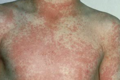
anterior cervical
pharyngeal edema
Scarlatiniform rash
Streptococcal Pharyngitis (strep Throat)
DIAGNOSIS:
Whom NOT to test:
Testing may not be necessary in children & adolescents with manifestations clearly suggestive of viral illness (eg, rhinorrhea, conjunctivitis, cough, hoarseness, anterior stomatitis, discrete ulcerative lesions or vesicles, diarrhea).
In children <__ years of age, routine testing is ____ recommended because both GAS pharyngitis and its complication, Acute rheumatic fever, are ____ in this age group.
3
NOT
rare
Streptococcal Pharyngitis (strep Throat)
Whom to test
Testing for GAS is indicated in children>/= 3 years & adolescents with evidence of acute tonsillopharyngitis (fever, erythema, edema, &/or tonsillar exudates) or scarlatiniform rash on physical examination
o AND _____ __ multiple signs & symptoms of viral infections
Exposure to an individual with ____ at home/school or high prevalence of GAS in the community & symptoms of GAS.
If ≥3 Centorcriteria[(1) ____≥38°C, (2) absence of ____, (3) swollen ____ ____ _____,(4_____ exudates/swelling (1 point for each)].
Suspected acute rheumatic fever (ARF) or poststreptococcal glomerulonephritis.
absence of
GAS
fever
cough
anterior cervical lymph nodes
tonsillar

STREPTOCOCCAL PHARYNGITIS
is strep likely if there is a cough?
No
Streptococcal Pharyngitis (strep Throat)
Testing options:
_____ ______ detection test (RADT): _____ _____ test.
o if negative, throat cultures should be obtained, especially in children 5-15y).
Throat ______; ______ diagnosis (criterion standard). Will show all the other organisms
rapid antigen
best initial
culture; definitive
MANAGEMENT - Streptococcal Pharyngitis (strep Throat)
Analgesics eg, NSAIDs, Acetaminophen, or Aspirin for pain. Anesthetics: Lozenges, throat sprays.
Penicillin _____-____ antibiotic (eg, Penicillin V, Penicillin G, or ______) if testing is positive.
Penicillin allergy:
Cephalosporins (eg, ______), ________ (eg, Azithromycin), Clarithromycin, or Clindamycin
First-line; Amoxicillin
Cefuroxime
Macrolides
Streptococcal Pharyngitis (strep Throat)
COMPLICATIONS
______ _____(preventable with antibiotics). Severe pharyngitis (give IM Dexamethasone)
Acute ___________ (not preventable with antibiotics). Peritonsillar abscess.
_______: Pediatric autoimmune neuropsychiatric disorders associated with streptococcus.IF LEFT UNTREATED
Rheumatic fever
glomerulonephritis
PANDAS
Peritonsillar Abscess (Quinsy) and Peritonsillar Cellulitis
Abscess between the palatine tonsil & the pharyngeal muscles, resulting from a complication of _____ _____, tonsillitis or pharyngitis. Most common in adolescents & young adults 15-30 years of age.

strep throat
___________
ETIOLOGIES
Often polymicrobial the predominant species include Group A Streptococcus (S. pyogenes),
Staphylococcus aureus, and respiratory/oral anaerobes (eg, Fusobacterium necrophorum).
Peritonsillar Abscess (Quinsy) and Peritonsillar Cellulitis
Peritonsillar Abscess (Quinsy) and Peritonsillar Cellulitis
CLINICAL MANIFESTATIONS
Ill-appearing, severe unilateral pharyngitis, _____, odynophagia, high fever, malaise, ear pain
Muffled "hot potato" _____, difficulty handling oral secretions (_____), _____ (lockjaw).
dysphagia
voice
drooling
trismus
Peritonsillar Abscess (Quinsy) and Peritonsillar Cellulitis
Physical examination
Swollen or fluctuant tonsil, causing uvula _____ to the ______ side, bulging of the posterior soft palate near the tonsil with fluctuance, inferior and medial displacement of the infected tonsil(s), ______ rise of the uvula, & tender anterior cervical lymphadenopathy.
_______ Cellulitis: similar symptoms but not associated with fluctuance or deviation.
deviation
contralateral
asymmetric
peritonsillar
Peritonsillar Abscess (Quinsy) and Peritonsillar Cellulitis
DIAGNOSIS
Primarily a ______ diagnosis without need for imaging or labs if classic. ______ ultrasound if unsure.
????? - imaging of choice if imaging is needed to differentiate Cellulitis vs. Abscess or complications.
clinical
intraoral
CT Neck w/ IV contrast
MANAGEMENT - Peritonsillar Abscess (Quinsy) and Peritonsillar Cellulitis
______ (Aspiration or I&D or tonsillectomy) + antibiotics (if cellulitis)
o Drainage: needle aspiration (preferred) or incision & drainage (I&D).-caution of carotid arteryAbx
o Oral (____ or _____)
o Parenteral (Ampicillin-sulbactam or Clindamycin), MRSA suspected: IV Vancomycin; Linezolid is an alternative._________
o usually reserved for patients who fail to respond to drainage
o PTA with complications (egimpending airway obstruction)
o prior episodes of PTA, or recurrent severe pharyngitis.
Drainage
amox clav; clindamycin
Tonsillectomy
right behind the peritonsillar abscess is the _____ _____ which is why it is INCREDIBLY important to be extermely careful
carotid artery
PREVENTION Peritonsillar Abscess (Quinsy) and Peritonsillar Celluli :Prompt treatment of _____ infections.
streptococcal
Retropharyngeal Abscess
Deep neck space infection located in the potential space behind the ______ _____ ____.
Most common in children ____ years. In adults often a result of ______ trauma (eg, chicken or fish bones, instrumentation, dental procedures, or dental infection).
posterior pharyngeal wall
2-4
penetrating
Retropharyngeal Abscess
MICROBIOLOGY:
• Similar to Peritonsillar abscess often _______ (eg, Group A Streptococcus, Staphylococcus aureus, & respiratory anaerobes).
polymicrobial
CLINICAL MANIFESTATIONS - RETROPHARYNGEAL ABSCESS
_____
_______, _______, drooling, sore throat, chest pain, muffled "hot potato" voice, trismus.
Neck: neck _____ especially with neck extension, ______ (unwilling to move the neck secondary to pain and spasms).
Chest pain with mediastinal extension
Stridor is uncommon.
Fever
odynophagia; dysphagia
stiffness; torticollis
Retropharyngeal Abscess
DIAGNOSIS
• ??????? imaging of choice & preferred imaging if suspicion is high.
Lateral neck _____; may be performed if ____ suspicion
o increased prevertebral space >50% of the width of adjacent vertebral body (5-7 cm prevertebral widening at the 2nd cervical vertebra). CAVEAT: will not RULE OUT diagnosis
• In smaller children with respiratory distress, evaluation is often performed in the operating room.
CT Neck w/ IV contrast
radiograph; low
________ ABSCESS
FIRST THINGS FIRST: ____ ____
ENT consultation - Referral to specialist
Large (>/=___cm) abscess on CT:
o surgical ____ & _____ in the Operating Room
o Antibiotics
Small abscesses (<2.5 cm) & children with no _____ compromise may be observed for 24-48 hours with ______ therapy and no drainage.
Retropharyngeal
Secure Airway
2.5
incision; drainage
airway; antibiotic
Retropharyngeal Abscess
MANAGEMENT
Antibiotics: IV _______-______ or _______ (similar to PTA). IV Vancomycin if no response."
Ampicillin-sulbactam
Clindamycin
Retropharyngeal Abscess
COMPLICATIONS - Airway obstruction, Mediastinitis (due to spread of the infection), sepsis, atlantoaxial dislocation
Acute epiglottis (supraglottitis)
Causes
Foreign immigrants or ______ –mc causeH influenza
Vaccinated –mc cause ______
unvaccinated
streptococcus
Acute epiglottis (supraglottitis)
Clinical manifestations –"clinically ____ and in respiratory _____"
Acute – Drooling, _____ & distress
Inspiratory _____
Dysphagia, odynophagia,
Muffled "hot" potato voice
______ or sniffing position
Respiratory distress –retractions (using ______ muscles to breathe), choking sensation, dyspnea
ill; distress
dysphagia
stridor
tripoding
Acute epiglottis (supraglottitis)
Diagnosis
Clinical diagnosis
_________ DIRECT VIEW OF EPIGLOTTIS–definitive diagnosis –visualization of an _______ ____ ___ epiglottis

Laryngoscopy
edematous cherry red
thumbprint sign on xray indicates what
Acute epiglottis
avoid using WHAT for acute epiglotitis
tongue depressor