biol 300 exam 2 my flashcards
 Call Kai
 Learn
 Practice Test
 Spaced Repetition
 Match
 Flashcards
 Knowt Play
1/52

Earn XP
Description and Tags
CAMPANALE FALL 2026
53 Terms
Three levels of chromatin organization
1) 10 nm chain of nucleosomes (beads on a string)
2) 30 nm fiber: helical array of nucleosomes
3) Further folding and compacting of 30 nm fibers
Levels of chromatin compaction diagram

What is a nucleosome?
fundamental structural unit of chromatin in all eukaryotes (not prokaryotes/bacteria)
Are nucleosomes found in euchromatin or heterochromatin?
occurs in both euchromatin (loosely packaged) and heterochromatin (tightly packaged)
Molecular components of nucleosome
~200 bp DNA; depends on if the H1 histone is present or not
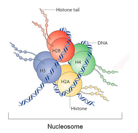
Histone octamer
Nucleosome diagram

When chromatin is released from the nucleus and stretched, what do we see?
Is the thread resistant to nuclease digestion? Why?
What about the particles? Why?
Discrete particles connected by thread is visible when chromatin is released from the nucleus and stretched
The thread connecting particles are susceptible to nuclease digestion (why? likely because contain little proteins).
The particles are resistant to nuclease digestion due to protein binding
How do we test for the structure of the nucleosome?
(Diagram/Process)
Treat chromatin with MNase nuclease
Why? MNase has low seq specificity and is both an endo and exonuclease that prefers AT rich regions
The particles are not digestable by nuclease (but thread is!)
Run on gel
DNA from particles are identical in size (the beads of the beads on a string)
There was incomplete digestion
Which means MNase didn’t get every single linker (thread) region, so the DNA fragment mixture contains: 1 nucleosome, 2 nucleosomes, etc. etc. That’s why we see a “ladder” pattern
205 bp = fragment of 1 nucleosome (on the gel)
405 bp = fragment of 2 nucleosomes right next to each other (on the gel)

How is DNA in a nucleosome defined?
What are the two types?
Length?
Bound by?
Resistance to nuclease digestion?
Sequence specificity? Why?
By susceptibility to nuclease
Core DNA + Linker DNA
Core DNA: 145-147 bp
needed to form stable nucleosome
bond by core histone H2A, H2B, H3, and H4
relativley resistant to nuclease digestion
In general, no sequence specificity
Why? Because histone proteins bind to the DNA backbone (not base-pair sequences)
Linker DNA:
the rest of the repeating unit
some bond to H1
susceptible to nuclease digestion
What are nucleosomes made of?
What are the core histone(s)? (How many of each?)
What are the linker histone(s)?
What proteins make them up? What do they do?
What are the histone tails? What type of sites are they?
Histone octomers:
4 types of core histones, 2 proteins for each type (4×2= 8 “octa”)
each type corresponds to one histone family
H2A, H2B, H3, H4
1 linker histone: H1
Small basic proteins:
~100 aa
arginine or lysine (basic aa)
neutralize the (-) on DNA backbone to facilitate compaction
Core histone tails:
flexible amino or carboxyl-terminal regions of the core histones
extend beyond surface of nucleosome
extensive posttranslational modication sites
Are histones conserved?
Yes,
Orthologs (arise due to speciation, among different species) are highly conserved in eukaryotes
especially H3 and H4 (>90% across species)
originated from Archaea
What are the core histone variants? Do they have their own gene families?
Are they paralogs? DO canonical histones distribute to the entire genome?
What special functions do each varient typically have?
What does H3 do?
Is there seq homology between families?
Has own gene family?
Yes: H2A, H2B, H3
NO: H4
Paralogs: one canonical histone (within the same species) with several variants
canonical histones (H3, H2A, H2B, and H4) distribute to the entire genome
Each variant has a special function
often related in epigenetic regulation of specific locations
ex: Centromere H3 (CenH3) is responsible to maintain centromere identity and is a variant
NO SEQUENCE HOMOLOGY BETWEEN FAMILIES
H3 and H4 are NOT homologs
H3 and CenH3 are paralogs
Do core histone share structural homology?
Do they share sequence homology?
Yes structural homology
3D structural motif: histone fold
different origin but evolved to have same function
NO sequence homology
nucleosome main structure
what does each subcomplex do?
octamer with (2) subcomplexes
(1x) H3-H4 tetramer
can organize DNA in vitro into particles
these particles display properties of the core particles (H3, H4 more conserved)
(2x) H2A-H2B dimer
two dimers formed
Entire octamer interacts with DNA
mainly DNA backbone + minor grooves
What does the 5th histone (H1) do?
Is it required for nucleosome formation?
Do all nucleosomes have linker histones?
Are they required for higher-order chromatin structure?
Are they conserved?
Is H1 single histone or a group of histones?
Is there specificity? Towards what?
**What are nucleosomes with the H1 linker called?
It’s a linker between nucleosomes
NO
NO
YES
YES, Highly conserved!
A group of closely related histones
YES, tissue and species specificity
**CHROMATOSOMES
How are histone proteins modified?
Which terminal tails does this occur on?
Small modifications
Larger modifications
Are modifications maintained or transient?
How can they be maintained?
Are they removeable?
Covalently
Both N and C terminal tails
Methylation, acetylation, phosphorylation
ADP-ribosylation, monoubiquitylation, sumylation
Can be both.
Through multiple cell division (= epigenetic, DNA is not the only thing replicated)
YES
Methylation
Adds what groups?
What groups is it done on? Does it affect the charge on these? Which aa? What charges?
Mono, di, or tri methylation?
Addition of -CH3 groups (neutral)
happens on NH3+ groups of Arginine, Lysine residues ((+) aa)
DOES NOT effect NH3+ (+) CHARGE
Lysine: mono, di-, tri methylation
Arginine: mono, di- methylation
Acetylation
Adds what groups?
What groups is it done on? Does it affect the charge on these? Which aa? What charges?
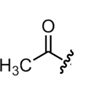
Adds -CO-CH3 groups
happens on NH3+ groups of Lysine residues ((+) aa)
NEUTRALIZES (+) CHARGE OF LYSINE
adds bulky group
Phosphorylation
Adds what groups?
What groups is it done on? Does it affect the charge on these? Which aa? What charges?
-PO4 groups added (-) CHARGED
happens on hydroxyl groups of Serine and Theronine (polarized aa)
INTRODUCES (-) CHARGE
ALL histone post translational modications (table)
Answer this for (METHYLATION , ACETYLATION, PHOSPHORYLATION):
added chemical groups
amino acids modified
effect on charges
other notes

What do histone modifications do?
Change gene expression
activate or repress expression of assoiated genes
Can happen on diff histones or diff aa of a histone
ex: K (lysine), R (arginine), S (serine), or T (theronine)
What is the transcriptional activity of a chromatin region ultimately decided by?
What is the "histone code"?
What type of proteins "interpret" the histone code?
What does it mean to "interpret" a histone code? (Give an example mentioned on the slide).
How do histone modifications affect chromatin structure and function (what are the two ways)?
By the combined effect of multiple modifications that can occur at a chromatin region.
A collection of multiple modifications at a particular site.
Non-histone proteins.
To take a biological action based on a certain marking.
Ex: Changing chromatin accessibility based on histone codes
Ex2: recruiting a specific DNA repair complex suitable for DNA damage at the site.
Some histone modifications can directly affect chromatin structure.
Most function by providing binding sites for non-histone proteins, which in turn change chromatin structures.
Two types of nucleosome assembly?
replication:
need newly synthesizes histones
replicated-coupled pathway
transcription and dna repair:
no newly synthesized histone
replication-independent pathway
What does nucleosome assembly during replication require?
Synthesis comes from which histone genes?
How to make a large amount efficiently?
How to make an equal amount of 4 types of histones?
newly synthesized histones
ALL four types: H2A, H2B, H3, H4 (canonical histones)
canonical histone genes are organized in clusters (Histone variants do no; each histone is from a diff family rather than being formed through duplication from a single ancestor gene)
[COME BACK BLANK]
How are canonical histone genes organized, and do histone variants do this?
Are these clusters formed through duplication from a single ancestor gene? What are their family relationships?
Is cluster organization evolutionarily conserved?
What does clustering ensure regarding histone expression (what are the two points)?
How does the cell meet the high demand for histones during replication?
Canonical histone genes are organized in clusters, whereas histone variants do not.
No, this is not a cluster of a gene family formed through duplication from a single ancestor gene; each histone is from a different family.
Yes, cluster organization is evolutionarily conserved.
Clustering ensures the coordinated expression of histones:
Similar amounts for each histone.
Same trigger: DNA synthesis.
There are multiple copies of such a cluster, sometimes in tandem repeats, to meet the high demand during replication.

How nucleosomes disassemble from template DNA
What disrupts nucleosome structure during replication?
How do octamers disassemble during replication?
Where do these disassembled tetramers and dimers go?
What happens immediately following replication on the daughter DNA duplexes?
The replication fork disrupts nucleosome structure.
Octamers partially dissociate into H32-H42 tetramers and H2A-H2B dimers.
They enter the mixed pool of old and newly synthesized tetramers and dimers.
Replication is followed by immediate nucleosome formations on daughter DNA duplexes.

How nucleosomes reassemble on daughter DNAs:
Is there any discrimination during reassembly on daughter DNAs regarding:
New vs. old tetramers and dimers? How are they reassembled?
Histone variants? Are there any exceptions?
What facilitates reassembly, and how are these factors recruited?
What is the definition of a chaperone?
What is the order of entry during nucleosome reassembly?
In the replication diagram, what do the blue and orange colors represent?
* No discrimination between new and old tetramers and dimers: randomly mixed after disassembly and randomly reassembled in daughter DNA duplexes.
* No discrimination between variants except special regions like the centromere.Facilitated by histone chaperones, which are recruited by DNA replication machinery.
Chaperone: enzymes that help the folding/unfolding of other proteins.
H3-H4 tetramer enters first, followed by H2A-H2B dimers.
Blue: old histones; Orange: new histones.
When can nucleosome disassembly and reassembly occur outside of DNA synthesis (give two examples)?
How condensed are transcribed genes, and do they keep their nucleosome structure?
What does RNA polymerase do to histone octamers during transcription in vitro, and what happens as soon as it passes?
According to the diagram, what factor releases the H2A-H2B dimer?
What releases the H3-H4 core?
When DNA is not synthesized, such as during DNA repair and transcription.
Transcribed genes are less condensed, but most retain the nucleosome structure (→ disassembly and assembly).
RNA polymerase may displace histone octamers during transcription in vitro, but octamers re-associate with DNA as soon as the polymerase has passed.
FACT releases the H2A-H2B dimer.
Other factors release H3-H4.
What is one variation in the nucleosome assembly mechanism suggested by evidence?
Can the cell rely on DNA replication apparatus during replication-independent assembly, and what does it use instead?
According to the diagram, what is the fate of the displaced octamer?
In the diagram, what happens when DNA is displaced from the octamer as RNA polymerase advances?
What displaces the octamer, and where does it reinsert relative to the RNA polymerase?
Some evidence suggests that some nucleosomes never completely leave template DNA.
It cannot rely on DNA replication apparatus; it uses alternative mechanisms and histone chaperones.
"The displaced octamer never leaves DNA".
DNA is displaced from the octamer and forms a closed loop.
Torsion ahead of RNA polymerase displaces the octamer, which reinserts behind the polymerase.
Although histones immediately reassemble behind RNA Pol after it passes, what can transcription do?
What is the definition of nucleosome positioning?
In the diagram showing nucleosome movement:
Where is the nucleosome initially assembled?
Where does RNA polymerase bind, and how far does it transcribe?
What happens to the nucleosome's location after RNA polymerase transcribes?
Transcription can alter the nucleosome positioning.
Nucleosome positioning: which sequence is in the core and which sequence is in the linker.
Nucleosome assembled at specific location.
RNA polymerase binds at promoter and transcribes up to terminator.
Nucleosome is found at new position.
What is the nucleosome status and positioning in repressed genes (not actively transcribed)?
What is the nucleosome status and positioning in actively expressed genes (transcribed)?
What is the exception regarding heavily transcribed genes?
Repressed genes:
Nucleosomes are present at promoters.
Nucleosomes are positioned in phase throughout the gene.
Actively expressed genes:
Nucleosomes are absent from promoters.
Nucleosomes are out of phase.
Exception: Some heavily transcribed genes can be devoid of nucleosomes.
Chromatosomes are:
nucleosomes that contain linker histones
Fill in the blank. To isolate nucleosomes, chromatin is digested with the enzyme ______________
MNase
True or False? During DNA replication, histone octamers are removed from DNA, remain intact, and are repositioned on the newly replicated DNA.
False
A researcher mutates a histone acetyltransferase (HAT) enzyme in yeast. Predict how global transcription levels might change and why.
Loss of HAT reduces histone acetylation, leading to more compact chromatin and decreased transcription globally.
WHY:
HATs add acetyl groups to lysine residues
turning (+) → (neutral) charge
neutral = less affinity towards DNA (since DNA backbone is negative)
if less affinity, histone won’t bind, and chromatin opens up (decondenses into euchromatin)
BUT when MUTATED acetyl groups aren’t placed, and chromatin remains compact ( + → 0 , histone stays positive and bound to DNA)
Discuss how nucleosome positioning and histone modifications work together to regulate gene expression. In your answer, describe the interplay between chromatin remodeling complexes, epigenetic marks, and the balance between euchromatin and heterochromatin.
Nucleosome positioning controls DNA accessibility.
repressed genes: nucleosomes are tightly positioned in-phase and block promoters
actively expressed genes: nucleosome-depleated region at the promoter with out-of-phase positoning
Epigenetic histone marks (ex: acetylation, methylation, phosphorylation)
may change charges → changes DNA affinity of histones → changes whether euchromatin or heterochromatin
Histone modifications act as signals through the “histone code” (collection of multiple markings interpreted by non-histone proteins) to recruit remodeling complexes or repressors.
Together, they establish dynamic chromatin states balancing transcriptional activation (euchromatin) and repression (heterochromatin).
Draw and label a nucleosome. Your drawing should include the DNA, all of the histones and show the N and C termini of each.
In what ways can histones be modified, and what parts of histones are usually modified?
The lysines of histone tails are most often modified by acetylation, phosphorylation, mono-ubiquitylation, sumoylation, and ADP-ribosylation.
In what ways can histones be modified, and what parts of histones are usually modified?
Histones can be modified via covalent post-translational modifications that typically occur on both the N- and C-terminal tails (as well as specific residues like Lysine [K], Arginine [R], Serine [S], and Threonine [T]).
The major types of modifications include:
Methylation: Occurs on lysine (mono-, di-, tri-) and arginine (mono-, or di-methylation).
Acetylation: Occurs on lysine, neutralizing its positive charge.
Phosphorylation: Occurs on serine and threonine, introducing a negative charge.
Larger modifications: Such as mono-ubiquitylation, sumoylation, and ADP-ribosylation.
Does the cell cycle control replication?
Yes
How many rounds of replication per cell cycle?
One and only one
Consequences of uncoupling of replication and cell cycle?
If # replication < # of cell division
divide before replication is finished → loss of genetic materials
If # replications > # of cell divisions
Endocycle → making polyploid cells (liver cells do this)
Do prokaryotes have slow or fast cell cycle?
What are the consequences of this?
Rate limiting factors?
Fast Cell cycle
Not concerned w/ errors (some level of quality control)
FAST replication LESS regulation
Rate limiting factors:
External factor: mainly nutrient availibility
Instrinsic limiting factors: C constant (~40 mins required to replicate entire E. coli genome) and D constant (~20 mins required between completion of replication and cell division)
What are the two constants (cannot be changed by nutrient/environment) that limit the length of the cell cycle?
C: ~40 min required to replicate (SYNTHESIZE) the entire E. Coli genome (polymerase 50kbp / min)
D: ~20 min required between completion of replication and cell division.
PREPPING & CELL DIVISION
Likely for assembly of cell division apparatus
How can bacteria proliferate faster than 60 min?
Normal: replication for division 2 occurs after division 1.
To speed up: replication for division 2 occurs before division 1.
one round of 60 min overlap with previous round of 60 min = replication starts before the cell cycle starts
IN OTHER WORDS: DNA is already half made for the next division
What is the fastest E. Coli can divide? Show diagram of the cell cycle
20 minutes
before daughter genome can finish, granddaughter genome is being made

How can E. Coli start replication for round 2 before round 1 finishes? Show diagram
must start DNA replication for next division before the first division is done

What is septum formation related to, and why is rod shape important?
What three genes control rod shape, and what are their specific functions?
What two main processes are involved in chromosome separation during bacterial division?
According to the diagram, where are the origin and terminus located in newborn cells, and what happens as origins replicate?
Septum formation is the same as the bacteria envelope (peptidoglycan). Rod shape is important because it is controlled by specific shape-determining genes.
The genes controlling rod shape are:
MreB: actin-like.
PBP2: peptidoglycan synthesis.
RodA: crosslinks glycans.
Chromosome separation is driven by Topoisomerase and Recombination.
In newborn cells, origin and terminus are at mid-cell; newly replicated origins move apart, a cell septum divides the cell at mid-cell, and finally origins and termini reorient as the cell divides.
How to identify genes with a certain function?
What technique is used to generate mutant strains to identify genes involved in specific functions?
On average, how many mutations does each strain contain, and how do mutations occur across genes?
How do you identify which specific strain/mutation affects cell division?
Mutagensis to generate mutant strains.
On average, each strain contains a mutation, and mutations occur in a random fashion (meaning there is likely more than one strain for each gene).
By using phenotypes related to the studied function to screen through the strains.

What problem does this diagram show?
Failure of cell division
long enough to divide but septum is not centered → you get 1 small cell and 1 big cell
What is the consequence of a division loss-of-function (LOF) mutation in cells?
Why can diploid cells carry a LOF division allele and survive, whereas bacteria cannot?
What is the solution used to study LOF division mutations in monoploid bacteria?
How do temperature sensitive (TS) alleles work at the structural level (amino acid sequence → 3D structure → protein function)?
At what temperature do TS strains appear wild-type, and at what temperatures do they show mutant phenotypes?
failure in cell division and cell death.
Diploid cells (heterozygous +/m) can carry the LOF allele yet still survive because they have a second functional copy. Bacteria are monoploid, so a cell with any LOF division mutation cannot survive and cannot be studied under normal conditions.
Temperature sensitive (TS) alleles.
An altered amino acid sequence leads to a mutant 3D structure where the mutant protein unfolds and loses its normal structure/conformation at high temperatures, thereby losing protein function.
TS strains appear wild-type at normal (permissive) temperature, but exhibit mutant phenotypes at high or low (restrictive) temperatures.
What phenotype and defect characterize minicells, and what genes are mutated?
What phenotype and defect characterize multinucleated filaments, and what genes are mutated?
What phenotype and defect characterize anucleate cells, and what genes are mutated?
According to the diagrams and micrographs on the right:
What appearance do wild-type cells show at normal temperature compared to high temperature? (What does this study?)
What color does the DNA stain in the bottom right image?
Minicells:
Defect: Misplaced septum.
Genes: Genes involved in septum positioning.
Multinucleated filament:
Defect: Failure in septum formation.
Genes: Genes involved in septum formation.
Anucleate cells:
Defect: Failure in chromosome separation.
Genes: Genes involved in chromosome separation.
At normal temperature, cells show a wild type phenotype. At high temperature, temperature-sensitive mutants display the mutant phenotype (to study minicell formation).
DNA stains blue.
What genes/proteins control the location of the septum in bacteria?
What specific factor determines the number and location of septa?
How does the dynamic movement of Min proteins regulate Z-ring assembly across the cell?
What is the specific function of SlmA/Noc proteins during cell division?
According to the diagram showing the oscillation of Min proteins in E. coli:
Which protein forms a ring at the edge of MinD?
What action does the MinE ring promote?
Where does MinD diffuse and bind after dissociating from one pole?
minC, -D, and -E and by noc/slmA.
ratio of MinE/MinCD.
Sets up a pattern where inhibition of Z-ring assembly is highest at the poles and lowest at mid-cell.
prevent septation from occurring in the space occupied by the bacterial chromosome.
MinD binds to the membrane at one pole, MinC binds to MinD, and a MinE ring forms at the edge of MinD.
The MinE ring promotes dissociation of MinD from the membrane.
MinD diffuses and binds to the opposite pole, where the MinE ring dissociates and reforms at the edge of the new MinD zone.