B&C Psych Lecture 7 Biology of visual perception
 Call Kai
 Learn
 Practice Test
 Spaced Repetition
 Match
 Flashcards
 Knowt Play
1/52

There's no tags or description
Looks like no tags are added yet.
53 Terms
The general overview of how our brain converts light into meaningful perception (6 steps)
Stimulus: light reaches the eye
Sensation: photoreceptors detect light
Transduction: light produces electrical changes
Coding: neural activity represents visual features
Projection: information travels to the visual cortex
Perception: higher-level areas interpret visual information

Sensation vs perception
Sensation is the detection of physical stimuli by specialized sensory receptors, which convert environmental energy into signals that can be processed by the nervous system.
Perception is the conscious experience and interpretation of sensory information by the central nervous system.
The distinction is important:
Sensation allows the nervous system to detect light.
Perception allows us to recognize what we are looking at, where it is, and what it is doing.
The different sensory modalities and what they detect
Sensory modality | What it detects |
|---|---|
Vision | Light |
Audition | Sound |
Somatosensation | Touch, pain, temperature, body position, and internal bodily sensations |
Gustation | Taste |
Olfaction | Smell |
Vestibular senses | Balance |
What are the different levels of visual processing, and what information is processed at each level?
The visual system processes information at different levels, from basic visual features to higher-order perception.
1. Basic visual features:
Colours
Lines and angles
Brightness
2. Higher-order visual features:
What: Object, face and identity recognition.
Where: Spatial locations of objects.
Depth: Relative distances between objects.
Movement: Motion and its direction.
Associations: Relationships between objects and events.
Key distinction: Basic processing extracts visual features, while higher-order processing integrates information to interpret objects, locations, movement and events.
Lecture fact: Approximately 25% of the brain's processing capacity is devoted to vision, according to the lecture.
How do basic and higher-order visual processes work together to produce meaningful perception?
Visual perception involves integrating basic sensory features into meaningful representations of objects and events.
Example — Football scene (slide 2):
1. Basic feature processing:
Detects colours, lines, angles and brightness.
2. Higher-order processing:
What: Recognises the player, ball and goal.
Where: Determines their spatial locations.
Depth: Judges their relative distances.
Movement: Tracks the ball's movement and direction.
Associations: Interprets relationships between the players, ball and goal to understand the action.
Key principle: Recognising individual objects is not the same as understanding their spatial relationships, movement and interactions.

What is light, which wavelengths can humans perceive, and how are wavelength and frequency related?
Light is a form of electromagnetic radiation detected by photoreceptors in the retina.
Human visible spectrum: Approximately 380–760 nanometres (nm).
Wavelength: Distance between successive peaks of a light wave.
Frequency: Number of wave cycles passing a point per second.
Relationship:
Shorter wavelength → higher frequency (towards violet/blue).
Longer wavelength → lower frequency (towards orange/red).
Key principle: Wavelength and frequency are inversely related.

What are the three perceptual dimensions of light, and which physical properties are they related to?
1. Hue — Perceived colour
Related primarily to wavelength.
Different wavelengths produce different perceived colours (e.g., blue versus red).
2. Brightness — Perceived lightness or darkness
Related primarily to light intensity.
Greater intensity generally produces a brighter appearance.
3. Saturation — Colour purity or vividness
Related to the mixture of wavelengths present.
More saturated = purer, more vivid colour.
Less saturated = more washed-out colour.
Memory cue:
Hue = Which colour?
Brightness = How light or dark?
Saturation = How pure or vivid?

How does the mixture of wavelengths influence colour saturation, and how does this relate to white light?
Saturation describes the perceived purity of a colour.
- Highly saturated colour:
Associated with light concentrated around a particular wavelength.
Appears vivid and intense in colour.
- Less saturated colour:
Contains a broader mixture of wavelengths.
Appears paler or more washed out.
- White light:
Contains a broad mixture of visible wavelengths.
Has no dominant perceived hue and is considered achromatic.
Key distinction: Saturation describes colour purity, not brightness.
Note: A single wavelength produces maximally pure spectral light, but perceived saturation also depends on visual context and how the light stimulates the visual system.
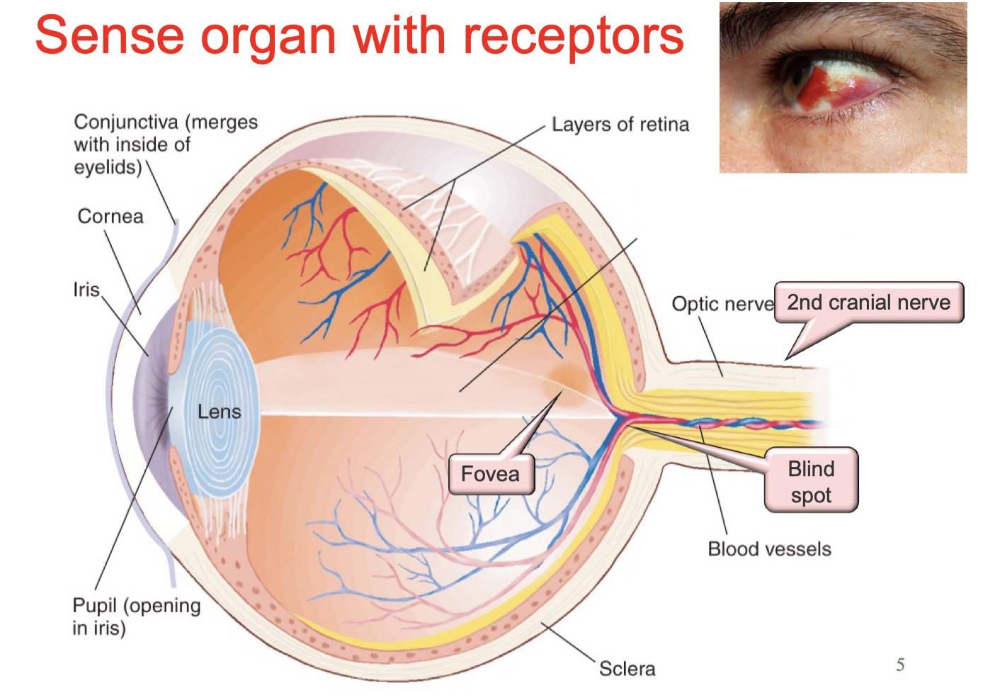
What are the main anatomical structures of the eye, and what are their functions in vision?
The eye contains structures that regulate and focus incoming light onto the retina, where visual sensation begins.
Cornea: Transparent outer surface that allows light to enter and provides much of the eye's focusing power.
Iris: Controls the size of the pupil.
Pupil: Opening through which light enters.
Lens: Changes shape to focus near or distant objects onto the retina.
Retina: Contains photoreceptors and neurons that begin visual processing.
Fovea: Central retinal region responsible for particularly sharp vision.
Optic disc (blind spot): Where the optic nerve exits; contains no photoreceptors.
Optic nerve (CN II): Carries signals from retinal ganglion cells towards the brain.
Key idea: The eye focuses light onto the retina, which converts visual stimulation into neural information.
What is accommodation, and how does the eye adjust its focus for near and distant objects?
Accommodation is the process by which the lens changes shape to focus images from different distances onto the retina.
- Near objects:
Ciliary muscles contract.
Lens becomes more rounded, increasing its focusing power.
- Distant objects:
Ciliary muscles relax.
Lens becomes flatter, reducing its focusing power.
Key principle: Accommodation adjusts the lens's curvature to keep objects at different distances in focus.
Memory cue: Near = rounder lens; Far = flatter lens
What causes the blind spot in the retina, and why do we normally not notice it?
The blind spot (optic disc) is the region where retinal ganglion-cell axons leave the eye to form the optic nerve.
- Cause:
The optic disc contains no rods or cones.
Light falling on this region cannot be directly detected.
- Why it is usually unnoticed:
The brain perceptually fills in missing information using surrounding visual information.
During binocular vision, the other eye can often provide information about the corresponding part of the visual field.
Key distinction: The blind spot is caused by an absence of photoreceptors, not by malfunctioning receptors.

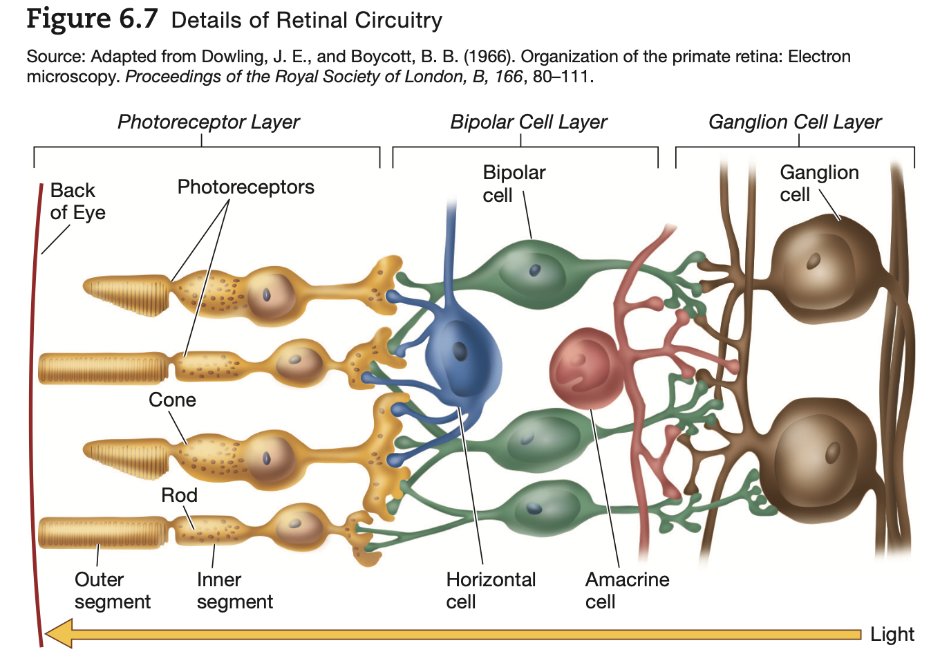
How does visual information travel through the retina, and what roles do the main retinal neurons play?
The retina contains layers of neurons that convert and process visual information before it reaches the brain.
Main signalling pathway:
Photoreceptors (rods and cones)
Detect light and convert it into changes in electrical activity (phototransduction).
Bipolar cells
Relay signals from photoreceptors towards ganglion cells.
Ganglion cells
Generate action potentials.
Their axons form the optic nerve, transmitting information towards the brain.
Additional retinal neurons:
Horizontal cells: Connect retinal pathways laterally and help modulate photoreceptor-to-bipolar signalling.
Amacrine cells: Modulate signalling between bipolar and ganglion cells.
Important anatomical detail: Light passes through the inner retinal layers before reaching rods and cones near the back of the retina.
Memory cue: Photoreceptors → Bipolar cells → Ganglion cells → Optic nerve.
+++Next slide pcituresssss


How do rods and cones differ in their functions, sensitivity, distribution and photopigments?
The retina contains approximately 120 million rods and 6 million cones.
1. Rods — Dim-light vision
Light sensitivity: Highly sensitive; function especially in dim light.
Colour: Do not support normal colour discrimination.
Visual detail: Lower spatial acuity.
Distribution: Most prevalent outside the central retina.
Photopigment: Rhodopsin.
2. Cones — Bright-light and colour vision
Light sensitivity: Require more light than rods.
Colour: Support colour vision through three cone pigment types.
Visual detail: Higher spatial acuity.
Distribution: Particularly concentrated in the fovea.
Photopigments: Three types with different spectral sensitivities.
Memory cue: Rods = sensitive but less detailed; Cones = colour and detail.

How does photoreceptor convergence explain the difference in visual acuity between central and peripheral vision?
Convergence refers to multiple photoreceptors contributing signals to the same downstream retinal pathway.
1. Foveal (central) vision
Relatively little convergence:
Individual cones generally connect to fewer downstream neurons.
Produces high spatial acuity because information from nearby locations remains more distinct.
2. Peripheral vision
Greater convergence:
Many photoreceptors can contribute signals to the same ganglion cell.
Produces lower spatial acuity, but greater sensitivity to weak light.
Why? Combining signals improves light sensitivity, but reduces the ability to distinguish exactly where light originated.
Key trade-off: Less convergence = greater precision; More convergence = greater sensitivity.
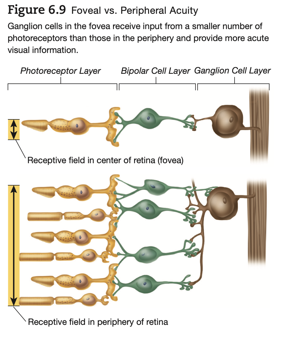
Why does the fovea provide particularly sharp vision compared with the peripheral retina?
The fovea is the central region of the retina specialised for high-acuity vision.
Two important reasons:
1. High cone concentration
Cones are densely packed in the fovea.
Support detailed visual processing under adequate illumination.
2. Low convergence
Foveal cones generally contribute to relatively separate retinal pathways.
Allows precise localisation of visual information.
Peripheral retina:
Contains many rods and generally greater convergence.
More sensitive to dim light but less precise in resolving fine details.
Key principle: Foveal vision prioritises spatial detail, while peripheral vision is better suited to detecting weak illumination.
What is visual transduction, and how does it allow the nervous system to process light?
Visual transduction (phototransduction) is the conversion of light energy into changes in the membrane potential of retinal photoreceptors.
Location: Rods and cones in the retina.
Stimulus: Light (electromagnetic radiation).
Initial response: Light absorption triggers intracellular processes that change the photoreceptor's membrane potential.
Neural consequence:
Changes in photoreceptor glutamate release influence bipolar cells.
Bipolar cells subsequently influence retinal ganglion-cell firing.
Key principle: Light energy → electrical changes in photoreceptors → altered neural signalling towards the brain.
What are photopigments composed of, and which photopigments are found in rods and the three types of cones?
Photopigments are light-sensitive molecules located in the membrane structures (lamellae) of photoreceptors.
Each photopigment contains:
Opsin: A protein that helps determine wavelength sensitivity.
Retinal: A light-sensitive molecule derived from vitamin A.
Four photopigments:
Blue-sensitive cones: Cyanolabe + retinal.
Green-sensitive cones: Chlorolabe + retinal.
Red-sensitive cones: Erythrolabe + retinal.
Rods: Rod opsin + retinal = rhodopsin.
Key idea: Different opsins produce different wavelength sensitivities, allowing the visual system to respond to different parts of the visible spectrum.

What happens during phototransduction, from light absorption to changes in retinal ganglion-cell activity?
Phototransduction converts light into neural signals through the following sequence:
1. Light absorption
Light reaches rods or cones and is absorbed by photopigments.
2. Photopigment activation
Light alters the retinal component of the pigment, triggering an intracellular signalling cascade.
In rods, this activates the rhodopsin-mediated pathway.
3. Photoreceptor hyperpolarization
The signalling cascade reduces the inward current through light-sensitive channels, making the membrane potential more negative.
4. Reduced glutamate release
Hyperpolarization decreases neurotransmitter release from the photoreceptor.
5. Bipolar-cell response
ON and OFF bipolar cells respond differently to the reduction in glutamate.
6. Ganglion-cell response
Changes in bipolar-cell signalling influence ganglion-cell action-potential firing.
Ganglion-cell axons form the optic nerve, carrying visual information towards the brain.
Memory cue: Light → Pigment activation → Hyperpolarization → ↓ Glutamate → Bipolar cells → Ganglion cells.

How do photoreceptor membrane potential and glutamate release differ in darkness compared with light?
1. Darkness
Photoreceptors are relatively depolarized.
They continuously release relatively large amounts of glutamate.
2. Light
Light activates phototransduction.
Photoreceptors hyperpolarize (become more negative).
Glutamate release decreases.
Important distinction:
Photoreceptors communicate mainly through graded membrane potentials and neurotransmitter release.
Retinal ganglion cells generate action potentials that travel towards the brain.
Memory cue:
Dark = depolarized → more glutamate.
Light = hyperpolarized → less glutamate.
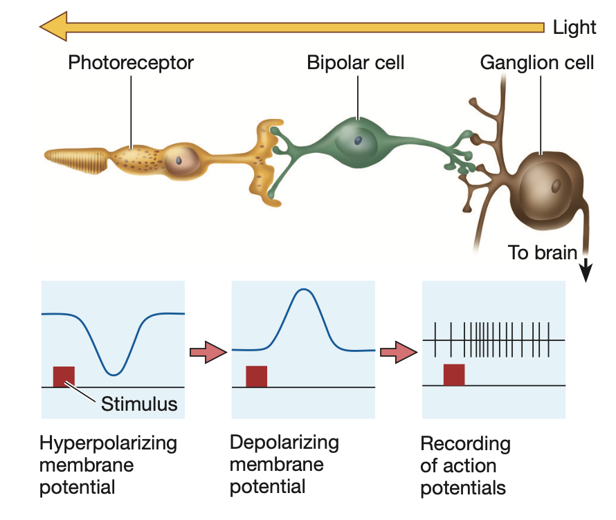
How do ON and OFF bipolar cells respond differently to glutamate, and what happens to each when light reaches photoreceptors?
ON and OFF bipolar cells respond oppositely to changes in photoreceptor glutamate release.
1. ON bipolar cells
Glutamate: Causes hyperpolarization.
During light: Photoreceptor glutamate release decreases.
ON bipolar cells depolarize.
2. OFF bipolar cells
Glutamate: Causes depolarization.
During light: Photoreceptor glutamate release decreases.
OFF bipolar cells hyperpolarize.
Functional significance:
ON pathway: Responds to increases in illumination.
OFF pathway: Responds to decreases in illumination.
Memory cue: Light ON → ON bipolar depolarizes; OFF bipolar hyperpolarizes.

How do photoreceptors, bipolar cells and ganglion cells differ in their electrical signalling during visual processing?
The retina uses both graded potentials and action potentials to process and transmit visual information.
1. Photoreceptors (rods and cones)
Respond to light with graded changes in membrane potential.
Regulate glutamate release according to illumination.
2. Bipolar cells
Receive glutamate signals from photoreceptors.
Respond primarily through graded membrane-potential changes.
Transmit information towards ganglion cells.
3. Ganglion cells
Integrate retinal input.
Generate action potentials.
Their axons form the optic nerve, carrying information towards the brain.
Key distinction: Photoreceptors and bipolar cells mainly use graded signals; ganglion cells convert retinal processing into action-potential output.

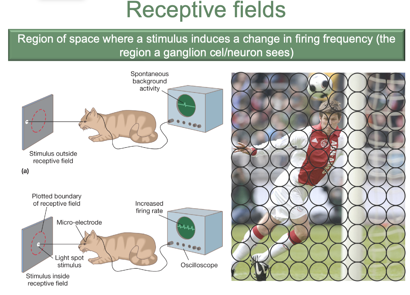
What is a receptive field, and what is its function in retinal visual processing?
A receptive field is the region of sensory space where stimulation influences a neuron's activity.
For a retinal ganglion cell:
Its receptive field is the region of the visual field where changes in illumination affect its firing rate.
Different locations within the receptive field can produce different responses:
Excitation: Increased firing rate.
Inhibition: Decreased firing rate.
Functional significance:
Allows the visual system to encode where changes in illumination occur.
Provides the basis for detecting spatial patterns, contrast and edges.
Key idea: A receptive field describes where a stimulus can influence a neuron, not simply where the neuron is physically located.
How do retinal ganglion cells encode differences in light intensity through action-potential frequency?
Retinal ganglion cells can encode light intensity through changes in their firing rate (action potentials per unit of time).
Example — ON-centre ganglion cell:
Low illumination of the receptive-field centre:
Relatively little excitatory stimulation → lower firing rate.
Greater illumination of the centre:
Stronger excitatory stimulation → higher firing rate.
OFF-centre cells:
Respond in the opposite direction to changes in illumination of their centres.
A reduction in centre illumination can increase their firing rate.
Important: Firing-rate changes depend on the type of ganglion cell and where light falls within its receptive field.
Key principle: Light intensity is represented by patterns of neural activity, rather than all retinal neurons increasing their firing whenever illumination increases.

How do ON-centre and OFF-centre retinal ganglion cells differ in their centre-surround receptive-field organisation?
Retinal ganglion cells can have centre-surround receptive fields, in which the centre and surrounding region influence firing in opposite directions.
1. ON-centre / OFF-surround cell
Centre illuminated: Excitation → increased firing.
Surround illuminated: Inhibition → decreased firing.
2. OFF-centre / ON-surround cell
Centre illuminated: Inhibition → decreased firing.
Surround illuminated: Excitation → increased firing.
Key principle: Centre and surround have antagonistic effects, allowing cells to detect local differences in illumination.
ADED STUFF ON NEXT SLIDE


How do ON-centre and OFF-centre retinal ganglion cells differ in their centre-surround receptive-field organisation?
Why is centre-surround receptive-field organisation important for detecting contrast and edges?
Centre-surround antagonism enables retinal ganglion cells to respond to local differences in illumination, rather than simply overall light intensity.
How it works:
1. Uniform illumination:
Both centre and surround receive similar stimulation.
Their opposing effects partially cancel, producing a relatively limited net response.
2. Uneven illumination:
Centre and surround receive different amounts of light.
Excitation and inhibition become unbalanced, producing a stronger response.
Functional significance:
Detects contrast between neighbouring regions.
Emphasises light–dark boundaries.
Supports edge detection and object perception.
Key idea: The retina is especially sensitive to changes in brightness across space.
How do centre-surround receptive fields allow retinal ganglion cells to detect the boundary between light and dark surfaces?
Edge detection occurs because ganglion-cell responses depend on how illumination is distributed across their receptive fields.
At a light–dark boundary:
A ganglion cell's receptive field overlaps the edge.
Its centre and surround receive unequal illumination.
Their opposing effects no longer cancel to the same extent.
The cell produces a stronger response than it would under uniform illumination.
Example — ON-centre cell:
Bright centre + dark surround → strong excitation.
Bright centre + bright surround → surround inhibition reduces the response.
Conclusion: Cells near brightness boundaries can respond more strongly, enhancing the neural representation of edges.

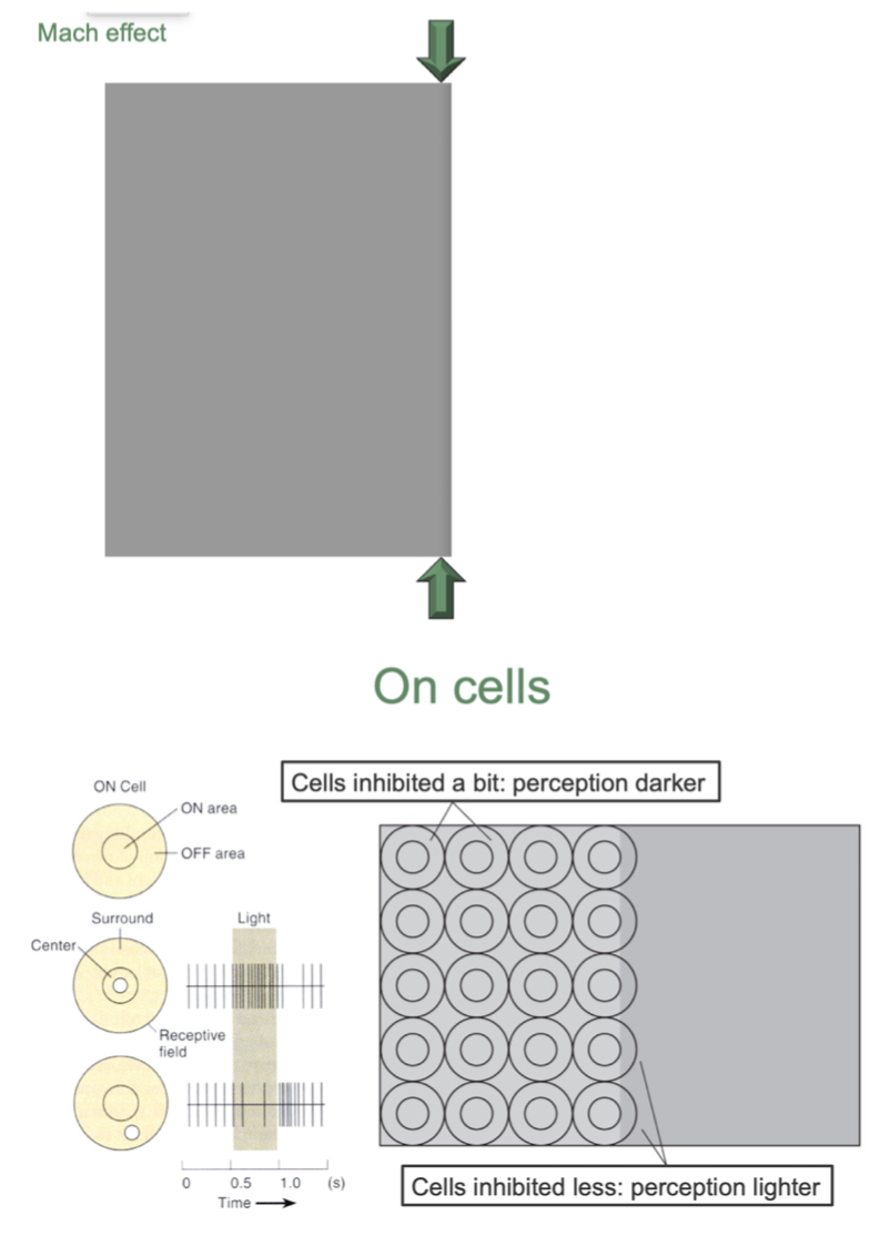
What is the Mach effect, and how can centre-surround inhibition explain the exaggerated brightness differences near an edge?
The Mach effect (Mach bands) is a visual illusion in which regions near a boundary between different brightness levels appear lighter or darker than their actual physical illumination.
Explanation — Centre-surround inhibition:
1. Near a brightness boundary:
Ganglion-cell receptive fields receive unequal illumination in their centres and surrounds.
2. Different amounts of inhibition:
Cells on one side of the boundary receive relatively less surround inhibition.
Cells on the other side receive relatively more inhibition.
3. Enhanced perceived contrast:
The bright side appears even brighter near the edge.
The dark side appears even darker near the edge.
Key principle: Inhibitory interactions exaggerate differences at brightness boundaries, helping the visual system emphasise edges.
What is trichromatic theory, and how do the three types of cones enable colour perception?
Trichromatic theory proposes that colour is initially encoded through the relative activity of three types of cone photoreceptors.
S-cones (short wavelengths): Most sensitive to the blue/violet region.
M-cones (medium wavelengths): Most sensitive to the green region.
L-cones (long wavelengths): Most sensitive to the yellow-green region, often called red-sensitive cones.
How colour is encoded:
Different wavelengths stimulate the three cone types to different degrees.
The visual system compares their relative activation patterns to distinguish colours.
Their sensitivity curves overlap, so each cone type responds to a range of wavelengths.
Key principle: Colour perception depends on combinations of cone responses, not a separate receptor for every colour.

Why must the visual system compare the activity of different cone types to determine colour?
Individual cones cannot uniquely identify the wavelength of incoming light because their responses depend on both wavelength and intensity.
A single cone type may respond similarly to:
A dim light near its preferred wavelength.
A brighter light at a less preferred wavelength.
Different wavelengths produce different patterns of activation across S-, M- and L-cones.
Therefore:
Comparing cone activity helps the brain distinguish wavelengths and perceived colours.
This explains why three broadly sensitive cone types can support a wide range of colour experiences.
Key idea: The pattern across cones carries colour information, not the activity of one cone alone.
What is opponent-process theory, and how do opponent-colour cells encode colour information?
Opponent-process theory proposes that colour information is encoded through neural channels with opposing responses to different colours.
Two principal colour-opponent pairs:
1. Red versus green
One opponent-cell type is excited by red-related signals and inhibited by green-related signals.
2. Yellow versus blue
One opponent-cell type is excited by yellow-related signals and inhibited by blue-related signals.
Other cells may have the opposite response direction.
Functional significance:
The same channel cannot simultaneously signal both of its opponent colours in opposite directions.
Helps explain why reddish green and yellowish blue are not normally perceived as single simultaneous hues.
Key distinction: Cone responses encode wavelength combinations; opponent cells compare and contrast those signals.

How do trichromatic and opponent-process mechanisms work together to produce the perception of yellow?
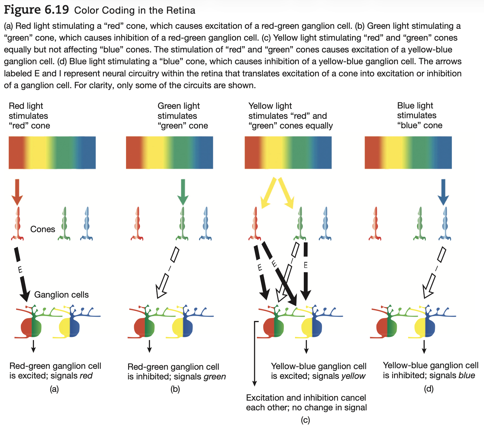
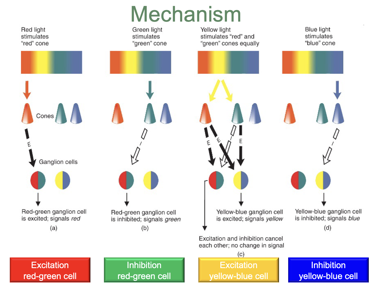
Yellow perception demonstrates how cone signals are combined into opponent-colour channels.
1. Cone activation
Yellow light stimulates L-cones (red-sensitive) and M-cones (green-sensitive).
2. Red–green opponent channel
The L- and M-cone contributions approximately balance, producing little net red-versus-green signal.
3. Yellow–blue opponent channel
Combined L- and M-cone signals favour the yellow response relative to the blue response.
4. Colour perception
The visual system interprets the resulting opponent-channel activity as yellow.
Key principle: Yellow does not require a separate yellow cone; it emerges from combinations of cone signals.

What is a negative afterimage, and what does it reveal about visual processing?
A negative afterimage is a temporary visual experience in which brightness or colours appear reversed after prolonged exposure to a stimulus.
Two forms:
1. Brightness afterimages
Previously light regions may appear dark.
Previously dark regions may appear light.
2. Colour afterimages
A colour may produce an afterimage resembling its opponent.
Example: Green → reddish afterimage.
Explanation:
Prolonged stimulation changes the responsiveness of visual neurons through adaptation.
After the stimulus disappears, the temporary imbalance can produce an opposite percept.
Key idea: Perception reflects the state of the visual system, not just the light currently entering the eye.

How can adaptation and rebound in opponent-colour cells explain why green stimulation produces a red afterimage?
Example: Green → Red afterimage
1. Green stimulation
Green-related input inhibits a red-excited/green-inhibited opponent cell.
2. Adaptation
Prolonged stimulation alters the cell's responsiveness.
3. Stimulus removal
When green stimulation stops, the opponent response may temporarily rebound above its normal activity.
4. Afterimage
The increased red-related signal produces a reddish afterimage, often seen against a neutral background.
Key principle: Adaptation can temporarily shift the balance of opponent-colour activity, producing perception of the opposite colour.
This is the opponent-cell explanation presented in your lecture; afterimages also involve adaptation within earlier visual pathways, including photoreceptors.
How do trichromatic and opponent-process coding work together, and what does each mechanism explain?
Colour perception involves two complementary stages of neural coding.
1. Trichromatic coding — Cone photoreceptors
Mechanism: Relative activation of S-, M- and L-cones.
Function: Encodes the spectral composition of incoming light.
Explains: How different wavelengths can produce different colour signals.
2. Opponent-process coding — Retinal and later visual pathways
Mechanism: Opposing responses in red–green and yellow–blue channels.
Function: Compares cone signals to encode colour differences.
Explains: Opponent-colour relationships and negative afterimages.
Relationship:
Cone activation → Comparison of cone signals → Opponent-colour channels → Colour perception.
Key conclusion: These theories describe different, interacting stages of colour processing, not mutually exclusive explanations.