Anatomy and Radiography Review: Digits, Hand, and Wrist
 Call Kai
 Learn
 Practice Test
 Spaced Repetition
 Match
 Flashcards
 Knowt Play
1/28

Earn XP
Description and Tags
Vocabulary flashcards covering anatomy review and radiographic positioning procedures for digits, hand, and wrist.
29 Terms
Phalanges (Hand)
The 14 bones present in each hand, numbered 1 to 5 from lateral to medial and classified as long bones.
Metacarpals
The 5 long bones in each hand that compose the palm.
Carpals
The 8 wrist bones located in each hand, arranged in proximal and distal rows.
Proximal Carpal Row
The bottom row of carpals consisting of the scaphoid, lunate, triquetrum, and pisiform (remembered by 'Steve Left The Party').
Distal Carpal Row
The top row of carpals consisting of the trapezium, trapezoid, capitate, and hamate (remembered by 'To Take Carol Home').
Interphalangeal (IP) Joints
Freely movable hinge joints (synovial/diarthrotic) that form the articulations between phalanges.
Metacarpophalangeal (MCP) Joints
Articulations situated between the heads of the metacarpals and the proximal phalanges.
Carpometacarpal (CMC) Joints
Gliding type joints formed by the articulations between the bases of the metacarpals and the carpal bones.
Intercarpal Joints
Articulations located between individual carpal bones.
Radiocarpal Joint
The primary articulation joint between the radius and the carpal bones.
Source-to-Image Distance (SID) for Digits, Hand, and Wrist
A standard distance set to 40 inches for all X-rays of digits, hand, and wrist.
Lap Shielding
A mandatory radiation protection measure placed over the patient's lap during hand, digit, and wrist radiography.

PA View: Digits 2-5
Central Ray: Should be on PIP
Collimation: 1 inch on all sides of digit, including 1 inch beyond the MCP joint
Instruct patient to hold still w/ affected digit flat on IR

Lateral View: Digits 2-5
Flex MCP joints/hold other fingers out of anatomy of interest
Central Ray: Should be on PIP
Collimation: 1 inch on all sides of digit, including 1 inch beyond the MCP joint

PA Oblique View: Digits 2-5
Hand resting flat on IR/Sponge
Rotate digit of interest 45 degrees
Central Ray: Should be on PIP
Collimation: 1 inch on all sides of digit, including 1 inch beyond the MCP joint
PA Oblique Digits 2-5 Evaluation
Identified when a concave aspect is seen on one side of the finger, starting wide and dipping back in.

AP View: First Digit (Thumb)
Central Ray: Should be on MCP of thumb
Collimation: 1 inch on all sides of digit, including 1 inch beyond the CMC joint w/ thumb fully extended/flat on IR

Lateral View: First Digit (Thumb)
Thumb should be extended and resting on IR w/ other fingers out of the way
Central Ray: Should be on MCP of thumb
Collimation: 1 inch on all sides of digit, including 1 inch beyond the CMC joint
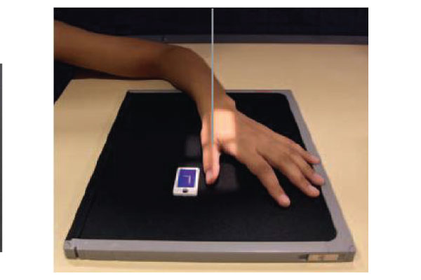
PA Oblique View: First Digit (Thumb)
Thumb should be resting on IR, w/ other fingers out of the way
Central Ray: Should be on MCP of thumb
Collimation: 1 inch on all sides of digit, including 1 inch beyond the CMC joint
45 degree rotation

PA View: Hand
Hand placed flat on IR,
Central Ray: Should be on the MCP of the third digit (middle finger)
Collimation: 1 inch on all sides of the hand, including 1 inch beyond the ulnar styloid w/ fingers extended and spread apart

PA Oblique View: Hand
Hand placed flat on IR/Sponge, MCP joints should be at a 45 degree angle to IR
Central Ray: Should be on the MCP of the third digit (middle finger)
Collimation: 1 inch on all sides of the hand, including 1 inch beyond the ulnar styloid w/ fingers extended and spread apart

True Lateral View (lateromedial): Hand
Hand should be positioned with ulnar side of hand/forearm resting on the IR, both styloids superimposed
Extend fingers and abduct the thumb
Central Ray: Should be on the MCP joint of the second digit (pointer finger)
Collimation: 1 inch on all sides of the shadow of the hand and thumb, including 1 inch beyond the ulnar styloid
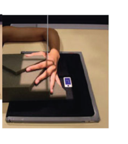
Fan Lateral View: Hand
separate extended digits and rest hand on sponge. If no sponge, have the hand make the “ok” sign with first and second digits almost touching
Central Ray: Should be on the MCP joint of the second digit (pointer finger)
Collimation: 1 inch on all sides of the shadow of the hand and thumb, including 1 inch beyond the ulnar styloid

PA View: Wrist
fingers should be flexed/cupped under hand, wrist flat on the IR
Central Ray: Should be on the midcarpals
Collimation: 2.5 inches beyond the wrist joint both distal (mid-metacarpals) and proximal (past radius/ulna), and 1 inch on the sides

Lateral View: Wrist
wrist should be resting on ulnar surface on IR, radial/ulnar styloid should be superimposed, fingers extended and aligned with each other
Central Ray: Should be on the center of the wrist
Collimation: 2.5 inches beyond the wrist joint both distal (mid-metacarpals) and proximal (past radius/ulna), and 1 inch on the sides (sides = dorsal and palmar surfaces)

PA Oblique View: Wrist
wrist should be rotated 45 degrees externally, supported with a wedge sponge
Central Ray: Should be on the center of the wrist
Collimation: 2.5 inches beyond the wrist joint both distal (mid-metacarpals) and proximal (past radius/ulna), and 1 inch on the sides

PA Ulnar Deviation: Wrist
hand should be flat on IR, turned outward toward ulna
Central Ray: Should be on the scaphoid bone in the wrist to visualize it clearly
Collimation: 2.5 inches beyond the wrist joint both distal (mid-metacarpals) and proximal (past radius/ulna), and 1 inch on the sides
PA Axial Scaphoid Projection
Special PA wrist view with CR on the scaphoid that elongates the scaphoid bone.
Gaynor-Hart Method
Tangential carpal tunnel special projection performed with the wrist hyperextended and CR on the radial styloid.