Neuro anatomy Q2
 Call Kai
 Learn
Learn Practice Test
 Spaced Repetition
 Match
Match Flashcards
 Knowt Play
1/128

There's no tags or description
Looks like no tags are added yet.
Name | Mastery | Learn | Test | Matching | Spaced | Call with Kai | Chat |
|---|
No analytics yet
Send a link to your students to track their progress
129 Terms

What does 'Medial' mean in the structure of the nervous system?
Medial means towards the midline of the body.


What does 'Lateral' mean in neuroanatomy?
Lateral means towards the side of the body.


What directions does 'Dorsal' refer to?
Dorsal refers to the back of the body and the top of the head.

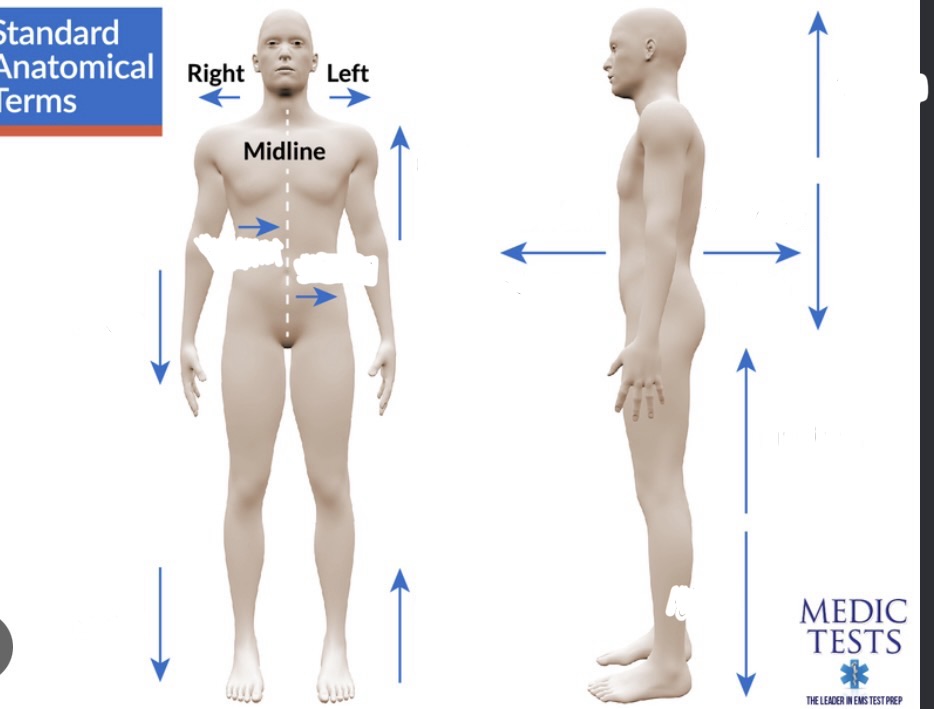
What areas does 'Ventral' refer to in the body and head?
Ventral refers to the front of the body and the lower head.


What location does 'Rostral' describe?
Rostral describes the front of the forehead and the top inside of the head or neck.


Anterior anatomical directio
Front of body


Posterior anatomical direction
Back of body


Proximal anatomical direction
Towards joint/center


Distal anatomical direction
Away from joint/ center

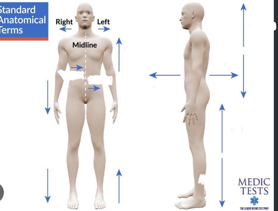
Superior anatomical direc
Up/top/above


Inferior anatomical direction
Down/bottom/bellow

From which embryonic layer does the nervous system derive?
The nervous system derives from the ectoderm, which is the outside of the cell.
What occurs during the early development of the nervous system from the neural plate?
The neural tube folds over and forms a groove.

What condition occurs if the telencephalon never forms due to the neural tube failing to close?
Anencephaly occurs, causing the brain to deteriorate and die.
What is Spina Bifida?
Spina Bifida is a condition where the back of the neural tube does not form, leaving the spinal cord open in the back and often causing a lack of feeling or movement in the feet.
What is Lissencephaly?
Lissencephaly is a smooth brain condition lacking normal sulci and gyri.
What does ipsilateral mean?
Ipsilateral refers to two structures located on the same side of the body.
What does contralateral mean?
Contralateral refers to two structures located on different sides of the body.
What is the midsagitta plane?
The midsagittal plane is a vertical slice down the very center of the body.
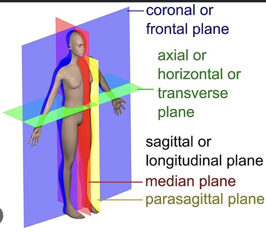
What is the sagittal plane?
The sagittal plane consists of sections parallel to the midsagittal plane.

What is the horizontal plane?
The horizontal plane consists of sections parallel to the ground.

What is the coronal plane?
The coronal plane divides the front and back of the body.

What is the cerebrum?
The cerebrum is the most rostral and largest part of the brain.
What separates the two cerebral hemispheres?
The two cerebral hemispheres are separated by the sagittal fissure.
What are the functions of the right and left cerebral hemispheres?
The right hemisphere receives sensation and controls movement of the left side of the body, while the left hemisphere receives sensation and controls movement of the right side.
What is the function of the brain stem?
The brain stem regulates breathing, consciousness, and body temperature, making it the most primitive and most important part for life.
Spinal cord
half a dozen tracts that run in columns that run up and down. Neurons in gray matter start analysis of sensory information, critical for coordinating movements and reflexes. The is encased by the bony vertebral column, attaches to the brain stem, and acts as a conduit for information between the body and the brain.
What are spinal nerves?
Spinal nerves are part of the PNS used by the spinal cord to communicate with the body, exiting through notches in the vertebrae and attaching via dorsal and ventral roots.
What is the difference between dorsal and ventral roots of the spinal cord?
The dorsal root brings sensory information into the spinal cord, and the ventral root takes motor information away from the spinal cord.

Somatic PNS
The somatic PNS controls all spinal nerves that innervate the skin, joints, and muscles that can be voluntarily controlled.
What are somatic motor neurons?
Somatic motor neurons control muscle contraction, are derived from motor neurons in the ventral spinal cord, and have somas in the CNS with axons in the PNS.
Where are the somas of somatic sensory neurons located?
The somas of somatic sensory neurons are located outside the CNS in dorsal root ganglia.
Autonomic Nervous System (ANS)
The autonomic nervous system consists of neurons that innervate internal organs, blood vessels, and glands.
What is the function of visceral sensory neurons?
Visceral sensory neurons bring information like pressure and oxygen content of blood to the CNS.
What do visceral motor neurons control?
Visceral motor neurons control the contraction and relaxation of smooth muscles, the rate of cardiac muscle contractions, and the secretory function of glands.
What are afferent axons?
Afferent axons carry information towards the subject.
What are efferent axons?
Efferent axons carry information away from the subject.
Cranial Nerves
Cranial nerves consist of 12 pairs from the brain stem that mostly innervate the head, with some being part of the somatic PNS and autonomic PNS.
What are the meninges?
The meninges are three membranes that protect the CNS from coming in direct contact with bone.
What is the arachnoid membrane?
The arachnoid membrane is located under the dura and looks like a spider web.

Dura mater
Forms tough, inelastic bad that surrounds the brain and spinal cord

What is a subdural hematoma?
A subdural hematoma is blood collected between the dura and arachnoid caused by a ruptured blood vessel, which disrupts brain function by compressing parts of the CNS.
Pia mater
the thin membrane that adheres closely to the surface of the brain. Along pia runs blood vessels that go to brain. It is separated from the arachnoid by the subarachnoid fluid filled space.

What is the subarachnoid space?
The subarachnoid space is a fluid-filled space separated from the arachnoid and filled with cerebrospinal fluid.

Cerebrospinal fluid (CSF)
Cerebrospinal fluid is a clean, salty liquid that fills the space between arachnoid membranes, suspending the brain.
What is the ventricular system?
The ventricular system consists of cerebrospinal fluid-filled caverns and canals inside the brain.
What is the choroid plexus?
The choroid plexus produces cerebrospinal fluid and is located in the ventricles of the cerebral hemispheres.
What are arachnoid villi?
Arachnoid villi are structures containing blood vessels that absorb cerebrospinal fluid. If the flow they provide for CSF is disrupted, there will likely be brain damage.
CLARITY
A technique for studying the brain by replacing light-absorbing lipids with a water-soluble gel to make the tissue transparent. This makes it easy to see flourescent tag
X-ray
An imaging method that uses radiation to view structures, though it is limited for the brain due to varying radiopacity levels.
Computed tomography (CT)
A brain imaging technique that rotates an X-ray source around the head to generate a radiopaque slice of the brain.
Magnetic resonance imaging (MRI)
An imaging technique superior to CT that uses a strong magnetic field to detect electrical magnetic signals emitted by hydrogen atoms.
Diffusion tensor imaging
An imaging method that visualizes axons in the brain by comparing the positions of hydrogen molecules.
Functional magnetic resonance imaging and Position emission tomography
Functional brain imaging methods that detect changes in regional blood flow and metabolism in the brain.
Gray matter
A collection of neural cell bodies located in the central nervous system.
Cortex
A collection of neurons that forms a thin sheet, typically located at the brain's surface.
Nucleus
A mass of neurons deep in the brain, distinct from a cell nucleus.
Substantia
A group of related neurons deep in the brain containing less distinct neuron groupings than nuclei.
Locus
A small, well-defined group of cells, such as the locus coeruleus which controls wakefulness and behavioral arousal.
Ganglion
A collection of neurons located in the peripheral nervous system.
Nerve
A bundle of axons located in the peripheral nervous system, with the optic nerve being the only nerve in the central nervous system.
White matter
A collection of axons located within the central nervous system.
Tract
A collection of central nervous system axons that share a common origin and destination.
Bundle
A collection of axons that run together but do not share the same origin and destination.
Capsule
A collection of axons that connect the cerebrum with the brain stem.
Lemniscus
A neural tract that loops through the brain like a ribbon, such as the medial lemniscus carrying touch information.
Commissure
Any collection of axons that connect one side of the brain to the other side.
Endoderm
The embryonic layer that becomes the lining of many internal organs.
Mesoderm
The embryonic layer that becomes bones and muscle.
Ectoderm
The embryonic layer that becomes the nervous system and skin.
Neural groove
A valley that appears on the surface as the ectoderm develops.
Neural folds
The walls that fold together to form the neural groove.
Neural tube
The structure formed from the folding of the neural disk, from which the entire nervous system develops.
Neural crest
Ectoderm that pinches off during the folding process and lies laterally from the neural tube.
Somites
Bulges formed by the neural crest that eventually develop into 33 individual vertebrae.
Neurulation
The developmental process by which the neural plate becomes the neural tube, occurring 22 days post-conception.
Differentiation
The developmental process by which structures become more complex and functionally specialized. In the brain, it causes 3 primary vesicles to form.
Prosencephalon
The rostral-most primary vesicle of the embryonic brain, also known as the forebrain.
Mesencephalon
The primary brain vesicle located behind the prosencephalon, also known as the midbrain.
Rhombencephalon
The primary brain vesicle that connects with the caudal neural tube and becomes the spinal cord, also known as the hindbrain.
Diencephalon
The center part of the forebrain that remains after secondary vesicles have formed.
Optic vesicles
Structures that grow and invaginate to form optic stalks and optic cups, which become the optic nerves and retinas.
Telencephalic vesicles
Structures that grow posteriorly to lie over and lateral to the diencephalon.
Olfactory bulbs
Structures that sprout off the ventral surface of the cerebral hemispheres. They receive information from cells that sense chemicals in the nose and relay this information to the cerebral cortex.
Third ventricle
The fluid-filled space located in the center of the diencephalon.
Thalamus
The thalamus is a relay center specifically for hearing, sight, and touch.
Internal capsule
serves as a relay center for thalamic neurons to send axons to the cortex and for cortical neurons to send info back to the brain stem. This is where signals from the left side of the body switch to be interpreted by the right side of the brain.
Basal ganglia
a collection of cells in the basal telencephalon. Damage to this area inhibits initiation of voluntary movement.
Amygdala
Located in brain stem. Involved with fear and emotion.
Hypothalamus
Located below thalamus in the forebrain’s telencephalon. Performs primitive functions of the autonomic nervous system like heart rate, blood flow, and temperature.
Forebrain
Top part of brain, Most diverse section of the brain that is critical for complex cognition and voluntary motor controls.
Midbrain
Located in brain stem. Critical for motor movement (eye) and sensory processing (hearing and sight). The midbrain houses descending white matter tracts like the corticospinal tract, as well as ascending tracts.
Tectum
Dorsal surface/roof if the midbrain
Tegmentum
Floor of midbrain
Cerebral aqueduc
CSF-filled space between the tectum and tegmentum. Connects rostrally with the third ventricle of the diencephalon.
Optic tectum
Also superior colliculus, tructure of tectum that receives direct input from the eye
Inferior Collicu
Structure of tectum that receives sensory information from the ear. Relay station for auditory information en route to the thalamus.
Hindbrain
Important for information passing from forebrain to spinal cord and vise versa. Neurons here also contribute to processing sensory info, control voluntary movement, and regulating the autonomic nervous system.
Pons
Located in the rostral hindbrain (the metencephalon). Connects the cerebral cortex to the cerebellum and houses the p-dunkles.
Medulla
Develops from the caudal half (myelencephalon) portion of the hindbrain. Also communicates for auditory, touch and taste. Also relays somatic sensory info from spinal cord to thalamus. controls involuntary functions such as breathing, digestion, and heart rate.