ECG Interpretation: Steps 4 (MEA) & 5
 Call Kai
 Learn
 Practice Test
 Spaced Repetition
 Match
 Flashcards
 Knowt Play
1/11

There's no tags or description
Looks like no tags are added yet.
12 Terms
mean electrical axis (MEA)
average path of electrical activity during ventricular depolarization


right axis shift
depolarization wave takes more time to travel through the right heart
RV enlargement
R bundle branch block
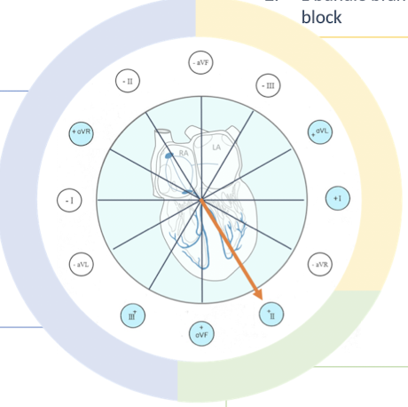
left axis shift
depolarization wav takes more time to travel through the left heart
LV enlargement
L bundle branch block

normal MEA
species dependent
dogs: +40° to +100°
cats: 0 to +150°

determine MEA: choose the largest QRS deflection

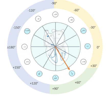

circle the QRS in each of 6 leads
compate and find the largest deflection from baseline
in the largest QRS, determine id the wave is traveling towards the + or - pole
assign the degree associated with that lead and electrode
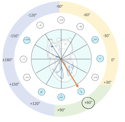

Which lead has the largest QRS deflection?
Is the wave directed towards the + or - pole?
What degree is associated with this pole of the lead?
Interpret the MEA
Lead II
(-) pole
MEA = -120°
right axis shift → RV enlargement or R bundle branch block
determine MEA: isoelectric method

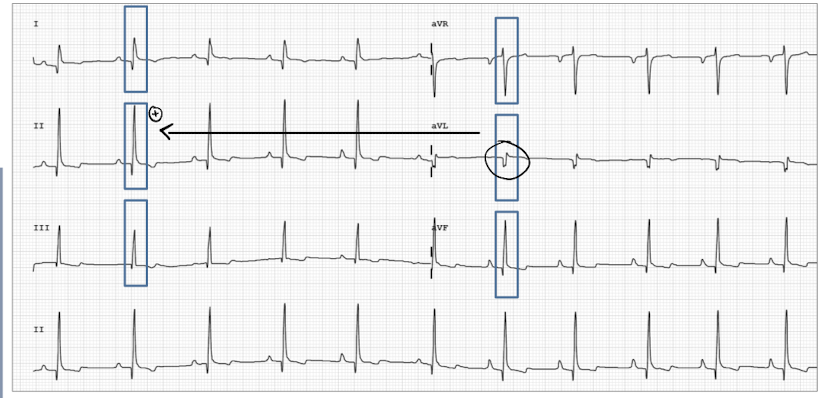
circle the QRS in all 6 leads
compare and find the “isoelectric lead”
smalles deflection
the + and - parts of the QRS are about equal in size (sum is 0 = almost no electrical activity)
find the lead perpendicular to the isoelectric lead and determine if the wave is traveling toward the + or - pole of this lead
assign the degree associated with that lead and electrode


Which lead is the isoelectric lead?
Which lead is perpendicular to the isoelectric lead?
Is the depolarization wave moving towards the + or - pole of the perpendicular lead?
MEA = ?
Interpret the MEA
aVL
lead II
(-) pole
-120°
right axis shift → RV enlargement or R bundle branch block
low heart rate
Sinus bradycardia
second or third degree AVB
atrial stanstill
sinus node dysfunction
normal heart rate
sinus rhythm
first degree AVB
sinus with ventricular ectopic (VPC or escape)
sinus with supra ventricular premature complexes (SVPC)
high heart rate
sinua tachycardia
ventricular tachycardia
supra ventricular tachycardia
atrial fibrillation
a right axis shift may be present on an ECG for a patient with which of the following diseases?
mitral valve regurgitation
pulmonary hypertension
systemic hypertension
sub aortic stenosis
pulmonary hypertension makes the R heart work harder → R side enlargement
mitral valve regurgitation, systemic hypertension, and sub aortic stenosis → L side enlargement