Anatomy Lab Quiz #4
 Call Kai
 Learn
 Practice Test
 Spaced Repetition
 Match
 Flashcards
 Knowt Play
1/79

Earn XP
Description and Tags
Lower Extremity & Special Senses
80 Terms

dorsal root ganglia
collection of sensory neurons that transmit sensory info

dorsal root
afferent pathway → carries sensory info from the periphery to the CNS

ventral root
efferent pathway that carries motor & autonomic signals away from the CNS

dorsal ramus
→ posterior branch of the mixed spinal nerve
→ innervates muscles of the back
→ carries sensory signals from the skin of the posterior head, neck, & back

ventral ramus
→ branch of mixed spinal nerve that exits through the intervertebral foramen
→ innervates the anterolateral parts of the trunk, body wall, and upper & lower limbs
spinal nerve
→ mixed nerve that carries motor, sensory, & autonomic signals between the spinal cord & the rest of the body
→ 31 pairs: 8 cervical, 12 thoracic, 5 lumbar, 5 sacral, 1 coccygeal
difference between a neuron & a nerve
→ neuron: 1 cell, information is unidirectional
→ nerve: composed of mnay neurons, usually bidirectional
difference between a root & ramus (rami)
→ root: fibers that form spinal nerves
→ ramus: branch off from spinal nerves
epineurium
tough fibrous sheath around nerve
perineurium
coarse connective tissue, bundles fibers into fascicles
endoneurium
loose connective tissue, encloses axons & myelin sheaths
monosynaptic reflex
→ impulse only has to cross 1 synapse
→ afferent fiber synapses with efferent fiber in the spinal cord
efferent fiber runs to effector (extension) & antagonists (relaxation)
what is the goal of testing reflexes?
the reflex intensity is the same on both sides
inequality indicates lesion at that particular level of the spinal cord
pelvis
formed by 3 fused regions
→ iliac (ilium)
→ pubic (pubis)
→ ischial (ischium)
sacroiliac joint
→ articulation between sacrum and pelvis
→ synovial joint
pubic symphisis
→ articulation between pubic bones
→ cartilaginous joint
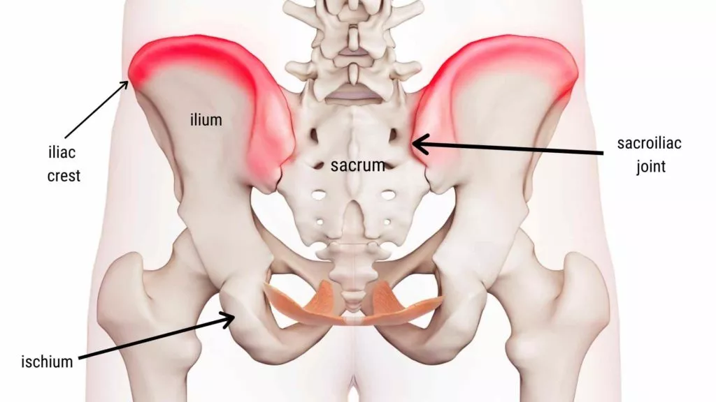
iliac crest
narrow ridge along the ilium

ilium
largest and uppermost part of the pelvis
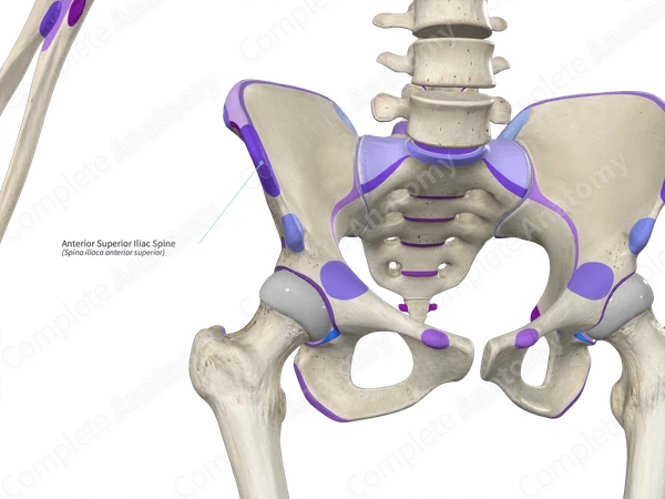
anterior superior iliac spine
upper pointy bit on the front of the ilium
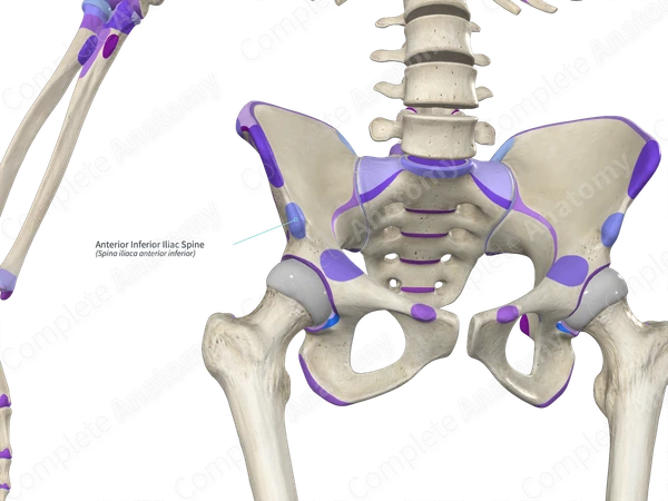
anterior inferior iliac spine
lower pointy bit on the front of the ilium

acetabulum
→ cup-shaped socket on the lateral side of the pelvis, composed of all 3 bony regions
→ articulation between pelvis & head of femur to form ball-and-socket joint
→ synovial joint
obturator foramen
hole in pelvis
→ oval in males, wider in females
posterior inferior iliac spine
lower pointy but on the back of the ilium
greater sciatic notch
large indentation for sciatic
ischial spine
pointy part on ischium
less sciatic notch
small indentation for sciatici
ischial tuberosity
bumpy part of ischium
→ weight-bearing area when seated
iliac fossa
scooped out area on the ilium
auricular surface
“ear-shaped” surface
articulates with the sacrum & creates sacroiliac joint
articular surface of the pubis
part of pubic symphysis
ischial ramus
bar of ischial bone
female pelvis vs. male pelvis
female: great pelvic angle (> 90), sacrum tilted back, bigger pelvic outlet
male: narrow pelvic angle (< 90), sacrum tilted forward, small pelvic outlet
how can you tell medial from lateral views of the pelvis?
the acetabulum is always facing laterally
femur
largest & strongest bone in the body
length ~ ¼ of a person’s height
articulates proximally w/ acetabulum of hip & distally w/ tibia & patella
how can you tell medial from lateral views of the femur?
the head of the femur always points medially
how can you tell anterior from posterior views of the femur?
the smooth patellar surface faces anteriorly
greater trochanter of femur
large, bumpy surface
lesser trochanter of femur
smaller bumpy surface
head of femur
articulate at the acetabulum
fovea capitis
depression/pit on the head of femur
adductor tubercle of femur
bump created from the adductor muscles
medial epicondyle of femur
bone on top of the condyle
later epicondyle of femur
bone on top of the condyle
patellar surface of femur
smooth, articular surface for the patella
intertrochanteric crest of femur
ridge between greater & lesser trochanters
medial condyle of femur
rounder articular surface
lateral condyle of femur
rounded articular surface
intercondylar fossa of femur
scooped out area between condyles
what covers femoral condyles & articular surface of the patella?
hyaline cartilage
what stabilizes the knee joint?
fibrocartilage meniscus
tibia
medial leg bone, receives weight of body from femur and transmits to foot
connected to fibula by interosseous membrane
fibula
not weight-bearing, no articulate with femur
several muscles originate from fibula
articulates proximally & distally with tibia
how can you tell anterior from posterior on the tibia/fibula?
the tibial tuberosity always face anteriorly
how can you tell medial from lateral on the tibia/fibula?
the tibia is always medial & the fibula is always lateral
tibial tuberosity
lumpy part of tibia
lateral condyle of tibia
smooth articular surface
medial condyle of tibia
smooth articular surface
intercondylar eminence of tibia
sticks up between medial & lateral condyles
medial malleolus
inner ankle bone, on tibia
lateral malleolus
outer ankle bone, on fibula
tarsus
7 tarsal bones form posterior half of the foot
body weight carried primarily by talus & calcaneus
other tarsal bones: cuboid, navicular, medial/intermediate/lateral cuneiform
metatarsals
5 metatarsal bones
enlarged head of metatarsal 1 form ball of foot
phalanges
14 bones of the toes
digit 1: hallux (big toe) has 2 bones with no middle phalanx
digits 2-5: have 3 bones (distal, middle, proximal phalanx)
talar dome
articulates with the distal tibia/fibula to create ankle joint
ball-and-socket joint of the hip
made of acetabulum & head of femur
multiaxial (flexion/extension, abduction/adduction, rotation)
knee joint
made of distal femur & tibia
hinge joint (flexion/extension, minimal rotation)
ankle joint (2 movements)
made of distal tibia/fibula & talus
hinge joint (plantar flexion/dorsiflexion)
made of talus & calcaneous
inversion/eversion
interphalangeal (IP) joint
hinge joint, uniaxial, flexion/extension
metatarsophalangeal joint
condyloid joint, biaxial, flexion/extension & abduction/adduction
distal interphalangeal (DIP) joint
hinge joint, uniaxial, flexion/extension
proximal interphalangeal (PIP) joint
hinge joint, uniaxial, flexion/extension
anterior muscles crossing hip & knee joints
flex femur at hip, extend leg at knee
posterior muscles crossing hip & knee joints
extend thigh, flex leg
anterior compartment of thigh
muscles anterior to femur
anterior compartment of leg
muscles anterior to tibia
posterior compartment of thigh
muscles posterior to the femur
posterior compartment of leg
muscles posterior to tibia
medial compartment of thigh
muscles on medial aspect of femur
lateral compartment of leg
muscles on the fibula-side