Lab 3 Microbio
 Call Kai
 Learn
Learn Practice Test
 Spaced Repetition
 Match
Match Flashcards
 Knowt Play
1/68

There's no tags or description
Looks like no tags are added yet.
Name | Mastery | Learn | Test | Matching | Spaced | Call with Kai | Chat |
|---|
No analytics yet
Send a link to your students to track their progress
69 Terms
How do you calculate total magnification, and what are the usual lens powers?
Objective power × eyepiece power. Objectives: 4x, 10x, 40x, 100x (oil). Eyepiece: usually 10x.
What is the total magnification with a 10x eyepiece and a 40x objective?
10 × 40 = 400x
Lab Q1: What is the total magnification when you view a bacterial slide with the 100x oil immersion lens?
10 (eyepiece) × 100 = 1000x
What is a bright field microscope?
A compound light microscope. The specimen is lit from below and viewed from above. Contrast comes from the sample absorbing light (in dark field, contrast comes from the sample scattering light).
What is the eyepiece?
The part you look through. It holds the ocular lens, usually 10x to 15x.
What is the nosepiece?
Holds the objective lenses. It rotates to change magnification.
What are the objective lenses?
Usually 3 or 4 lenses: 4x (scanning), 10x (low power), 40x (high dry), 100x (oil immersion). The power is etched on each one.
What do the stage and stage clips do?
The stage is the flat platform that supports the slide. The stage clips hold the slide in place.
What does the diaphragm do?
Controls the intensity and size of the cone of light on the specimen. The more transparent the specimen, the less light it needs.
What does the light source do?
Projects light upward through the diaphragm, slide, and lenses.
What do the base and arm do?
The base supports the microscope. The arm supports it when carried (use both hands and do not tilt it).
What does the condenser lens do?
Focuses the light onto the sample. It is most helpful with the highest objective.
What do the coarse and fine adjustment knobs do?
Coarse moves the stage up or down for rough focus. Fine makes small focus adjustments. Use only the fine knob with the oil immersion lens.
What is magnification, and why isn't it enough for a good image?
Magnification makes an object look larger. A good image also needs clarity (more specimen detail), which magnification alone doesn't give.
What is resolution (resolving power), and what affects it?
The ability to tell apart two images that are close together. It depends on optical quality and, most importantly, the wavelength of light used.
What happens to resolution as magnification increases, and how do we improve it at 100x?
Resolution decreases as magnification increases. At 100x we add special oil (oil immersion) to increase resolution (clarity).
Lab Q2: Define resolution of a microscope. What is the role of the oil used at 100x?
Resolution is the ability to see two close-together points as separate (clarity of detail). Oil is used with the 100x objective to increase resolution (clarity). Shorter wavelengths of light give better resolution.
How does working distance change as magnification increases, and why does it matter?
It gets shorter as magnification goes up (longer at low power). At 40x and 100x the lens is very close to the slide, so you can damage the specimen. Oil immersion has the shortest working distance, so be extra careful.
What can you see with stained, prepared slides?
Bacteria, chromosomes, organelles, protists or metazoans, smears, blood, negative-stained bacteria, and thick tissue sections.
What can you see with unstained wet mounts, and why is it harder?
Living things like pond water, living protists or metazoans, and plant cells such as algae. It's harder because small specimens are distorted and there is no natural pigment for contrast.
What can a light microscope NOT show, and what do you need instead?
Molecules, atoms, and viruses. You need an electron microscope.
How do you start and finish using the microscope?
Start: carry it with both hands, set the light intensity to zero before turning it on, and begin with the low power (10x) objective. Finish: turn the light off and intensity to zero, clean with lens paper and cleaning fluid only, and rotate the low power objective into place.
How do you focus when viewing bacteria in this lab?
Focus on your specimen with the low power (10x) objective, then go DIRECTLY to 100x oil (you usually don't need 40x for bacteria). Use only the fine adjustment knob with oil. (For Gram-stained slides the manual says to start at 4x instead.)
How do you use immersion oil correctly?
Put a drop of oil on the specimen, swing in the 100x objective, and focus with the fine knob only. Never put the 40x objective in oil.

What are the three main bacterial shapes?
Coccus (cocci), rod or bacillus, and curved forms (spirillum / spirochete).


What shape group are these, and what are they called?
Cocci. Diplococci = cocci in pairs. Neisseriae = coffee-bean shape in pairs.


What shape group are these, and what are they called?
Cocci. Tetrads = cocci in packets of 4. Sarcinae = cocci in packets of 8, 16, or 32.


What shape group are these, and what are they called?
Cocci. Streptococci = cocci in chains. Micrococci and staphylococci = large cocci in irregular clusters.


What is this?
Coccobacillus: A rod (bacillus) type: slightly compressed spheres or short, plump rods.


What are these bacteria called?
Mycobacteria (a type of rod/bacillus).


What are these bacteria called?
Corynebacteria (rods in a palisades arrangement).
(a type of rod)


What are these bacteria called?
Left: spore-forming rods. Right: Streptomycetes (mold-like, filamentous bacteria). Both are under rod / bacillus.


What are these bacteria called?
Vibrios (curved rods), under curved forms.


What are these bacteria called?
Spirilla (rigid spirals), under curved forms.


What are these curved bacteria called?
Spirochetes (tightly coiled), under curved forms.


Lab Q14: Label each

What is simple staining, and what is it useful for?
Uses only one stain to color bacterial cells. It doesn't show internal structures but shows cell shape and arrangement (morphology).
What is differential staining, and what can it show?
Uses two or more stains plus other reagents that react differently with different bacteria. It sorts bacteria into groups (Gram + vs Gram -, acid-fast vs non-acid-fast) and can show structures like endospores.
Simple vs differential staining: how do they differ?
Simple = one stain, shows shape and arrangement only. Differential = two or more stains and reagents, separates bacteria into groups. Hook: simple = shape, differential = groups.
Why do we stain bacteria, and which stains do we usually use?
Bacteria are very small and must be stained to see their morphology. We usually use basic stains (positively charged, they bind negatively charged molecules like proteins and nucleic acids): crystal violet, safranin (red), and methylene blue.
How do you make a bacterial smear from a solid culture?
Put a small drop of water on a clean slide, touch a SINGLE colony with a sterile loop, and mix into the drop. Don't scoop a whole colony. An opaque slurry means too many bacteria.
How do you dry the smear, and what must you NOT do?
Let it air dry. Do not heat it or blow on it, because that could force bacteria into the air and cause contamination or infection.
Lab Q12: What is the purpose of heat fixation?
Denatures bacterial proteins so the cells stick to the slide, and kills the bacteria so they are safe to handle in the following steps.
How do you do a simple stain after heat fixing?
Cover the smear with stain for 30 seconds, rinse with water, and gently blot (don't rub) dry with bibulous paper. No cover slip is needed because the cells are heat fixed.
Which two methods of differential staining are used for bacteria?
Gram staining (Gram + vs Gram -) and acid-fast staining (acid-fast vs non-acid-fast).
Lab Q6: Why is Gram staining important? What is the medical significance of identifying a pathogen as G+ or G-?
It quickly sorts bacteria into two groups based on cell wall composition. Clinically, it helps choose the right antibiotic treatment.
Lab Q8: What is the basis of Gram stain results?
Differences in cell wall structure: thick peptidoglycan (G+) vs thin peptidoglycan plus an outer membrane (G-).

What is this cell wall like, and what color does it stain?
Gram Postivite Cell Wall
Very thick, multi-layered peptidoglycan beyond the plasma membrane, with teichoic and lipoteichoic acids sticking out (negative charge). Stains purple/blue (keeps crystal violet).


What is this cell wall like, and what color does it stain?
Gram negative cell wall
thin (single-layer) peptidoglycan plus an outer membrane with lipopolysaccharide (LPS, an endotoxin that triggers the immune response). The space between membranes is the periplasm. Stains pink/red (takes up safranin).

Lab Q7: List the Gram stain reagents in the correct order.
1) Crystal violet (primary stain), 2) Gram's iodine (mordant), 3) Alcohol / 95% ethanol (decolorizer), 4) Safranin (counterstain).
Gram stain step 1: what is the primary stain and what does it do?
Crystal violet. Stains all cells purple (G+ purple, G- purple).
Gram stain step 2: what is the mordant and what does it do?
Gram's iodine. Forms a crystal violet-iodine complex that binds firmly in the peptidoglycan. Both G+ and G- stay purple.
Gram stain step 3: what is the decolorizer and what does it do?
Alcohol (95% ethanol). In G- cells it dissolves the outer membrane, and the thin peptidoglycan can't hold the crystal violet, so the dye washes out. In G+ cells the thick peptidoglycan dehydrates and traps the dye. G+ stay purple, G- become colorless.
Gram stain step 4: what is the counterstain and what does it do?
Safranin. Stains the colorless G- cells red/pink so they can be seen. G+ stay purple.
Lab Q10: Why are contrasting colors important in Gram staining?
Purple (G+) and red/pink (G-) are easy to tell apart, and the red counterstain makes the otherwise colorless G- cells visible.
How do you decolorize carefully in a Gram stain?
Hold the slide at a 45 degree angle and rinse with 95% ethanol one drop at a time. Stop as soon as no more blue color runs off, then rinse with water right away.
What happens if you over-decolorize or under-decolorize a Gram stain?
Over-decolorize: G+ cells lose crystal violet and look G-. Under-decolorize: G- cells keep crystal violet and look G+.
Lab Q9: Name two conditions needed for a reliable Gram stain.
1) The culture must not be older than 24 hours (18-24 h). 2) Decolorization must be done carefully (not over or under).
Lab Q11: Why is an 18-24 hour culture needed for Gram staining?
Gram staining depends on cell wall structure. In older cultures the walls break down, so G+ cells can stain as G-. (Manual: culture should not be older than 24 hours.)
Lab Q13: What are the advantages of Gram stain over a simple stain?
Simple stain shows only shape and arrangement. Gram stain also divides bacteria into G+ and G- based on cell wall, which helps identify them and guides treatment.
How do you view a Gram-stained slide under the microscope?
Start with the 4x scanning lens and work up to 100x (the manual says to aim for going straight from 4x to 100x). Use oil immersion at 100x and never put the 40x in oil.

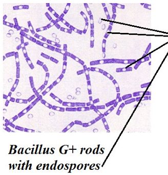
What do these Gram-stained bacteria look like?
Bacillus: Gram-positive (purple) rods in chains, with endospores.
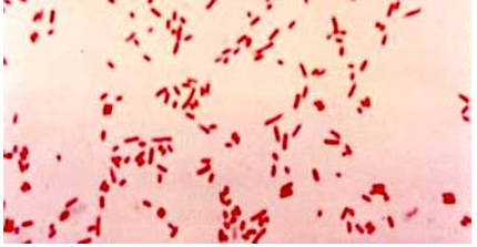
What do these Gram-stained bacteria look like?
E. coli: Gram-negative (pink/red) rod-shaped cells.


What do these Gram-stained bacteria look like?
Staphylococci: Gram-positive (purple) cocci in clusters.

Lab Q3: You see Gram-positive rods in chains with endospores. What is the identification?
Bacillus (Bacillus sp., e.g. Bacillus subtilis).
Lab Q4: Name one bacterium that appears as Gram-negative rods.
Escherichia coli (E. coli).
Lab Q5: Name one bacterium that appears as a Gram-positive coccus.
Staphylococcus (e.g. Staphylococcus epidermidis, used in lab).
What three organisms did you stain in the lab, and what are their Gram reaction and shape?
E. coli = G- rod. S. epidermidis = G+ cocci (clusters). Bacillus subtilis = G+ rod (with endospores).