Cutaneous Clues to Systemic Disease
 Call Kai
 Learn
 Practice Test
 Spaced Repetition
 Match
 Flashcards
 Knowt Play
1/101

There's no tags or description
Looks like no tags are added yet.
102 Terms
Skin Signs of Endocrine Abnormalities? (4)
Acanthosis Nigricans
Granuloma Annulare
Pre-tibial Myxedema
Vitiligo

Acanthosis nigricans
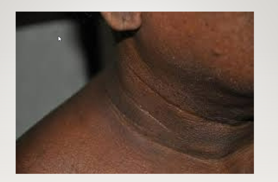
Acanthosis nigricans

ACANTHOSIS NIGRICANS

ACANTHOSIS NIGRICANS: AXILLA
Describe Acanthosis Nigricans (AN)
Lesion Characteristics (2)
Locations (4)
Disease Association: (3)
Tx: (2)
Acanthosis Nigricans (AN)
Lesion Characteristics
Hyperpigmented dark velvety papules
coalescing into plaques,
thickened skin in these areas
Appears Dirty
Locations
Axilla, Groin, Neck, Knuckles
Areas of flexure:
Disease Association:
Insulin Resistance,
Diabetes Type II,
Obesity
Tx:
Treat insulin resistance,
No cosmetic improvement in hyperpigmented areas.
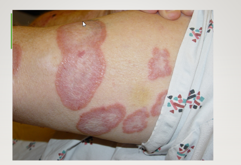
Granuloma Annulare:
Pink, annular plaques with central clearing , no scaling.

Granuloma Annulare: Reddish brown plaque with central clearing: Borders are palpable.
Note: Arcuate Pattern: The circle hasn’t completed itself.

Granuloma Annulare: Reddish plaque with central clearing with No scale. Annular Pattern (Dorsum of hand)
Describe Granuloma Annulare (GA)
Lesion Characteristics
Histology (3)
Mech:
Associated Disease (7)
Clinical Procedure: (2)
Tx (4)
Granuloma Annulare (GA)
Lesion Characteristics
Pink, reddish. brown plaques w/ central clearing
No scale.
Annular or Arcuate
Histology
Presence of palisading histiocytes around focus of necrobiosis + increased mucin deposition.
Mech:
Unclear
Associated Disease
Diabetes Type II
Insulin resistance,
Auto- immune thyroiditis,
Hyperlipidemia
Lymphoma (less so),
HIV
Solid tumors
Clinical Procedure:
Check ROS for pert +/- for potential associated malignancy
Fever, chills, weight loss, night sweats
For older pts: check for multiple myeloma
w/ SPEP & UPEP (serum/urine protein electrophoresis)
Tx
Difficult
Intralesional steroids,
calcineurin inhibitors (tacrolimus, pimecrolimus)
hydroxychloroquine
adalimumab

Vitiligo

Vitiligo

Vitiligo
Describe Vitiligo
What is it?
MOA
Associated disease (2)
Vitiligo
What is it?
Disfiguring skin disease -> loss of pigmentation (skin/hair)
MOA
Autoimmune-mediated destruction of melanocytes
Associated disease
autoimmune thyroid disease + thyroid cancer

Pretibial myxedema

Pre-Tibial Myxedema (PTM)
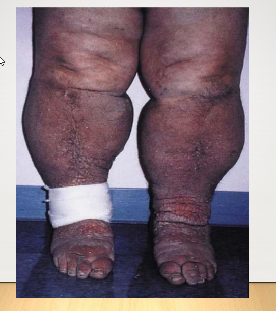
Severe Pretibial Myxedema: with severe lymphedema, and cobblestoning of shins and dorsum of feet.
Describe Pre-Tibial Myxedema (PTM)
Histopathology (2)
Associated Disease (1)
Presentation (5)
Tx (4)
Pre-Tibial Myxedema (PTM)
Histopathology
Mucin (glycosaminoglycans) deposition -> reticular dermis + subcutis-sparing papillary dermis
-> dermal edema
Collagen = splayed + decreased density
Associated Disease
Hypo/Hyper(Graves) thyroidism
Less common w/ hypo
Presentation
Skin thickening + induration in Pre-Tibial area
Yellow-red discoloration (“peau d’orange skin),
Hyperkeratosis,
Hyperpigmentation,
Hyperhidrosis.
Tx
Treat underlying thyroid
Compression stockings + topical steroids,
weight loss,
avoid tobacco.
List the Skin signs of HIV/AIDS
Kaposi Sarcoma
Bacillary Angiomatosis
Oral Hairy Leukoplakia
Syphillis
HPV
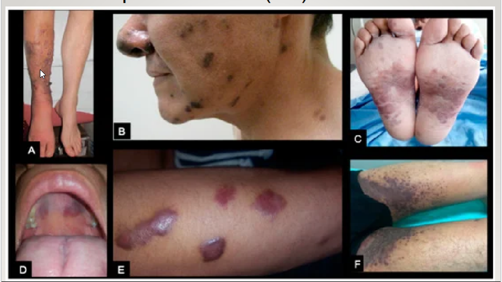
Kaposi Sarcoma (KS):
Exophytic and verrucous lesions limited to the right leg with lymphedema. (B) Dark plaques in face and nose.
) Erythematous plaques insoles. (D) Violaceous macules in the palate.(E) Violaceous and elevated plaques in the fore
) Bilateral groin infiltration with confluent violaceous plaques.

Kaposi sarcoma: Plantar area of the foot: purple plaques: from an overproliferation of blood vessels in the dermis.

Oral Kaposi sarcoma: Oral gingival presentation: These are extremely friable, and bleed easily

Oral Kaposi sarcoma of the upper palate:
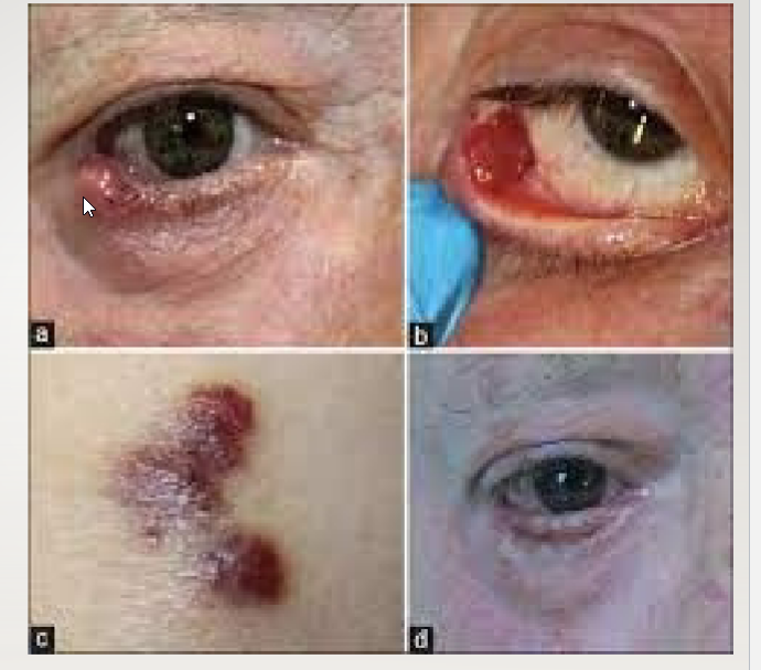
Kaposi Sarcoma of the eyelid
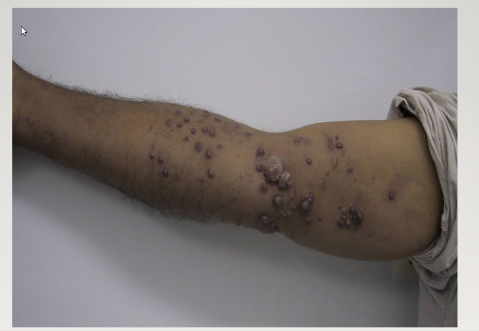
Kaposi sarcoma (KS):
Palpable Nodules: are a sign of a more advanced KS :requires more aggressive
Describe Kaposi sarcoma (KS)
What is it? (2)
Epidemiology:
Lesion Characteristics (2)
Histologically:
Kaposi sarcoma (KS)
What is it?
low grade vascular tumor
associated w/ Human herpes virus 8 (HHV-8)
Epidemiology:
Primary risk factor:
IMMUNE SUPPRESSION
Lesion Characteristics
Well demarcated DEEP PURPLE papules
coalescing into plaques
Can get GI involvement
Histologically:
Over proliferation of blood vessels (atypical) in dermis.
An infiltrative process.

Bacillary angiomatosis
Describe Bacillary angiomatosis
MOA
Transmission:
Lesion Characteristics (2)
Disease Association
Clinical Procedure
Bacillary angiomatosis
MOA
Infection: Bartonella henselae/quintana (Gram negative rods)
-> reactive proliferation of endothelial cells. (Vascular)
Transmission:
cat bites or scratches
Lesion Characteristics
Dome-shaped, smooth, red/purple papules/nodules
Ulcerate and bleed easily
Can be internal (like kaposi)
Disease Association
AIDS (uncommon)
Clinical Procedure
Always Biopsy
Looks similar to many dif types of lesions

Oral Hairy Leukoplakia
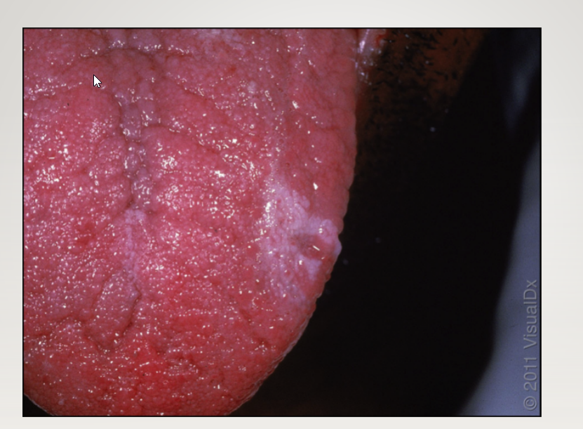
Oral hairy leukoplakia is indistinguishable from Oral leukoplakia which has malignant potential. Always biopsy these lesions
Describe Oral Hairy Leukoplakia
Associated disease (3)
Presentation
Clinical Procedure
Prognosis
Oral Hairy Leukoplakia
Associated disease
EBV
HIV (25%)
Immunosuppression
Presentation
Not an oral thrush so it cannot be scraped off with a tongue blade.
Clinical Procedure
Biopsy
Indistinguishable from SCC (oral leukoplakia)
Prognosis
NOT PRE CANCER
List the Additional Dermatologic Manifestations of HIV Infections
Additional Dermatologic Manifestations of HIV Infections
Bullous Impetigo
Folliculitis
Syphilis!
Viral infections: Herpes, HPV, EBV, Molluscum Contagiosum
Fungal infection (superficial and deep)
Psoriasis/Seborrheic dermatitis
Often see exaggerated forms
Pruritus

Primary syphilis: chancre: This is Painless chancre

Secondary syphilis: papulosquamous eruption
involving palms and soles, “Copper-Penny”

Also secondary syphilis…

Tertiary syphilis: “Gummas”
Describe Syphilis
What is it?
Stages/Description? (1,2,3)
Syphilis
What is it?
STD caused by T. Pallidum (spirochete)
Can -> HIV
Stages
Chancre (primary)
Painless (pt don’t know they have it)
3-6 weeks
Papulosquamous eruption (secondary)
Imitates other rashes ( Tinea, Eczema, Seborrheic dermatitis)
Copper-penny signs (palms/soles)
Neurosyphillis (tertiary)
Latent (several yrs later)
Associations: Neurology symptoms
Gummas
Wet, ulcerated plaques
Skin signs of other inflammatory conditions
LICHEN PLANUS (LP)
ERYTHEMA MULTIFORME (EM)
PORPHYRIA CUTANEA TARDA (PCT)
HIDRADENITIS SUPPURTIVA (HS)
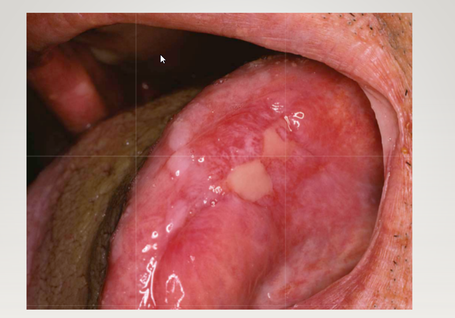
Oral Mucosa Lichen planus: Wickham Striae

Lichen planus
Describe Lichen planus (LP)
What is it?
Lesion Characteristics (2)
Location (3)
Epidemiology (2)
Dx
Disease Association
Tx (2)
Lichen planus (LP)
What is it?
Chronic, inflammatory, autoimmune disease
skin, oral mucosa, genital mucosa, scalp, and nails
Lesion Characteristics
6 P’s
planar [flat-topped]
purple,
polygonal,
pruritic,
papules,
Plaques
Wickham striae
Lacy, reticular, white lines on lesions
Location
flexor surfaces of wrists, forearms, and legs.
Epidemiology
0.1 to 4 percent of general population
MC in perimenopausal women
Most ages = btw 30-60yrs
Dx
Can be clinical but 4-mm punch biopsy = helpful
Disease Association
Hep C (mc in mucosal)
Tx
High-potency topical corticosteroids
Clobetasol; can also use topical tacrolimus (for vulvovaginal)
(can resolve spontaneously if not on mucous membranes)

Erythema multiforme
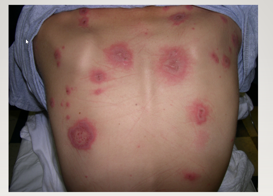
Erythema multiforme

Erythema multiforme

Erythema mutiforme
Describe Erythema Multiforme
Associated Disease (4)
What is it
Lesion Characteristics (3)
Erythema Multiforme
Associated Disease
Possible underlying infection?
Herpes (>90%)
Mycoplasma pneumonia = second MC
Medication reaction
Immunizations
Autoimmune diseases
What is it
immune-mediated reaction @ skin and mucosa
Lesion Characteristics
Target-like, bullous eruption
Isolated, recurrent, or persistent
Symmetrical on extremities
extensor surfaces
palms and soles
spread centripetally


Poryphyria cutanea tarda
Describe Porphyria Cutanea Tarda (PCT)
Presentation (2)
MOA
Triggers: (7)
Genetics
Porphyria Cutanea Tarda (PCT)
Presentation
Photo-distributed bullous eruptions on dorsal hands (typically)
Milia and scarring (unique feature)
MOA
Defect in (UROD)
uroporphyrinogen decarboxylase
Triggers:
estrogen,
alcohol,
renal failure,
hemochromatosis,
Iron overload disorders
NSAIDs (drug-induced variant)
HIV/Hepatitis C
Genetics
75-85 percent acquires it

Hidradenitis Suppurtiva
( Acne inversa)
Describe Hidradenitis Suppurtiva ( Acne inversa)
Presentation
MOA
Associated disease (2)
Genetics:
Tx (5)
Hidradenitis Suppurtiva ( Acne inversa)
Presentation
Relapsing, recurrent painful cysts or boils
axilla or groin
MOA
disorder of follicular occlusion
Associated disease
Obesity
Smoking
Genetics: AD
Tx
Depends on Hurley Stages
Warm Compresses,
topical antibiotics (topical or oral)
oral steroids,
Surgical excision,
Laser
List the Skin signs suggestive of underlying: MALIGNANCY
Skin signs suggestive of underlying: MALIGNANCY
PAGET’S DISEASE OF THE BREAST
SISTER MARY JOSEPH NODULE
SIGN OF LESER TRELAT
SWEET SYNDROME

Paget’s disease of the breast; Left = Affected

Pagets Disease of the Breast
Describe Paget’s Disease
What is it?
Presentation
Associated diseases
Epi
Histo (3)
Paget’s Disease
What is it?
rare form of breast cancer
Presentation
epidermal migration (of malignant cells) -> eczematous eruption
Nipple -> areola
Associated diseases
Underlying ductal breast cancer
In situ or invasive
Epi
1 - 4.3 of all breast cancers
Histo
Paget cells are large cells with clear cytoplasm and atypical nuclei w/in epidermis of nipple
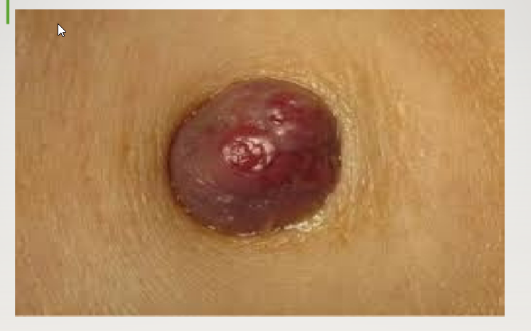
Sister Mary Joseph Nodule
Describe Sister Mary Joseph Nodule
Associated Cancers (2)
Presentation
Sister Mary Joseph Nodule
Associated Cancers
GI (35–65%) and GU Cancers (12–35%)
Other
lung, pancreas, liver, gallbladder, lymphoma, breast, kidney, penis, prostate, and testicles.
Presentation
periumbilical mass
NOTE
A periumbilical mass is not always a Sister Mary Joseph nodule. Other conditions that can cause a palpable periumbilical mass include umbilical hernia, infection, and endometriosis. Medical imaging, such as abdominal ultrasound, may be used to distinguish a Sister Mary Joseph nodule from another kind of mass
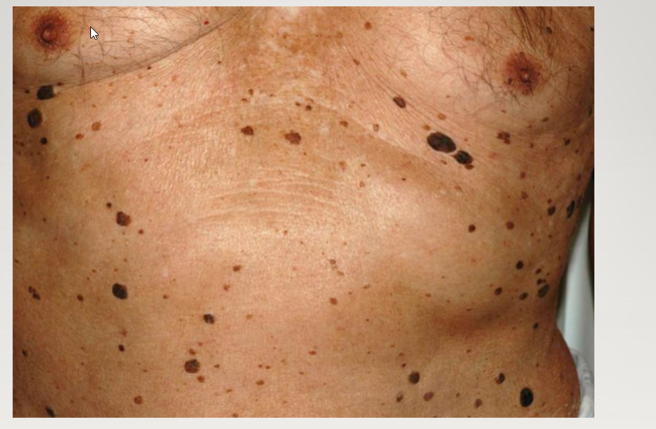
Leser Trelat= multiple seborrheic keratoses
Describe Sign of Leser Trelat
Associated Disease (2)
MOA
Dx:
Sign of Leser Trelat
Associated Disease
Gastric adenocarcinoma >> breast adenocarcinoma
RARE
malignant acanthosis nigricans
MOA
Thought related to increase in secretion of growth factors from the malignancy
Dx:
Must be a true eruption of seborrheic keratoses (hundreds)

Sweet’s syndrome
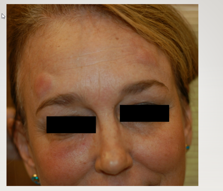
Sweet’s syndrome

Sweet’s syndrome
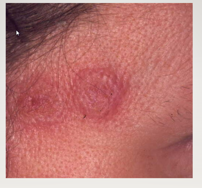
Sweet’s syndrome
Describe Sweet’s syndrome
AKA
Presentation (2)
Associations (7)
Locations (3)
Course
Sweet’s syndrome
AKA
Acute Febrile Neutrophilic Dermatosis
Presentation
Red/purple papules/nodules/plaques with mamillated “juicy” surface.
Very tender
Systematic Symptoms:
Fever, myalgias, joint aches, leukocytosis
Preceded by URI or flu-like illness
Associations
Hematologic Malignancies
AML
Solid tumors
Infections:
Strep, yersinia
Meds:
Granulocyte colony stimulating factor
Enteropathies:
Crohn’s disease
Ulcerative Colitis
Pregnancy
Locations
Upper extremities, face, neck
Course
Often resolves within 5-12 weeks

Sarcoidosis

Sarcoidosis: Subtype: Lupus Pernio (involvement area nasal ala): Look for lung involvemen

Sarcoidosis: Can mimic GA ( Both have histiocytic proliferation of the dermis.
Describe Sarcoidosis
What is it?
Epi
Skin Symptoms (1,4)
Sarcoidosis
What is it?
SYSTEMIC DISEASE + formation of NON- CASEATING GRANULOMAS in
LUNGS—EYES—HEART
HILAR LYMPH NODES
Epi
Often: African Americans
⅓ have skin involvement
Skin Symptoms
RED-BROWN PAPULES & PLAQUES
FACE,
NECK,
UPPER TRUNK,
EXTREMITIES AND SITES OF PRIOR TRAUMA,
(including scars and tattoos)

Left = Lepropsy; Right = GRANULOMA ANNULARE

Keloid scars from truncal acne can mimic Sarcoidosis : Difference is there
are NO Systemic Symptoms
Cutaneous signs of GI diseases and others?
Erythema Nordosum
Pyoderma Gangrenosum
Dermatitis Herpetiformis

Erythema nodosum
Describe Erythema nodosum
What is it
Presentation (2)
Disease Associations (7)
Erythema nodosum
What is it
A type of panniculitis
Presentation
ill-defined, tender, erythematous plaques and nodules
MC at pretibial areas
Systemic
Fever, malaise, and arthritis/arthralgia
Disease Associations
IBD:
Crohns > ulcerative colitis
Infections
(Streptococcus in children, tuberculosis, and coccidioidomycosis)
Pregnancy
Medications
(oral contraceptive pills)
Connective tissue diseases
Malignancy
Sarcoidosis

Pyoderma gangrenosum – note color of borders

Pyoderma gangrenosum – note undermining of borders

Pyoderma gangrenosum
Describe Pyoderma gangrenosum
What is it?
Associations (4)
Lesion Characteristics (3)
Tx (2)
Pyoderma gangrenosum
What is it?
Ulcerative, neutrophilic skin disease
(Despite neutrophils, it is not from infection!)
Associations
IBD
Hematologic malignancy
(myeloma and leukemia)
Connective tissue diseases
Drugs
(levamisole-contaminated cocaine)
Lesion Characteristics
tender skin nodules or pustules -> ulcerate
Ulcer borders = violaceous or dusky color + undermined
(overhang into ulcer bed)
Favors pretibial and peristomal areas
Tx
PROTECT THE PG LESION FROM TRAUMA
NO DEBRIDEMENT
Systemic anti-inflam. Meds
If local: topical steroids
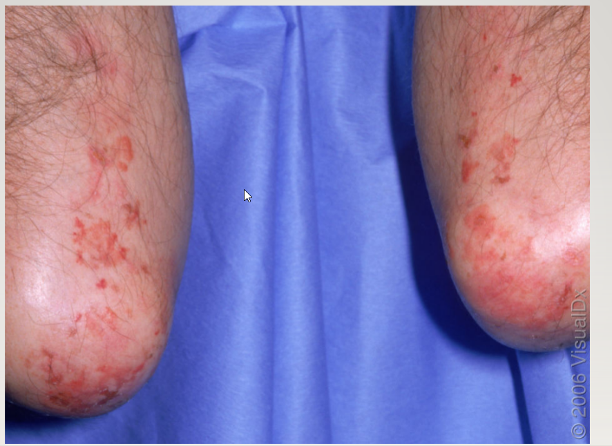
Dermatitis herpetiformis – note extensor elbow location

Dermatitis herpetiformis – note intact and burst vesicles
Describe Dermatitis herpetiformis
Associated disease?
MOA
Presentation
Dermatitis herpetiformis
Associated disease?
gluten-sensitive enteropathy
(including celiac disease)
Earliest Sign
MOA
Deposition of IgA Complexes in skin -> lesions
Autoabs targets transglutaminase 2
Presentation
pruritic papules and vesicles
elbows, knees, scalp, shoulders, and buttocks
Scratching = only erosions and excoriations
Cutaneous signs of cardiac disorders?
Janeway lesions
Oslers nodes
Xanthelasma palpebra

Janeway lesions
Describe Janeway lesions
Associated Disease
Presentation
Location (4)
Janeway lesions
Associated Disease
Subacute bacterial endocarditis
Presentation
Painless hemorrhagic macules which blanch on pressure
Location
Thenar, hypothenar eminences,
palms and soles

Osler nodes
Describe Osler nodes
Presentation
Associations
Osler nodes
Presentation
Painful red erythematous lesions
Pads of fingers/toes
Associations
Sign of subacute bacterial endocarditis

Xanthelasma palpebra
Describe Xanthelasma palpebra
Presentation
Association
Xanthelasma palpebra
Presentation
Well-circumscribed flat or slightly elevated yellowish growth
Contains ChL deposits
Typically around eyelids
Association
hyperlipemia
List the Nail abnormalities of systemic diseases
• Beau lines
• Clubbing
• Koilonychia
• Mees lines
• Muehrcke lines
• Splinter hemorrhages

BEAU LINES
Describe BEAU LINES
What is it?
MOA
Caused by: (4)
BEAU LINES
What is it?
Horizontal grooves in the nail plate
MOa
interruption of nail bed mitosis
Caused by:
High fever,
Pemphigus,
Chemotherapy,
Raynauds

TERRY’S NAILS
Describe TERRY’S NAILS
Presentation
Associations (5)
TERRY’S NAILS
Presentation
Nails= white + band of pink at top
Associations
Aging
liver disease,
kidney failure
CHF
Diabetes

NAIL CLUBBING
Describe NAIL CLUBBING
Presentation?
Pathogenesis
Associations (4)
NAIL CLUBBING
Presentation?
thickening of proximal end of nail bed soft tissue
Pathogenesis
Alt. vasculature
Associations
COPD
Cirrhosis
Celiac sprue
Smoking
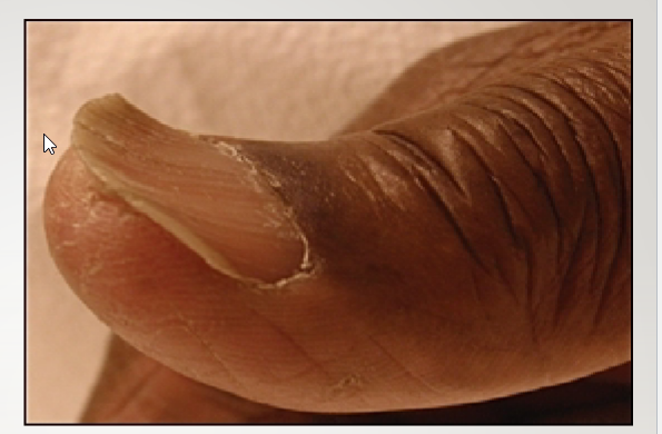
KOILONYCHIA
Describe KOILONYCHIA
Presentation
Associations (2)
KOILONYCHIA
Presentation
Nail = Concave
“Spoon Nail”
Associations
Iron deficiency Anemia
NORMAL IN INFANTS

MEES LINES
Describe MEES LINES
Associations (2)
Location:
MEES LINES
Associations
Arsenic Poisoning
Renal Failure
Location:
In NAIL PLATE no the bed

MUEHRCKE LINES
Describe MUEHRCKE LINES
What is it?
Patho:
Associations: (4)
MUEHRCKE LINES
What is it?
Pairs of transverse white lines
Patho:
Localized pathology within the nail bed.
Associations:
Hypoalbuminemia,
nephrotic syndrome,
liver disease,
malnutrition