RAD POS 1: Hand, Finger, Thumb, Wrist
 Call Kai
 Learn
Learn Practice Test
 Spaced Repetition
 Match
Match Flashcards
 Knowt Play
1/46

There's no tags or description
Looks like no tags are added yet.
Name | Mastery | Learn | Test | Matching | Spaced | Call with Kai | Chat |
|---|
No analytics yet
Send a link to your students to track their progress
47 Terms
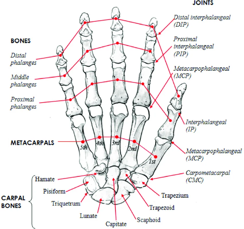
Bones of Hand pic
Fingers = phalanges (14); distal, middle, proximal (thumb only distal and proximal)
Bones between fingers + wrists = metacarpals (5)
Wrist = carpals (8)

Joint of hand + wrist
Distal interphalangeal joint (DIP) = between the distal and middle bones of the fingers (not thumb)
Proximal interphalangeal joint (PIP) = between the middle and proximal bones of the fingers (not thumb)
Interphalangeal joint joint (IP) = between the distal and proximal bones of the thumb (ONLY THUMB)
Metacarpophalangeal joint (MCP) = between the proximal and metacarpal bones (1-5 = all 5 fingers)
Carpometacarpal joint (CMC) = between metacarpals and carpal bones (1-5 = all 5 fingers)
Hand protocol views; Baystate + Textbook techniques (3+1 trauma views)
All tabletop TT SID of 40'“; use arrow to identify site of pain or injury on most relevant view
Technique for PA/OBL = Baystate 0.5-0.8 mAs @ 55-60 kVp; Handout 58-60 kVp
Technique for Lateral = 0.8-1 mAs @ 58-62 kVp
PA hand = CR 3rd MCP, marker on the lateral border along pinky, use arrow to indicate pain if relevant, wrist/forearm on the same plane
Eval. Criteria = entire hand + carpals shown, CR at 3rd MCP, no rotation of phalanges/metacarpals, proper exposure factors
PA oblique 45 degrees = CR 3rd MCP, separate fingers and no curling of them
Eval. Criteria = entire hand + carpals shown, CR at 3rd MCP, joints open, enough separation of phalanges/metacarpals, proper exposure factors
Fan lateral (straight lateral for trauma) = CR at 2nd MCP, pt make “okay” sign then spread fingers (use sponge/tape if needed)
Eval. Criteria = entire hand + metacrapals/carpals shown, CR at 2nd MCP, fingers separated + wrist no rotation, proper exposure factors
Straight lateral (trauma only) = “karate chop” position fingers superimposed, CR at 2nd MCP
Eval. Criteria = entire hand + carpals shown, CR at 2nd MCP, proper exposure factors
Baystate Finger Protocol (3 views)
All tabletop TT SID 40”; use arrow to identify site of pain or injury on most relevant view
Technique PA/OBL = 0.5-0.6 mAs @ 58 kVp
Technique Lateral = ADD
PA hand = to include all fingers
CR = PIP
Eval. Criteria = entire finger and minimum ⅓ of MCP demonstrated, CR at PIP joint, no rotation of phalanges, proper exposure factors
PA oblique (45 degrees) = of affected finger, digit parallel to IR
CR = PIP of affected finger
Eval. Criteria = entire phalanx and MCP joint demonstrated, IP and MCP joints open, CR at PIP, proper exposure factors
Lateral (of affected finger) = digit parallel to IR, reduce OID when possible, use sponges if needed
CR = PIP of affected finger
Eval. Criteria = Entire phalanx and MCP joint demonstrated, CR at PIP, true lateral position, digit parallel to IR, proper exposure factors
Baystate Thumb Protocol (3 + 1 trauma view) revise
All tabletop TT SID 40”; use arrow to identify site of pain or injury on most relevant view
Technique PA Hand (OBL thumb) = 0.5-0.8 mAs @ 58-60 kVp
Technique Lateral/AP = 0.5-0.6 mAs @ 58 kVp
Oblique
PA
AP
Lateral
Dislocation | Subluxation
Dislocation = displacement from joint
Subluxation = partial dislocation
Sprain | Contusion
Sprain = rupture or tearing of connective tissue
Contusion = bruise, without fracture
Fracture types (4)
Simple = closed fracture
Compound = open fracture that breaks through skin
Comminuted = fracture where bone is splintered or crushed
Impacted = fracture where fragments driven into each other
Types of Hand Fractures (4)
Displaced = the bones break into two or more pieces and becomes misaligned; there is a separation between the two fracture faces or the faces are no longer lined up
Non-displaced = the bone cracks or breaks but doesn’t move and maintains its proper alignment and position; the two fracture surfaces have not moved relative to each other
Comminuted = the bone is broken in several pieces in different places
Open fracture = the bone breaks through the skin; closed is when the bone breaks but there is no puncture or wound
Barton’s fracture
Fracture and dislocation of the posterior lip of the distal radius involving the wrist joint

Bennett’s fracture
Fracture of the base of the first metacarpal bone, extending into the carpometacarpal joint, complicated by subluxation with some posterior displacement

Boxer fracture
fracture of the metacarpal neck, 5th metacarpal most common boxer fracture

Colles fracture
Transverse fracture of the distal radius in which the distal fragment is displaced posteriorly; an associated ulnar styloid fracture is seen in 50% to 60% of cases
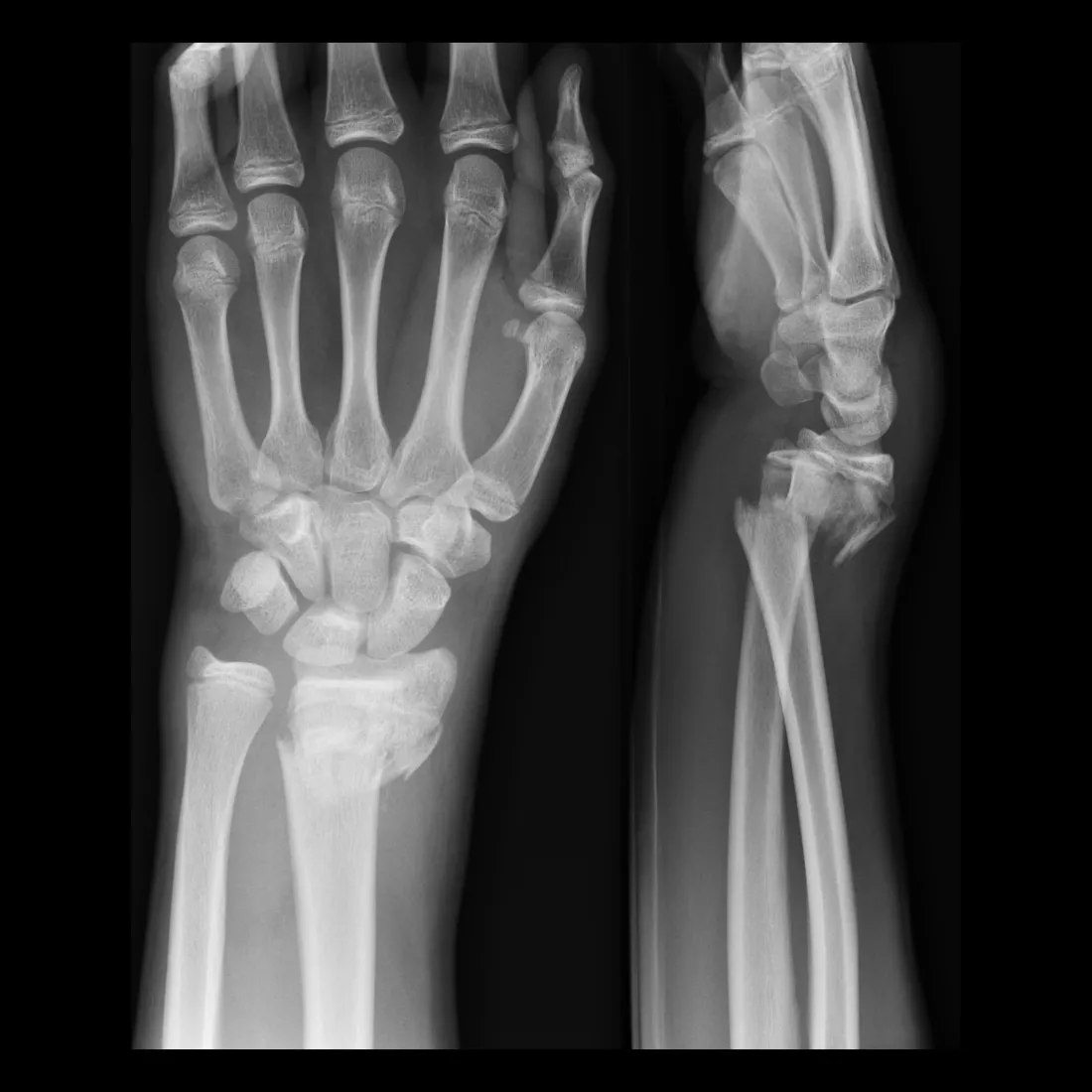
Smiths fracture
Reverse of Colles fracture, or transverse fracture of the distal radius with the distal fragment displaced anteriorly

Greenstick fracture
Break on one side only; the cortex on one side of the bone is broken, and the other side is bent

Skier’s Thumb Injury
sprain or tear of the ulnar collateral ligament of the thumb; appears as widening of inner MCP joint space of thumb and increase in degrees of angle of MCP line

Positioning considerations for upper limb (6)
40 in SID (100cm) minimum
Gonadal shielding
Four sided collimation when possible
Long axis of part to long axis of IR
Side marker visible on lateral border
Use arrow to identify pain
Positioning considerations (6)
Make sure your patient’s upper limb is on the same place, elbow is at right angle if possible
Do not have hand and wrist at an angle
If splint if possible removed
Increase exposure factors for cast/splint
Separate fingers
Remove rings if possible
AP oblique bilateral hands “ball catchers” view
used when looking for rheumatoid arthritis (not routine at clinical)
Pediatric bone age (3)
Left hand and wrist
For children with advanced or delayed growth
Radiologist will measure and estimate the maturity of a child's skeletal system
Wrist Protocol (4 views)
All tabletop TT 40” SID, 8×10 FOV; use arrow to indicate pain
Technique PA/OBL/ULDV = 0.8-1.2 mAs @ 60 kVp
Technique Lateral = 1-1.2 mAs @ 62 kVp
PA = CR midcarpal area, pt make loose fist to decrease OID, collimate appropriately (more rectangle then square), marker on lateral border along pinky side
Eval. criteria = distal radius + ulna + carpals, CR at field of midcarpals, no rotation
Oblique 45 degrees = CR at midcarpal area, fingers straight but thumb down when needed (thumb kickstand to stabilize)
Eval. criteria = distal radius + ulna + carpals + trapezium seen in entirety, CR at midcarpals
Lateral = CR to midcarpal area, hand/wrist in true lateral + SUPERIMPOSE radius/ulna (tilt further than u think)
Eval. criteria = distal radius + ulna + carpals demonstrated; radius/ulna SUPERIMPOSED; CR at midcarpals; no rotation
Ulnar deviation (age 10+ only) = CR at scaphoid, deviate wrist toward ulna 20 degrees without moving forearm
Eval. criteria = scaphoid elongated
Special Projections for Scaphoid (2)
PA axial with ulnar deviation = textbook: 15-20 degrees towards elbow; Baystate: 20 degrees tube angle toward elbow CR to scaphoid
Eval. criteria = scaphoid seen without superimposition and not foreshortened
Modified stretcher method = sponge elevates hand 20 degrees with tube not angeled
Eval. criteria = scaphoid seen without imposition
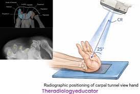
Special projection = Tangential (1) Carpal Canal
Carpal canal (tunnel) Inferosuperior Projection w/ Gaynor Hart Method = CR 1st inch distal to base of 3rd metacarpal 25-30 degrees to hand; hyperextend wrist while holding fingers back making the long axis of the metacarpals near vertical + rotate wrist 10 degrees toward radius
Eval. criteria = carpal sulcus demonstrated, pisiform + hamate separated + scaphoid in profile
Special projection = Tangential (2) Carpal Bridge
Carpal bridge = flex wrist CR 1 ½ inches proximal to wrist joint, angle CR 45 degrees
Eval. criteria = demonstrates dorsal (posterior) aspect

Wrist bones (8) + their Synonyms
Some = Scaphoid (navicular)
Lovers = Lunate (semilunar)
Try = Triquetrum (triangular or cuneiform)
Positions = Pisiform (none/pea shaped)
That = Trapezium (greater multangular)
They = Trapezoid (lesser multangular)
Can’t = Capitate (Os magnum)
Handle = Hamate (unciform)
Interphalangeal (IP) joint
Ginglymus (hinge) joint; Flexion and extension motion
Metacarpophalangeal (MCP) joint
Ellipsoid (condylar) joint; Flexion and extension, abduction, adduction, and circumduction motion
Carpometacarpal (CMC) joint (2)
articulations between the bases of the metacarpals and the carpal bones
1st finger (thumb) is a saddle (sellar) joint. Allows flexion, extension, adduction, abduction, opposition, and circumduction, and some rotation movement
2nd to 5th finger (plane/gliding jt); allows sliding/gliding movement
Intercarpal joints
Plane (gliding) joint; Sliding/gliding motion
Wrist Carpal joint
Ellispoid (condylar); Flexion and extension, abduction and adduction, and circumduction movement
Proximal radioulnar joint
Pivot (trochoid); Rotational movement
Distal radioulnar joint
Formed at the head of ulnar notch on the lower extremity of radius
Pivot joint; rotation movement
Elbow joint (humeroulnar and humeroradial)
Ginglymus (hinge); Flexion and extension movement
Wrist movements (2)
Ulnar deviation (scaphoid)
Radial deviation (lunate, triquetrum, and hamate)
Wrist fat stripes (2)
Scaphoid fat stripe (seen on PA and oblique)
Pronator fat stripe (seen on lateral wrist)
Bone metastases
A transfer of disease or cancerous lesions from one organ or part to another; appears as dark holes or bright white patches
Bursitis
Inflammation of bursae or fluid-filled sacs that enclose the joint; process involves formation of calcification in associated tendons; appears as fluid-filled joint
Carpal tunnel syndrome
Common painful disorder of the wrist/hand from compression of the median nerve as it passes through the center of the wrist, most common in middle-aged women; appears as fluid-filled joint space with possible calcification
Joint effusion
refers to accumulated fluid (synovial or hemorrhagic) in the joint cavity, sign of an underlying condition like a fracture, dislocation, soft tissue damage, inflammation; appears as fluid-filled joint cavity
Osteoarthritis
aka degenerative joint disease (DJD), is a noninflammatory joint disease where this is a gradual deterioration of the articular cartilage with hypertrophic (enlarged/overgrow) bone formation; appears as a narrowing of the joint space with periosteal growth on joint margins
Osteomyelitis
is a local or generalized infection of bone/bone marrow that may be caused by bacteria from trauma or surgery; appears as soft tissue swelling and loss of fat pad detail visibility
Osteoporosis
the reduction in quantity of bone or atrophy or skeletal tissue, commonly from fracture of bone that may lead to obliteration of marrow space; appears most visibly in distal extremities and joints as a decrease in bone image receptor exposure, long bones demonstrating in thin cortex
Paget’s disease
common chronic skeletal disease characterized by bone destruction followed by reparative process of overproduction of dense but soft bones that tend to easily fracture; appears as missed areas of sclerotic and cortical thickening along with radiolucent lesions creating a “cotton wool” appearance
Rheumatoid arthritis
chronic systemic disease with inflammatory changes throughout the connective tissue, the earliest change is soft tissue swelling that is most prevalent around ulnar styloid of the wrist, 3x more common in women than in men; appears as closed joint spaces with subluxation of MCP
Bone neoplasia
tumors that are most often benign but can be malignant; appearance based on type
Malignant bone tumors (4)
cancerous tumors
Multiple myeloma = most common primary cancerous bone tumor, affects ages 40-70, tumors occur in various parts of the body arising from the bone marrow/marrow plasma cells (not exclusively bone tumors); highly malignant + fatal; appears as multiple “punched-out” osteolytic (loss of calcium in bone) lesions throughout affected bone
Osteogenic sarcoma = aka osteosarcoma, second most common type of primary cancerous bone tumor affecting ages 10-20 but can happen at any age, may lead to Paget disease; appears as a bone lesion and mass in the metaphysis
Edwing’s sarcoma = common primary malignant bone tumor in children/young adults that arises from bone marrow; appears as new bone formation that looks like an “onion peel” pattern
Chondrossarcoma = slow growing malignant tumor of the cartilage; appearance similar to other malignant tumors but dense calcifications often seen within cartilaginous
Benign (2)
non cancerous bone tumors
Endochondroma = slow- growing benign cartilaginous tumor most often found in small bones of the hands and feet of adolescents and young adults; appears as well defined radiolucent appearing tumors with a thin cortex
Osteochondroma = most common type of benign tumor. Most common at the knee but also appear on scapula and pelvis on young adults or children. Arises from outer cortex with the tumor growing parallel to the bone, point away from the adjacent joint.