exam 2 ap info
 Call Kai
 Learn
Learn Practice Test
 Spaced Repetition
 Match
Match Flashcards
 Knowt Play
1/37

There's no tags or description
Looks like no tags are added yet.
Name | Mastery | Learn | Test | Matching | Spaced | Call with Kai | Chat |
|---|
No analytics yet
Send a link to your students to track their progress
38 Terms
What does the skeleton include and do?
bone and cartilages
supports internal organs
protects internal organs
Provides levers for movement by skeletal muscles
stores lipids and mineral
initial site of blood cell formation (hematopoiesis)
→ heme = blood
→ poiesis = forming
How may bones does the skeleton contain?
206
divided into axial and appendicular

The axial skeleton includes?
head
neck
torso/trunk
made up of bones that lie along the midline of the body
includes: skull, hyoid, vertebrae, ribs, sternum, and sacrum

The appendicular skeleton includes?
made up of bones that support the limbs and attach them to the skeleton.
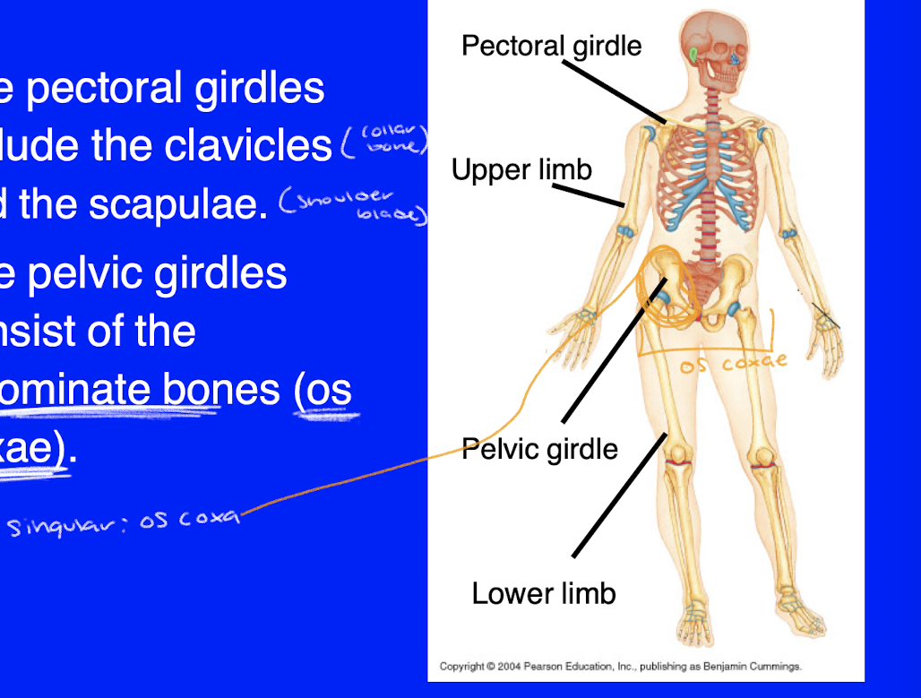
includes the pectoral girdles, upper limb, pelvic girdles, and lower limb.
What does the pectoral girdles include on the appendicular skeleton?
clavicles (collar bone)
scapulae (shoulder blade)
innominate bones (os coxae)
For the classification of bone, what are the two different forms of osseous tissue?
compact bone and spongy bone
Bones are also classified by shape. Name the 6 shapes.
F.I.L.S.S.
flat
irregular
long
short
sesamoid
sutural (AKA wormian)

Long bones
→ most append. skeleton is made up of long bones
examples of long bone:
femur
phalanges
metacarpals

What is the key characteristic structure of long bones?
epiphysis (at the ends)
diaphysis
→ two ends, called epiphysis and a shaft called diaphysis
Short bones
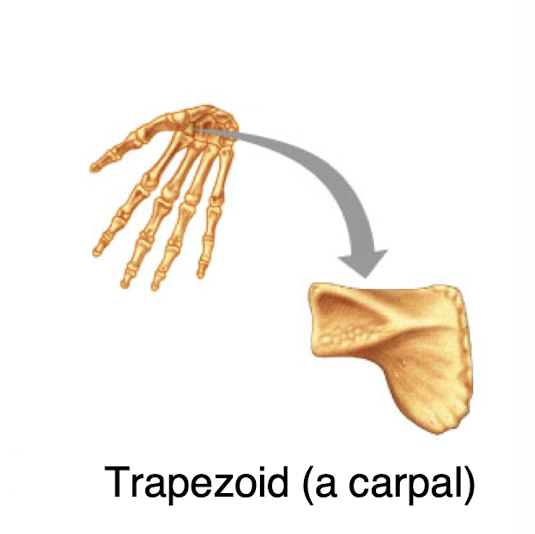
roughly cubical
they are primarily composed of spongy bone
short bones include:
→ carpals
→ tarsals

Flat bones
composed of two think layers of compact bone sandwiching a layer of spongy bone.
Flat bones include the bones of the skull, the ribs, the scapulae, and the sternum.

Irregular bones
Irregular bones are irregular in shape and do not fit into any other category.
They are often made up of several components fused during development.
the vertebrae and innominates are irregular bones.

Sesamoid bones
→ “seed shaped”
sesamoid bones form within tendons
the patella is the only sesamoid bone that is consistently present in all people.
sesamoid bones are sometimes considered to be a subcategory of short bones.

Wormian bones
→ “wormy shaped”
wormian (or sutural) bones form in the sutures of the skull.
they are not present in all people in the same locations.
they are sometimes to be considered a subcategory of flat bones.
Bone markings: What are the four categories?
projections for attachment of muscles or ligaments
projections that form joints
cavities
depressions or openings for the passage of blood vessels and nerves
→ projections for the attachment of muscles and ligaments will be important when we study the origins and insertions of muscles in exercise 15.
→ projections that form joints will be important when we study articulations in exercise 13.
We will examine cavities (specifically
sinuses) when we examine the skull in
Exercise 10.
• Depressions and openings will be important
when we study the nervous system in
Exercises 19 and 21, and the blood vessels
next semester in Exercise 32.
Structure of bones
individual bones are organs
an organ is a discrete structure composed of two or more tissue types that performs a specific function.
bones are composed of:
→ osseous connective tissue
→ dense irregular connective tissue
→ adipose connective tissue
→ and one or more types of cartilage
Structure of bones: Most bones contain both ____ and ____ forms of osseous connective tissue.
compact
spongy
Structure of bones: What is the function of compact bone?
provides strength along a single axis, bit is weak perpendicular to that axis
Structure of bones: What is the function of spongy bone?
provides limited strength in many directions and helps distribute forces
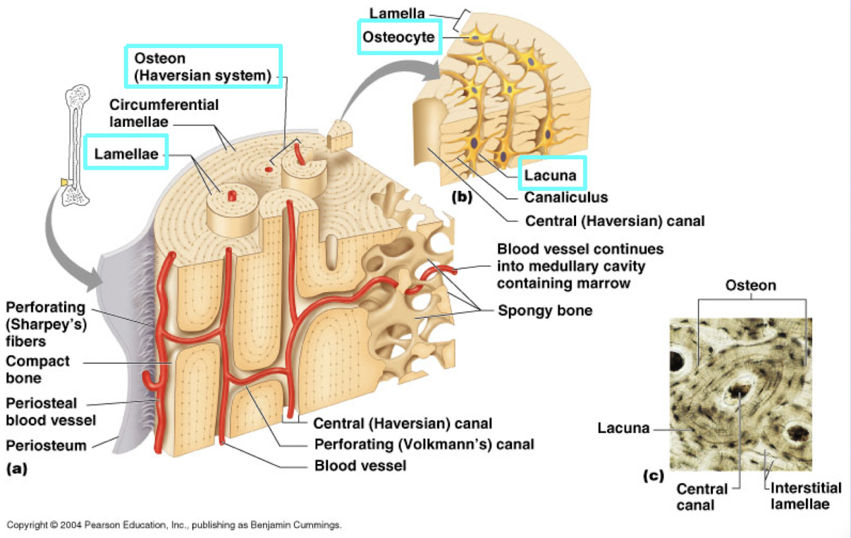
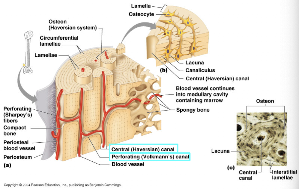
Compact bone is built from units called:
osteons

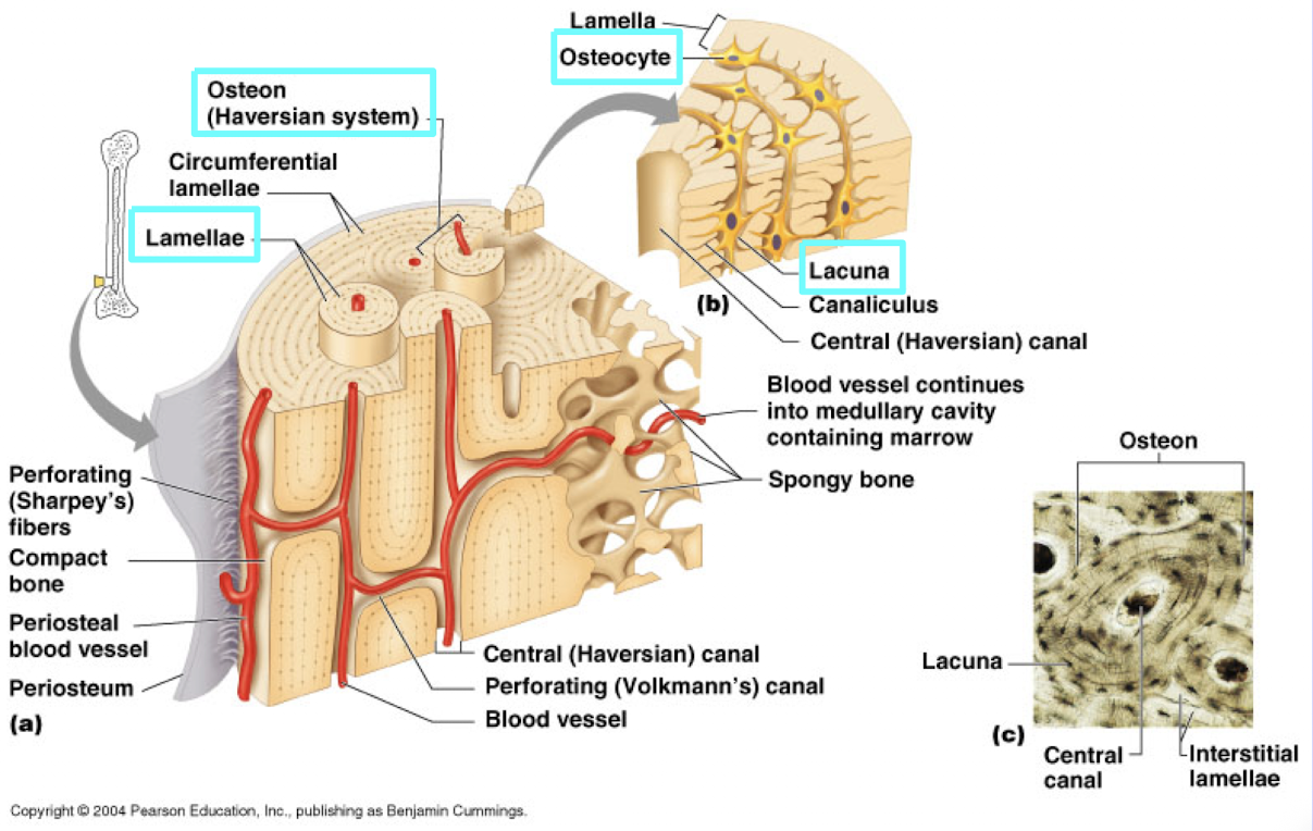
Compact bone:
An osteon (or Haversian system) is composed of a series of concentric rings called:
lamellae


Compact bone:
In between the lamellae, osteocytes sit within spaces called:
lacunae

Compact bone:
Osteocytes send cytoplasmic projections into the surrounding lamellae via ________.
Canaliculi
→ “little canals in the bone”
these projections allow osteocytes to take up and deposit material from the lamellae
they also allow osteocytes to receive nutrients form the blood supply of the bone

Compact bone:
Blood vessels and nerves travel through the __________ of the osteon.
central (or Haversian) canal of the osteon
→ up and down
Compact bone:
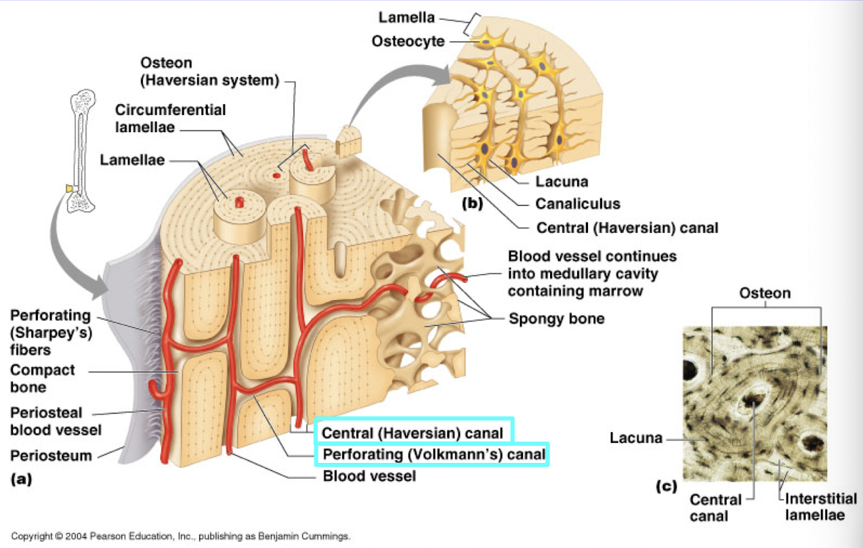
Blood vessels and nerves also travel perpendicular to the osteons via _________.
perforating (or Volkman’s) canal
→ L to R
Spongy bone is composed of bars of osseous tissue called ______.
trabeculae

Spongy bone:
Are trabeculae arranged like osteons?
NO
→ osteocytes sit within the lacunae inside the trabeculae and communicate with each other and the surface of the bar via canaliculi


Structure of long bones:
The typical long bones has a characteristic structure of two _______ separated by a ________.
epiphyses (at the end)
diaphysis (in the middle)


Structure of long bones:
The diaphysis is composed of compact bone surrounding a central ___________.
medullary cavity
→ medullary = for middle


Structure of long bones:
The epiphysis is composed of a spongy bone surrounded by a thin layer of ________.
compact bone


Structure of long bones:
The medullary cavity is filled with _______, an adipose tissue.
yellow marrow


Structure of long bones:
The spaces within the spongy bone of the epiphyses are filled with ________which is the site of hematopoiesis.
red marrow
→ hematopoiesis = new blood cells = red marrow
Structure of long bones:
The ______ line is the boundary between epiphysis and diaphysis.
epiphyseal line
marks the zone of growth in length of long bones

Cartilage (fibrous, hyaline, elastin)
All cartilage contains ______ within lacunae.
chondrocytes
→ chondro = cartilage
→ cyte = cell
the matrix is composed of ground substance and different types of protein fibers, depending on the particular variety of cartilage.
Cartilage:
What are the three types of cartilage?
Hyaline cartilage: has a uniform matrix without visible protein fibers
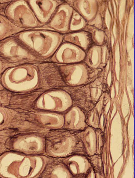
Elastic cartilage: has thick branched elastin fibers
Fibrous cartilage: has many parallel layers of collagen fibers

Cartilage:
Hyaline cartilage
provides uniform structure but not much strength
it forms the embryonic skeleton
the articular surfaces of the long bones
the costal cartilages (cartil. on rib)
laryngeal and tracheal cartilages


Cartilage:
Elastic cartilage
provides flexibility
forms the cartilage of the external ear and the structure of the epiglottis
Cartilage:
Fibrous cartilage
Fibrous cartilage provides great strength along a single axis.
It allows for cushioning under heavy compression forces.
It forms the intervertebral discs, the pubic symphysis, and the menisci of the knees.