2.1.3. Nucleotides and nucleic acids

0.0(0)
Studied by 0 people
call kaiCall Kai
learnLearn
examPractice Test
spaced repetitionSpaced Repetition
heart puzzleMatch
flashcardsFlashcards
GameKnowt Play
Card Sorting

1/45

encourage image

There's no tags or description

Looks like no tags are added yet.

46 Terms

1
New cards

how are nucleic acids formed, type of reaction, from what

Nucleotides (or mononucleotides) are monomers that are linked by condensation reactions to form nucleic acids (or polynucleotides; DNA and RNA).


2
New cards

what is DNA and RNA, what are their roles

Deoxyribonucleic acid (DNA) and ribonucleic acid (RNA) are information–carrying molecules found living cells. They are polymers of nucleotides.

  • DNA stores genetic information.

  • RNA has several roles,

  • 1. including transferring genetic information from DNA to ribosomes (mRNA), 2. combining with proteins to form ribosomes (rRNA),

  • 3. and being involved in protein synthesis (tRNA).


3
New cards

what are nucleotides, anatomy

Nucleotides (mononucleotides) consist of three primary components joined together by condensation reactions:

  • a pentose sugar, which is a 5-carbon sugar molecule

  • a phosphate group, which is attached to the 5′ carbon of the pentose sugar

  • a nitrogenous base (nitrogen-containing organic base), which is attached to the 1′ carbon of the pentose sugar.

By convention, the carbon numbers in the sugar are numbered 1′ to 5′.
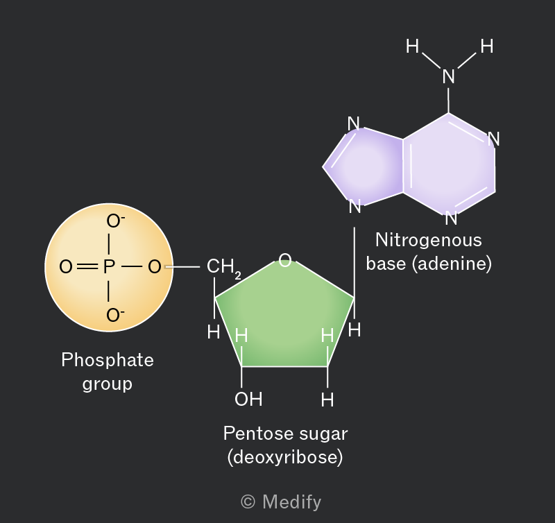
The image shows the structure of a nucleotide composed of three parts: a phosphate group, a pentose sugar (deoxyribose), and a nitrogenous base (adenine). The phosphate group is depicted as an orange circle with a chemical structure showing an oxygen atom double-bonded to a phosphorus atom, which is single-bonded to two additional oxygen atoms, one of which carries a negative charge. The pentose sugar is shown as a green pentagon labeled 'Pentose sugar (deoxyribose)', with the chemical structure indicating the presence of hydroxyl (OH) and hydrogen (H) groups. The nitrogenous base is represented as a purple double-ring structure labeled 'Nitrogenous base (adenine)', featuring nitrogen (N) atoms and hydrogen (H) atoms bonded to the rings.


4
New cards

DNA and RNA nucleotides differ in the type of pentose sugar they contain


  • DNA (deoxyribonucleic acid) contains the pentose sugar deoxyribose which lacks an oxygen atomon the 2′ carbon.

  • RNA (ribonucleic acid) contains the pentose sugar ribose which has a hydroxyl attached to the 2′ carbon.
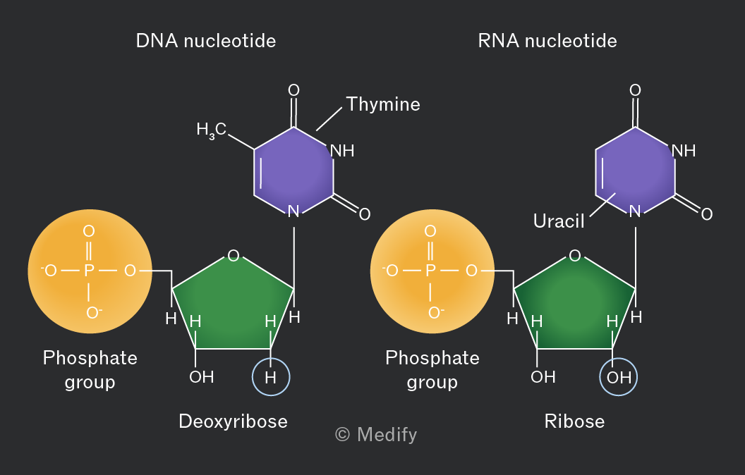
The image shows two nucleotide structures: a DNA nucleotide and an RNA nucleotide. The DNA nucleotide consists of a phosphate group, a deoxyribose sugar, and a thymine base. The phosphate group is illustrated as a circle with a chemical structure inside: O=P(O)(O-)O-. The deoxyribose sugar is shown as a pentagon with OH and H groups attached. The thymine base is a hexagon labeled 'Thymine' with a chemical structure including CH3 and NH groups. The RNA nucleotide consists of a phosphate group, a ribose sugar, and a uracil base. The phosphate group is similar to that in the DNA nucleotide. The ribose sugar is shown as a pentagon with OH groups. The uracil base is a hexagon labeled 'Uracil' with a structure including NH groups. Both nucleotides are labeled at the top: 'DNA nucleotide' and 'RNA nucleotide.'


5
New cards

DNA and RNA nucleotides differ in one of their nitrogenous bases (nitrogen-containing organic bases):


DNA and RNA polynucleotides both contain adenine, cytosine, and guanine.

The fourth nitrogenous base DNA contains is thymine, but in RNA it is uracil.


6
New cards

DNA and RNA have similar structures, but also several important differences.

length, strand, bases, location, pentose sugar
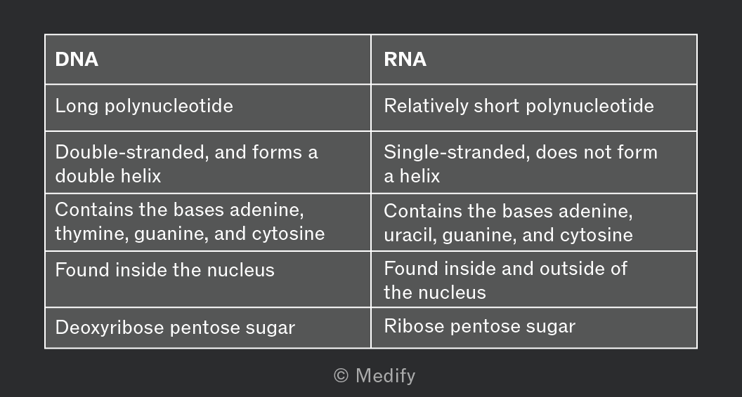

A comparison table between DNA and RNA. The table has two columns with headers: 'DNA' and 'RNA'. Under 'DNA': 1. 'Long polynucleotide', 2. 'Double-stranded, and forms a double helix', 3. 'Contains the bases adenine, thymine, guanine, and cytosine', 4. 'Found inside the nucleus', 5. 'Deoxyribose pentose sugar'. Under 'RNA': 1. 'Relatively short polynucleotide', 2. 'Single-stranded, does not form a helix', 3. 'Contains the bases adenine, uracil, guanine, and cytosine', 4. 'Found inside and outside of the nucleus', 5. 'Ribose pentose sugar'.


7
New cards

There are five nitrogenous bases in DNA and RNA nucleotides and they can be split into 2 groups:


  • Purines are larger, double carbon ring structures — adenine (A) and guanine (G).

  • Pyrimidines are smaller, single carbon ring structures — cytosine (C), thymine (T; only in DNA) and uracil (U; only in RNA).
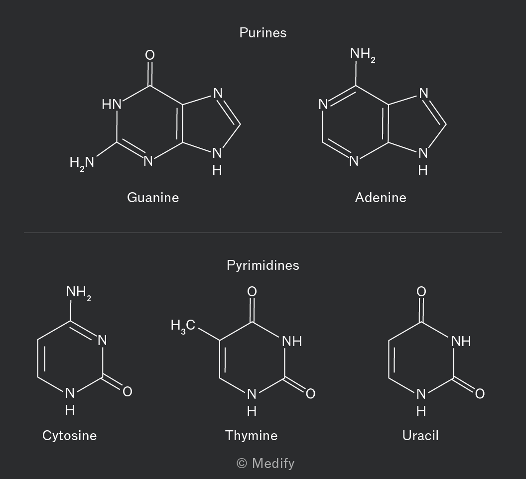
The image displays chemical structures of nucleobases divided into two categories: Purines and Pyrimidines. Under Purines, there are two structures: Guanine and Adenine. Guanine has a double-ring structure with an oxygen atom double-bonded to one ring and two amino groups (NH2) attached. Adenine also has a double-ring structure with an amino group (NH2) attached. Under Pyrimidines, there are three structures: Cytosine, Thymine, and Uracil. Cytosine has a single-ring structure with an amino group (NH2) and an oxygen atom double-bonded to the ring. Thymine has a single-ring structure with a methyl group (CH3) and two oxygen atoms double-bonded to the ring. Uracil has a single-ring structure with two oxygen atoms double-bonded to the ring.


8
New cards

what reaction causes what bond formation between nucleotides, how does the bond look like

Di- and poly nucleotides are formed when condensation reactions link nucleotides by phosphodiester bonds.

Phosphodiester bonds occur between;

  1. the phosphate group of one nucleotide

  2. and the hydroxyl group on the 3’ carbon of the pentose sugar of another nucleotide.

This linkage forms the sugar–phosphate backbone of DNA and RNA strands.
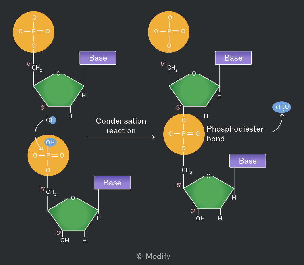
The image shows a diagram of a condensation reaction forming a phosphodiester bond between two nucleotides. On the left, two nucleotide structures are shown. Each nucleotide consists of a phosphate group (indicated by an orange circle with 'O - P = O' and two 'O' atoms bonded to 'P'), a pentose sugar (green pentagon), and a base (purple rectangle labeled 'Base'). The first nucleotide has a phosphate group labeled '5’', a sugar labeled '3’', and an 'OH' group. An arrow labeled 'Condensation reaction' points right toward the second nucleotide. The reaction results in a phosphodiester bond, indicated by the merging of the phosphate group with the sugar of the second nucleotide. The second nucleotide's structure is similar to the first, with a phosphate labeled '5’', a sugar labeled '3’', and a base. The right side shows the new bond formed, labeled 'Phosphodiester bond', and a byproduct '+H2O' indicated with an arrow. The bases remain unlabeled in detail but are represented by the purple rectangles. The structure and labels convey the process of nucleotide linkage in nucleic acids.


9
New cards

how does the phosphodiester bond break

The breakdown of polynucleotides occurs through the hydrolysis of phosphodiester bonds. This reaction uses water.
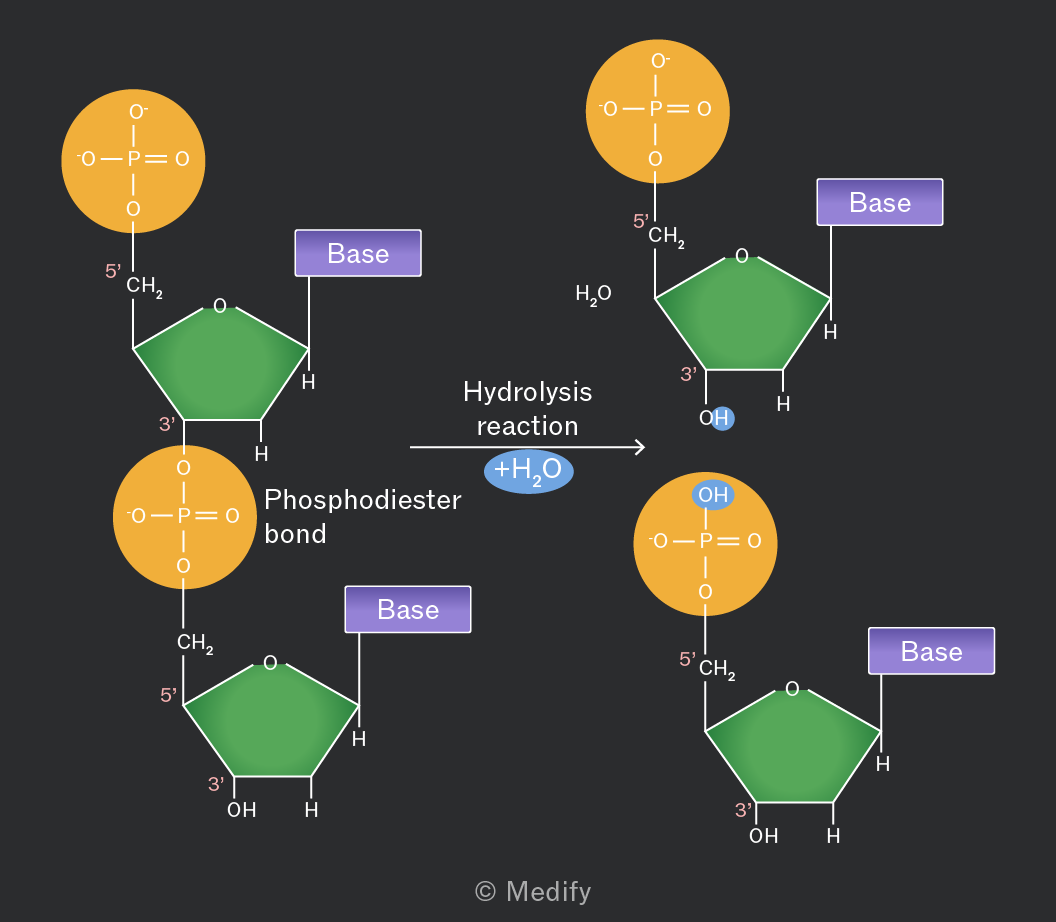
The image illustrates a hydrolysis reaction involving nucleotides. On the left, two nucleotide structures are shown linked by a phosphodiester bond labeled in yellow. Each nucleotide consists of a phosphate group (O-P=O with two -O^- groups), a sugar depicted as a green pentagon, and a purple rectangle labeled 'Base.' The top nucleotide has a 5' label near CH2 and a 3' label near the sugar. An annotation reads 'Phosphodiester bond.' An arrow pointing to the right indicates 'Hydrolysis reaction' with an added '+H2O' in blue. On the right, two separate nucleotide structures are shown after hydrolysis. Each nucleotide consists of a phosphate group, a sugar, and a base. The top nucleotide has a 5' label near CH2 and a 3' label near the sugar with an OH group. The bottom nucleotide has similar labels and also an OH group on the phosphate. An annotation shows 'H2O' near the top nucleotide. Both nucleotides have a 'Base' label attached to the sugar. The overall reaction shows the breaking of the phosphodiester bond by hydrolysis, resulting in two separate nucleotides.


10
New cards

what os atp and its anatomy

ATP (Adenosine triphosphate) is a phosphorylated nucleotide derivative that acts as the immediate energy source in cells. It consists of three key components:

  • Adenine: a nitrogenous base.

  • Ribose: a five-carbon (pentose) sugar.

  • Three phosphate groups: linked with high-energy bonds, which release energy when hydrolysed.


<p><span><strong>ATP</strong></span> (Adenosine triphosphate) is a phosphorylated <strong>nucleotide derivative</strong> that acts as the immediate energy source in cells. It consists of three key components:</p><ul><li><p><strong>Adenine</strong>: a nitrogenous base.</p></li><li><p><strong>Ribose</strong>: a five-carbon (pentose) sugar.</p></li><li><p><strong>Three phosphate groups</strong>: linked with high-energy bonds, which release energy when hydrolysed.</p></li></ul><p></p><p></p>
11
New cards

what is atp’s function, how is it achieved

ATP provides an immediate energy source for biological processes.

  1. ATP is continuously hydrolysed (by ATP hydrolase) to form ADP + Pi

  2. and resynthesised within cells


  3. to support metabolic activities such as active transport, muscle contraction and protein synthesis.

The phosphate released from the hydrolysis of ATP can be used to phosphorylate other compounds.
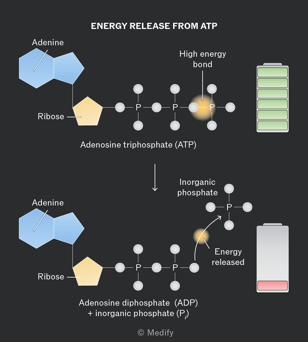
The diagram illustrates the process of energy release from ATP. At the top, Adenosine triphosphate (ATP) is shown with Adenine and Ribose structures on the left and three phosphate groups labeled 'P' connected in a chain. The third phosphate is highlighted as a 'High energy bond' with an adjacent full battery icon. Below, an arrow points downward to Adenosine diphosphate (ADP) with two phosphate groups and an inorganic phosphate (Pi) detached, labeled as 'Inorganic phosphate.' A curved arrow indicates 'Energy released,' pointing to the right where a depleted battery icon is displayed. Both ATP and ADP structures have Adenine and Ribose components. The diagram is labeled 'ENERGY RELEASE FROM ATP.'


12
New cards

what is the reaction of phosphorylating adp

ATP can be (re)synthesised by the condensation of ADP via a phosphorylation reaction requiring energy.

adp+ Pi → atp

Phosphorylation of ADP is catalysed by the enzyme ATP synthase and occurs in mitochondria, during aerobic respiration (electron transport chain) and in chloroplasts, during photosynthesis (light-dependent reactions).
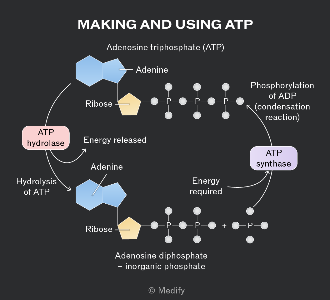
The diagram titled 'MAKING AND USING ATP' illustrates the cycle of ATP hydrolysis and synthesis. At the top, Adenosine triphosphate (ATP) is shown, consisting of adenine, ribose, and three phosphate groups. An arrow labeled 'Hydrolysis of ATP' points downward to a product labeled 'Adenosine diphosphate + inorganic phosphate', with a separate phosphate group shown. The process is facilitated by 'ATP hydrolase', with the release of energy noted. Below, the transition back to ATP is shown with an arrow labeled 'Phosphorylation of ADP (condensation reaction)', facilitated by 'ATP synthase', requiring energy. The cycle includes labels for adenine, ribose, and phosphate groups, and indicates the energy transitions involved.


13
New cards

how does the double helix form

The two bonded DNA strands twist to form a double helix structure.
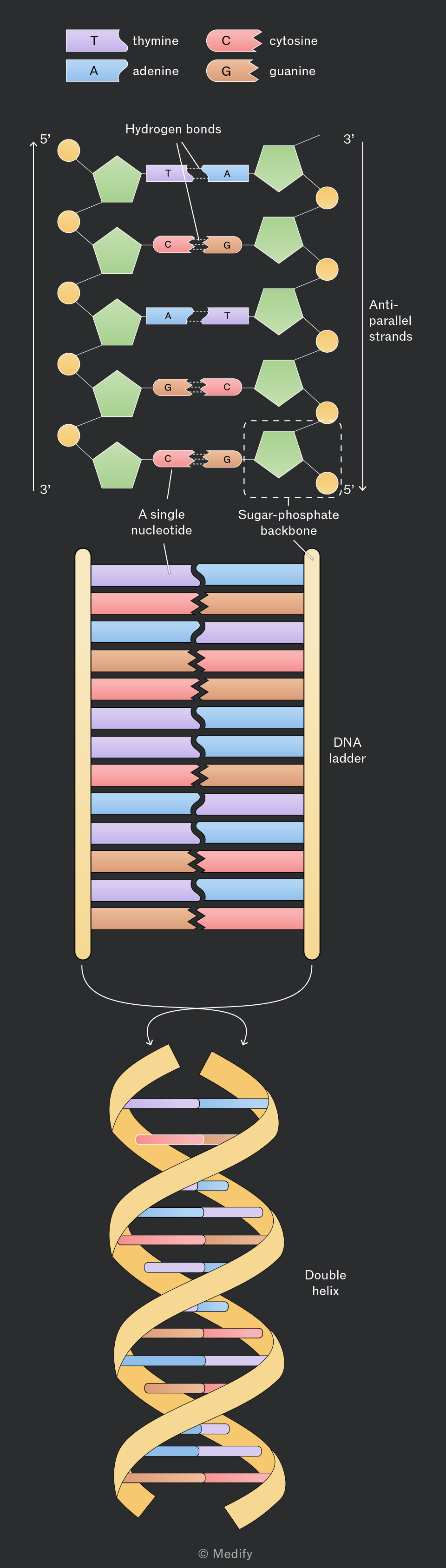
The image illustrates the structure of DNA. At the top, there is a legend showing base pairs: thymine (T) in purple, adenine (A) in blue, cytosine (C) in pink, and guanine (G) in orange. Below, a section of a DNA molecule is shown with two anti-parallel strands. The left strand is labeled 5' to 3' and the right strand is labeled 3' to 5'. The strands consist of green pentagons representing the sugar-phosphate backbone, and base pairs connected by hydrogen bonds: T-A and C-G. Annotations include 'Hydrogen bonds,' 'A single nucleotide,' and 'Sugar-phosphate backbone.' Below the strands, a simplified 'DNA ladder' is shown, with colored rungs representing base pairs. At the bottom, the DNA ladder is twisted into a 'Double helix.'


14
New cards

explain the process of dna extraction and purification, also the name?

DNA can be extracted from cells and purified using DNA precipitation.

This method involves a series of chemical treatments to isolate DNA from proteins, lipids, and other cellular components.

  1. To extract DNA from plant cells, first break down a sample in a pestle and mortar to destroy the cell walls. Transfer all the material to a test tube.

  2. Add a detergent to break down the cell membranes and release the cell contents.

  3. Add salt to break the hydrogen bonds between the DNA and water.

  4. Add proteases to break down the proteins bound to the DNA. histones

  5. Carefully add cold ethanol by pouring it down the side of the test tube.


DNA is insoluble in alcohol, so it precipitates as visible, white strands, which form as a layer between the plant tissue and the alcohol. The DNA can be removed from the test tube using a glass rod.
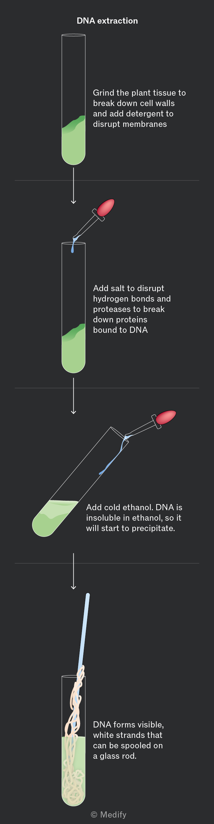
A step-by-step diagram of DNA extraction from plant tissue. The first step shows a test tube with green plant tissue and the label 'Grind the plant tissue to break down cell walls and add detergent to disrupt membranes.' An arrow points down to the second step, where a pipette adds liquid to a test tube with the label 'Add salt to disrupt hydrogen bonds and proteases to break down proteins bound to DNA.' Another arrow points to the third step, showing a tilted test tube with liquid being added from a pipette, labeled 'Add cold ethanol. DNA is insoluble in ethanol, so it will start to precipitate.' The final step shows a test tube with white strands and a rod, labeled 'DNA forms visible, white strands that can be spooled on a glass rod.'


15
New cards

what is dna replication, what is the mechanism like

DNA replication is the process by which DNA is copied.

  1. It follows a semi–conservative mechanism = each new DNA molecule consists of one original (parental) strand and one newly synthesised strand.

  2. This ensures that each daughter cell receives an exact copy of the DNA, maintaining genetic continuity between generations of cells.
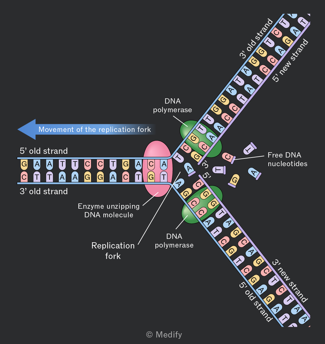
Diagram illustrating DNA replication. The image shows a replication fork with two strands: the 5' old strand and the 3' old strand. The enzyme unzipping the DNA molecule is labeled, and the DNA polymerase is shown on both the leading and lagging strands. Free DNA nucleotides A, T, C, and G are depicted near the replication fork. The 3' new strand and 5' new strand are being synthesized, with DNA polymerase facilitating the addition of nucleotides. An arrow labeled 'Movement of the replication fork' indicates the direction of replication. The DNA sequence includes bases such as G, A, T, C on the old strands and complementary bases on the new strands.


16
New cards

how does dna replication begin

DNA replication begins with

  1. DNA helicase unwinding the DNA double helix,

  2. separating the two strands by breaking the hydrogen bonds between complementary bases.

  3. This separation creates two single-stranded templates that can both be used for synthesising new DNA strands.

  4. One is called the leading strand and the other, the lagging strand (3 to 5 is lagging bc dna polymerase works in a 3 to 5)

The unwound DNA forms a Y–shaped structure called the replication fork.


17
New cards

how does dna polymerase work on the different strands

  1. DNA synthesis on the leading strand occurs continuously; The addition of nucleotides in the 5’ to 3’ direction means that the newly synthesised strand grows in the direction of the movement of the replication fork.


  2. DNA polymerase joins new adjacent nucleotides

  3. that are complementary to the template strand

  4. via condensation reactions

  5. which form phosphodiester bonds.


18
New cards

how does dna polymerase work on the lagging. strand

DNA synthesis on the lagging strand occurs discontinuously because DNA polymerase can only add nucleotides in the 5′ to 3′ direction. As a result, the lagging strand is synthesised in short sections called Okazaki fragments, which are later joined together. by dna ligase
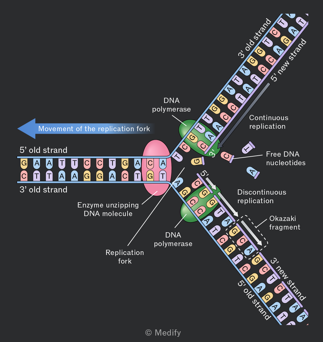
The image illustrates a DNA replication fork. The left side shows a 5’ old strand with bases G, A, A, T, T, C, C, T, G, and C, paired with a complementary 3’ old strand. An enzyme unzipping the DNA molecule is depicted in the center. The replication fork is labeled, with a blue arrow indicating the movement of the replication fork to the left. DNA polymerase is shown on both sides, synthesizing new strands. On the right, a 3’ old strand pairs with a 5’ new strand, showing continuous replication. Free DNA nucleotides C and A are shown as separate components. Below, a 5’ old strand pairs with a 3’ new strand, illustrating discontinuous replication with an Okazaki fragment labeled. The diagram includes text labels such as DNA polymerase, continuous replication, discontinuous replication, and Okazaki fragment.


19
New cards

what does errors in dna replication cause

Errors in DNA replication can cause random, spontaneous genetic mutations, which are changes to the base sequence of DNA within a gene. (t's the base part of the nucleotide that changes, rather than the whole nucleotide.)

They are often corrected by DNA repair mechanisms, but those that persist can lead to genetic diversity or, in some cases, diseases.

20
New cards

what is the genetic code, how is the genetic code written in

The genetic code is the set of rules by which the information encoded within DNA or RNA sequences is translated into proteins by living cells.

This code specifies how sequences of nucleotides correspond to specific amino acids, which are the building blocks of proteins.

The genetic code is written in triplet codes, which are sequences of three nucleotides and referred to as codons

21
New cards

what does it mean by non overlapping genetic code

the genetic code is non-overlapping, meaning that each nucleotide is part of only one codon.

  • One nucleotide = one base (A, T, C or G).

  • The bases are read in groups of 3 in a row.

  • Each group of 3 bases is called a codon.

  • Each base can only belong to one codon — it isn't shared between two codons.


22
New cards

why is an overlapping reading frame critical

the linear and non-overlapping reading frame is critical. A shift in the reading frame is known as a frameshift mutation.

This can lead to the placement of an incorrect amino acid in a polypeptide chain or the incorrect ‘reading’ of a stop codon, resulting in a truncated or non-functional protein.


23
New cards

what does it mean that the genetic code is degenerate, why’s it important

The genetic code is degenerate, which means that multiple codons can code for the same amino acid.
For example, the amino acid leucine is coded by six different codons (UUA, UUG, CUU, CUC, CUA, CUG).

This redundancy is beneficial because it provides a ‘buffer’ against mutations. A mutation in one nucleotide of a codon might not change the amino acid it codes for, thereby minimising the impact on the protein’s structure and function.

Methionine and tryptophan are the only amino acids that are coded by a single codon (AUG and UGG, respectively).

24
New cards

what does it mean that genetic code is universal, what does it suggest

The genetic code is universal, meaning that the same codons specify the same amino acids across almost all living organisms.

The universality of the genetic code suggests a common evolutionary origin and allows for the possibility of genetic engineering, where genes from one organism can be expressed in another (e.g., using bacterial cells to produce human insulin).


25
New cards

what are the 2 stages of protein synthesis

DNA contains the ‘instructions’ for making polypeptides, but is contained in the nucleus of cells. The ribosomes, which assemble polypeptides, are in the cytoplasm.

Protein synthesis, therefore, happens in two stages:

  • Transcription – messenger RNA (mRNA) is made from the DNA template. The mRNA takes the instructions from the nucleus to the ribosomes.

  • Translation – ribosomes join amino acids together in the order specified by the sequence of bases in the mRNA, forming a polypeptide.

1

26
New cards

describe the process of transcription

To start transcription, the DNA template of a gene must be exposed.

  1. At the start codon of a gene, the enzyme DNA helicase unzips the DNA double helix, breaking the hydrogen bonds between complementary base pairs.


The sense strand of DNA runs 5’ – 3’ and codes for proteins;

the antisense strand is complementary to it. 3 to 5 acts as the template


During transcription, the DNA sequence of a gene is copied into messenger RNA (mRNA):

  1. To copy the base sequence held on the sense strand, the mRNA is complementary to the antisense strand, which acts as a template. bc the new strand is made in a 5 to 3 way

  2. Free RNA nucleotides match up with their complementary base pairs on the exposed antisense strand.3 to 5

  3. RNA polymerase joins the RNA nucleotides in condensation reactions, forming phosphodiester bonds.

  4. Transcription stops at the end of a gene, and the mRNA molecule leaves the nucleus through a nuclear pore.
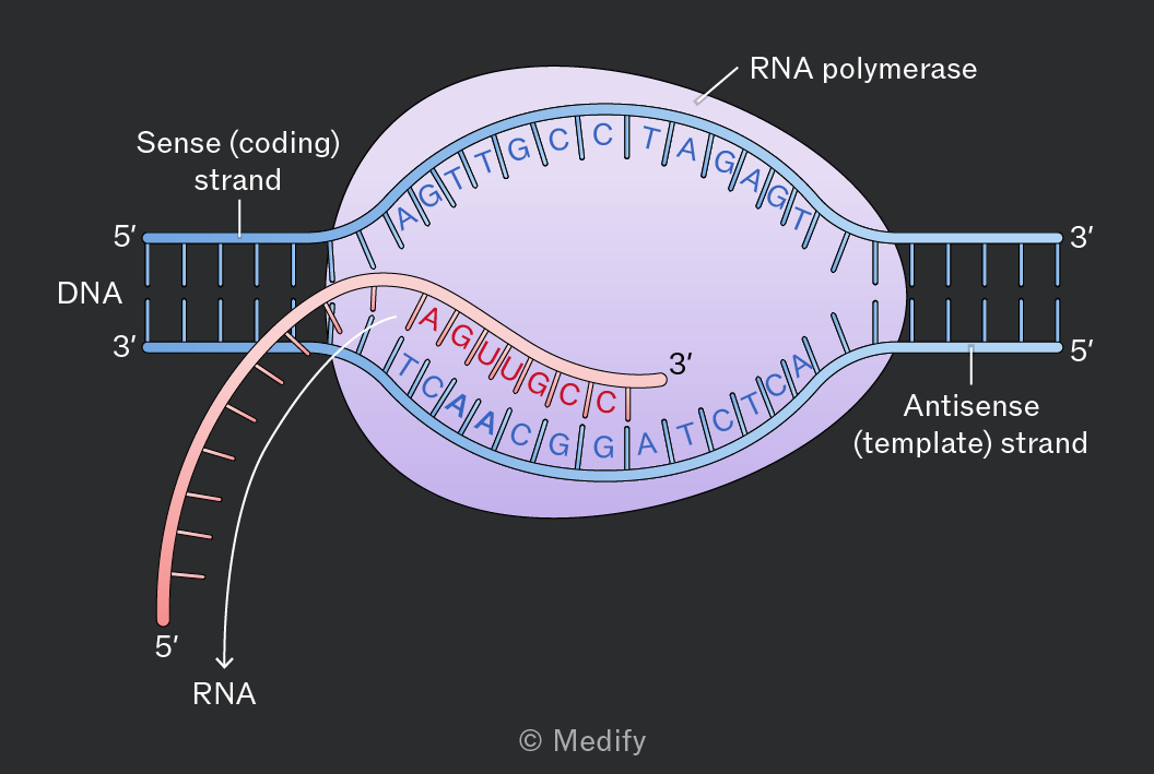
Diagram illustrating DNA transcription. The DNA is shown with two strands: the upper 'Sense (coding) strand' labeled with 5' and 3' ends, and the lower 'Antisense (template) strand' labeled with 3' and 5' ends. The DNA strands are separated in the center where RNA polymerase is binding. The RNA strand is being synthesized from the template strand, shown with 5' to 3' direction. The RNA sequence is AGUUGCC, and the DNA template sequence it pairs with is TCAACGG. The RNA extends outwards from the transcription bubble. Labels include 'RNA polymerase', 'Sense (coding) strand', 'Antisense (template) strand', and 'RNA'.


27
New cards

what occutes during translation

During translation, ribosomes join amino acids together in the order specified by the sequence of bases in the mRNA codons, forming a polypeptide and determining how it will fold and function.

Each codon specifies a particular amino acid, which is delivered to the ribosome by a transfer RNA(tRNA) molecule.

ATP provides energy for an enzyme to join amino acids together by peptide bonds.
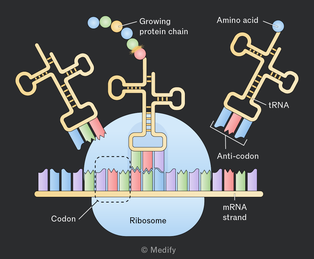
The diagram illustrates the process of protein synthesis. At the center is a ribosome depicted as a blue structure interacting with an mRNA strand shown as a sequence of colored bars labeled 'mRNA strand'. Above the ribosome, a 'Growing protein chain' is shown as a series of colored spheres. On the right, a tRNA molecule labeled 'tRNA' is depicted with an 'Amino acid' attached at one end and an 'Anti-codon' at the other. The tRNA is shown interacting with the mRNA strand. On the left, another tRNA is shown entering the ribosome with a different anti-codon. The mRNA strand has colored segments labeled 'Codon', indicating the interaction with anti-codons on the tRNA. The diagram is attributed to © Medify.


Transfer RNA, tRNA is made of RNA nucleotides. It is a small, single-stranded molecule that folds into a clover leaf shape due to complementary base pairing within its single strand.

Each tRNA has an anticodon region that pairs with a complementary codon on the mRNA.

At the other end of the molecule from the anticodon, tRNA molecules have an amino acid attachment site. Each tRNA can only bind to one specific amino acid which is determined by its anti-codon.
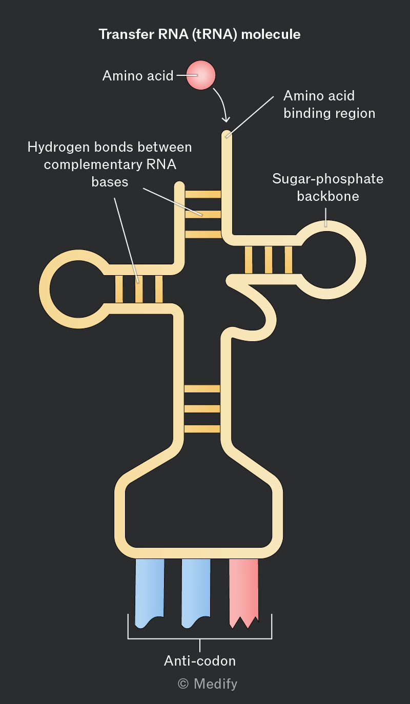
Diagram of a Transfer RNA (tRNA) molecule. The structure includes an amino acid depicted as a red sphere connected to the tRNA at the top, labeled 'Amino acid'. An arrow points to the 'Amino acid binding region' on the tRNA. The backbone of the tRNA is labeled 'Sugar-phosphate backbone'. There are regions with parallel lines labeled 'Hydrogen bonds between complementary RNA bases'. At the bottom, there are three colored segments, two blue and one red, labeled collectively as 'Anti-codon'.


28
New cards

During translation, ribosomes join amino acids together in the order specified by the sequence of bases in the mRNA codons, forming a polypeptide and determining how it will fold and function.

Each codon specifies a particular amino acid, which is delivered to the ribosome by a transfer RNA(tRNA) molecule.

ATP provides energy for an enzyme to join amino acids together by peptide bonds.

The diagram illustrates the process of protein synthesis. At the center is a ribosome depicted as a blue structure interacting with an mRNA strand shown as a sequence of colored bars labeled 'mRNA strand'. Above the ribosome, a 'Growing protein chain' is shown as a series of colored spheres. On the right, a tRNA molecule labeled 'tRNA' is depicted with an 'Amino acid' attached at one end and an 'Anti-codon' at the other. The tRNA is shown interacting with the mRNA strand. On the left, another tRNA is shown entering the ribosome with a different anti-codon. The mRNA strand has colored segments labeled 'Codon', indicating the interaction with anti-codons on the tRNA. The diagram is attributed to © Medify.

1

Add to favourites

Transfer RNA, tRNA is made of RNA nucleotides. It is a small, single-stranded molecule that folds into a clover leaf shape due to complementary base pairing within its single strand.

Each tRNA has an anticodon region that pairs with a complementary codon on the mRNA.

At the other end of the molecule from the anticodon, tRNA molecules have an amino acid attachment site. Each tRNA can only bind to one specific amino acid which is determined by its anti-codon.

Diagram of a Transfer RNA (tRNA) molecule. The structure includes an amino acid depicted as a red sphere connected to the tRNA at the top, labeled 'Amino acid'. An arrow points to the 'Amino acid binding region' on the tRNA. The backbone of the tRNA is labeled 'Sugar-phosphate backbone'. There are regions with parallel lines labeled 'Hydrogen bonds between complementary RNA bases'. At the bottom, there are three colored segments, two blue and one red, labeled collectively as 'Anti-codon'.


29
New cards
30
New cards

During translation, ribosomes join amino acids together in the order specified by the sequence of bases in the mRNA codons, forming a polypeptide and determining how it will fold and function.

Each codon specifies a particular amino acid, which is delivered to the ribosome by a transfer RNA(tRNA) molecule.

ATP provides energy for an enzyme to join amino acids together by peptide bonds.

The diagram illustrates the process of protein synthesis. At the center is a ribosome depicted as a blue structure interacting with an mRNA strand shown as a sequence of colored bars labeled 'mRNA strand'. Above the ribosome, a 'Growing protein chain' is shown as a series of colored spheres. On the right, a tRNA molecule labeled 'tRNA' is depicted with an 'Amino acid' attached at one end and an 'Anti-codon' at the other. The tRNA is shown interacting with the mRNA strand. On the left, another tRNA is shown entering the ribosome with a different anti-codon. The mRNA strand has colored segments labeled 'Codon', indicating the interaction with anti-codons on the tRNA. The diagram is attributed to © Medify.

1


31
New cards

what is t rna, made from

Transfer RNA, tRNA is made of RNA nucleotides

It is a small,

single-stranded molecule

that folds into a clover leaf shape due to complementary base pairing within its single strand.

Each tRNA has an anticodon region that pairs with a complementary codon on the mRNA.

At the other end of the molecule from the anticodon, tRNA molecules have an amino acid attachment site. Each tRNA can only bind to one specific amino acid which is determined by its anti-codon.

Diagram of a Transfer RNA (tRNA) molecule. The structure includes an amino acid depicted as a red sphere connected to the tRNA at the top, labeled 'Amino acid'. An arrow points to the 'Amino acid binding region' on the tRNA. The backbone of the tRNA is labeled 'Sugar-phosphate backbone'. There are regions with parallel lines labeled 'Hydrogen bonds between complementary RNA bases'. At the bottom, there are three colored segments, two blue and one red, labeled collectively as 'Anti-codon'.


32
New cards

explain the process of translation what

Translation is a multistep process involving mRNA, ribosomes, tRNA and amino acids:

  1. mRNA binds to the small subunit of a ribosome at its start codon.

  2. A tRNA molecule with the complementary start anticodon binds to the mRNA and the ribosome, bringing with it an amino acid.

  3. A tRNA molecule with an anticodon complementary to the next codon along on the mRNA molecule and an amino acid binds next.

  4. Part of the ribosomal RNA, rRNA of the ribosome, the enzyme peptidyl transferase catalyses the formation of a peptide bond, joining the two amino acids together.

  5. The ribosome moves along the length of the mRNA molecule, repeating the above process until reaching a stop codon. There is no complementary tRNA anticodon for the stop codon, so the polypeptide chain is released.

Multiple ribosomes can simultaneously synthesise multiple polypeptide chains from a single mRNA molecule by following each other along the mRNA.
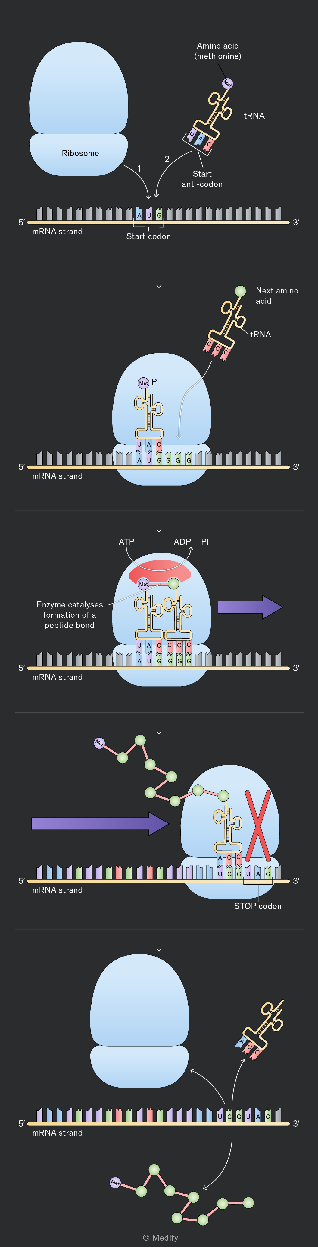
Five-panel schematic of translation on an mRNA strand. Panel 1 shows a blue ribosome above a horizontal mRNA labeled "mRNA strand," with 5′ on the left and 3′ on the right. A highlighted triplet on the mRNA is labeled "Start codon" and contains the letters A, U, and G. Two curved arrows labeled "1" and "2" point toward the start codon. At right is a tRNA labeled "tRNA" carrying an "Amino acid (methionine)" and showing a triplet labeled "Start anti-codon." Panel 2 shows the ribosome assembled around the mRNA. Inside the ribosome, a tRNA carrying "Met" is paired with the AUG start codon; the letter "P" appears near the tRNA. Another labeled "tRNA" carrying the "Next amino acid" approaches from the upper right. The mRNA still shows 5′ and 3′ and the AUG codon is visible, followed by additional visible bases including GGG. Panel 3 shows two tRNAs inside the ribosome and the text "Enzyme catalyses formation of a peptide bond." Above the ribosome are labels "ATP" and "ADP + Pi." The left tRNA carries "Met" and a growing connection to the next amino acid; the mRNA below shows visible codons including AUG followed by GGG. A large purple arrow points to the right. Panel 4 shows a longer chain of linked amino acids emerging from the ribosome. A large red X overlays a tRNA at a highlighted codon labeled "STOP codon." The mRNA remains labeled 5′ to 3′ and a visible triplet near the stop is UAG. A large purple arrow points toward the ribosome from the left. Panel 5 shows the blue ribosome separated above the mRNA, with curved arrows indicating release. A tRNA is shown leaving at right, the mRNA remains labeled "mRNA strand" with 5′ on the left and 3′ on the right, and the visible triplet near the right end is UAG. A free polypeptide chain is shown below. Copyright text at the bottom reads "© Medify."


33
New cards

what does insertion/ deletion mutations cause

an insertion of a nucleotide or deletion (hence of a base) causes a frame shift mutation bc there is a change in every successive codon from that point of mutation

34
New cards

What is a silent mutation?

There is no change in the protein being synthesis

No change in the phenotype

It results in a change to the primary structure, but no change in the overall structure/function of the protein

35
New cards

What is a nonsense mutation?

A codon becomes a stock resulting in a shorten protein being coded for

It's gonna have detrimental effect on the phenotype

36
New cards

What is a missense mutation?

There is an in cooperation of an incorrect amino acid into the primary structure during protein synthesis

The mutation can be silent, beneficial, or harmful


A conservative mutation is when the amino acid change leads to the change protein to have similar properties as original protein, and we'll have less likely in effect on the phenotype.

37
New cards

What are some causes of mutations?

Mutations occur spontaneously during DNA replication

The rate of mutation can increase by mitogens( e.g. Oxidizing agents= free radicals, can affect the structure of nucleotides


Physical agents also can cause this such as ionizing radiation


38
New cards
39
New cards
40
New cards
41
New cards
42
New cards
43
New cards
44
New cards
45
New cards
46
New cards



