chapter 37, part 1: disorders of gastrointestinal function
 Call Kai
 Learn
 Practice Test
 Spaced Repetition
 Match
 Flashcards
 Knowt Play
1/34

There's no tags or description
Looks like no tags are added yet.
35 Terms
GI tract digests:
food and absorbs nutrients
motility, secretion, digestion, and absorption work together
nutrients enter bloodstream or lymph after crossing intestinal wall
large intestine prepares waste for elimination

upper part (part of GI tract)
mouth, esophagus, and stomach
an intake source and parts in which initial digestive processes take place
middle portion (part of GI tract)
duodenum, jejunum, ileum
most digestive and absorptive processes occur
lower segment (part of GI tract)
cecum, colon, and rectum
storage channel for elimination of waste
common symptoms of many GI disorders
common symptoms: anorexia, nausea, and vomiting
serve as protective mechanisms that help body detect and respond to disease
anorexia is loss or decrease of:
appetite
emotional stress, medications, and illnesses cna reduce appetite
anorexia frequently precedes nausea and vomiting
anoreaxia and anorexia nervosa and diff conditions
an is an eating disorder characterized by fear of gaining weight
nausea is the unpleasant, conscious sensation of:
urge to vomit
results from stimulation of medullary vomiting center in brain
nausea often precedes vomiting
common causes: motion sickness, dizziness, migraine headaches, gastroenteritis, food poisoning, severe pain
retching
rhythmic, spasmodic contraction of diaphragm, chest, and abdominal muscles
usually precedes or alternates w/ vomiting
vomiting (emesis)
sudden, forceful expulsion of stomach contents through mouth
vomitus is expelled stomach contents
protective function removes harmful substances from stomach and upper small intestine
mechanism of vomiting
vomiting center: located in medulla oblongata
vomiting reflex involves:
deep inspiration
closure of airway (glottis)
relaxation of gastroesophageal
forceful contraction of diaphragm and abdominal muscles
expulsion of stomach contents through mouth
breathing temporarily stops
diarrhea
excessive frequency of stools may be acute or chronic
prolonged diarrhea may lead to dehydration, electrolyte imbalance, acidosis, malnutrition
large-volume (secretory or osmotic) diarrhea
watery stool resulting from increased secretions into intestine from plasma
often related to infection
limited reabsorption due to reversal of normal carriers of sodium and/or glucose
small-volume diarrhea
frequent passage of small amount of stool
often due to inflammatory bowel disease
stool may contain blood, mucus, pus
may be accompanied by abdominal cramps
constipation
less frequent bowel movements than normal
result from decreased intestinal peristalsis, leading to prolonged transit time and increased reabsorption of water from stool
periods of constipation may alter w/ period of diarrhea
common causes of constipation:
weakness of smooth muscle due to age or illness
inadequate dietary fiber
inadequate fluid intake
neurologic disorders
drugs - opioids (slows down digestive tract) and iron supplements
obstructions caused by tumors or strictures
blood in stool may occur with:
diarrhea, constipation, tumors, or inflammatory conditions
hematochezia (frank blood): red blood - indicating bleeding from lower GI tract
usuaally from lesions in sigmoid colin and rectum
occult blood: small hidden amounts, detectable w/ stool test
may be caused by small bleeding ulcers
melena: dark colored tarry stool
may result from significant bleeding in upper digestive tract
fibromuscular tube that connects the lower part of: (esophagus structure and function)
the pharynx to stomach
length in adults: 8-10 inches
anatoical relations: lies posteriorly to larynx and trachea as it descends through mediastinum btwn lungs
intersects diaphragm at lvl of t122 or t12 vertebra and passes through
involuntary, “wave-like” muscle contractions properl its contents downwards - peristalsis
musculature and nerve supply (esophagus structure and function)
upper 1/3rd - skeletal muscle fibers innervated by glossopharyngeal (CN IX) and vagus (CN X) nerves
lower 2/3rds - smooth muscle fibers innervated by vagus (CN X) nerve
sphincters at either end of esophagus:
upper esophageal - prevents reflux from esophagus into pharynx
lower esophageal - prevents reflux from stomach to esophagus
both sphincters remain closed except during swallowing

mastication (chewing)
begins in digestive process
saliva contains enzymes that break down complex molecules
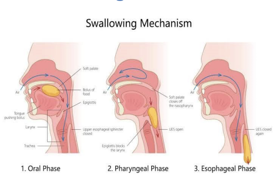
deglutition (swallowing) phases
buccal phase: food/drink is pushed upwards and backwards into pharynx by tongue
pharyngeal phase: epiglottis closes the entrance to larynx, and upper esophageal sphincter relaxes to let food/drink into esophagus
esophageal phase: food/drink moves downwards through esophagus into stomach
swallowing mechanism from deglutition
dysphagia
difficulty in swallowing
common causes:
xerostomia: due to reduced salivary secretions w/ age, dehydration, or poorly controlled diabetes
esophageal narrowing: due to scar tissue formation after chronic acid reflux or radiation therapy
brainstem injury: due to trauma, infections, or tumors
signs and symptoms of dysphagia
difficulty swallowing foods or liquids
choking or coughing during meals
sensation of food sticking in throat or chest
odynophagia — painful swallowing
achalasia
disorder in which lower esophageal sphincter (LES) fails to relax
caused by dmg to enteric nervous system and vagus nerve
when LES fails to relax:
food cannot enter stomach causing enlargement of esophagus above LES
food remains in esophagus for prolonged periods
retained food in esophagus cause:
regurgitation
aspiration into lungs and pneumonia
within achalasia, early diagnosis (endoscopy, barium studies) and appropriate:
treatment help to prevent complications
hiatal hernia
condition in which part of stomach protrudes through esophageal hiatus of diaphragm into chest cavity
occurs when opening in diaphragm becomes enlarged or weakened
types of hiatal hernia:
sliding: most common, often associated w/ acid reflux
gastroesophageal junction and upper part of stomach slide above diaphragm
paraesophageal: less common but carries higher risk of serious complications
part of stomach pushes through esophageal opening gastroesophageal junction remains in its normal position

GERD (gastroesophageal reflux disease)
condition in which gastric contents reflux abnormally into esophagus, oral cavity
one of the most common gastrointestinal disorders worldwide - !!
causes and mechanism of GERD:
weak or incompetent LES allows gastric contents to reflux
delayed gastric emptying increases gastric pressure and volume, promoting reflux
refluxed gastric acid irritates the esophageal lining
diagnostic criteria for GERD:
occurs more than twice per week or several weeks
specific symptoms of GERD:
heartburn: burning sensation behind sternum
regurgitation: backflow of gastric contents into mouth or throat
symptoms commonly occur 30-60 mins after meals:
worsened by bending forward or lying down
relieved by sitting or standing upright
additional symptoms: belching and chest pain: epigastric or retrosternal
complications of GERD:
barrett esophagus: replacement of norm squamous epithelium w/ abnorm columnar epithelium
presence of tan tongues of epithelium interdigitating w/ more proximal squamous epithelium is typical of barrett esophagus