Vet Med Term - Ch 8
 Call Kai
 Learn
 Practice Test
 Spaced Repetition
 Match
 Flashcards
 Knowt Play
1/283

There's no tags or description
Looks like no tags are added yet.
284 Terms
Functions of cardiovascular system
delivers oxygen, nutrients, and hormones to various body tissues
Transports waste products to appropriate waste removal systems
Cardiovascular system is sometimes called circulatory system which is divided into
systemic circulation
Pulmonary circulation
Systemic circulation
Blood flow to all parts of body expect lungs
Pulmonary circulation
Blood flow out of heart through lungs and back to heart
Structures of cardiovascular system
Heart
Blood vessels
Blood
Cardiovascular
Pertaining to heart and vessels (blood vessels in this context)
Heart
A hollow muscular organ that provides the power to move blood through the body
Cardi/o
Heart
Heart is located
Inside thoracic cavity
Mediastinum
Between lungs

Pericardium
Double-walled membrane surrounding the heart
Two layers of pericardium
Fibrous pericardium
Serous layer
Fibrous pericardium
Tough external layer of pericardium
Serous layer
Inner layer of pericardium that is divided into 2 parts
Parietal layer
Visceral layer
Parietal layer
Serous layer that lines the fibrous pericardium
Parietal in Latin
Belonging to the wall
Visceral layer (epicardium)
Serous layer that lines the heart
Viscus in Latin
Organ
Pericardial space
Between the serous layers
Pericardial fluid
Liquid in pericardial space that prevents friction between heart and the pericardium when the heart beats

Layers of heart wall
Epicardium
Myocardium
Endocardium

Epicardium (visceral layer of pericardium)
External layer of heart

Myocardium
Middle and thickest layer of heart
the actual heart muscle
My/o
Muscle
Epi-
Upper

Endocardium
Inner layer of heart
lines heart chambers and valves
Endo-
Within
Coronary arteries
Arteries that serve the heart
resemble a crown
Coron/o
Crown
Coronary veins
Remove waste products from myocardium
Coronary occlusion
May cause disruption of blood flow to myocardium causing it not to function
may lead to ischemia
Ischemia
Deficiency in blood supply to an area
may lead to necrosis
Necrosis
Tissue death
Infarct or infarction
Area of necrosis due to ischemia
Heart is divided into
Left and right sides
The left and right sides of the heart is subdivided into
Chambers
Mammalian and avian hearts are
4-chambered
Most reptile hearts have
3 chambers

Atria
Craniodorsal chambers of heart
all vessels coming into heart enter here
Atrium
Singular form of atria
Arti/o
Atria
Interatrial septum
Separate the left and right atria
Septum
Separating wall or partition

Ventricles
Caudoventral chambers of heart
pumping chambers of heart and all vessels leave heart via these
Ventricul/o
Ventricle
Interventricular septum
Separates ventricles

Apex (cardiac apex)
Narrow tip of heart
Blood flow through the heart - step 1
The right atrium receives blood from all tissues, expect the lungs, through the cranial and caudal venae cavae. Blood flows from the right atrium through the tricuspid valve into the right ventricle. (This is systemic circulation)
Blood flow through the heart - step 2
The right ventricle pumps blood through the pulmonary semilunar valve and into the pulmonary artery, which carries it to the lungs. (This is pulmonary circulation)
Blood flow through the heart - step 3
The left atrium receives oxygenated blood from the lungs through the 4 pulmonary veins. The blood flows through the mitral valve into the left ventricle. (This is pulmonary circulation)
Blood flow through the heart - step 4
The left ventricle receives blood from the left atrium. From the left ventricle, blood goes out through the aortic semilunar valve into the aorta and is pumped to all parts of the body expect the lungs. (This is systemic circulation)
Blood flow through the heart - step 5
Blood is retuned by the venae cavae to the right atrium, and the cycle continues
Valves
Controls blood flow through heart
Valve
Membranous fold
Valv/o
Valve

4 heart valves
Right atrioventricular valve
Pulmonary semilunar valve
Left atrioventricular valve
Aortic semilunar valve
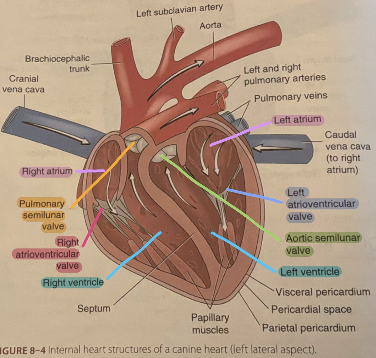
Right atrioventricular valve (right AV valve) (tricuspid valve (bc it has 3 points))
Controls opening between right atrium and right ventricle

Pulmonary semilunar valve (pulmonary valve)
Located between right ventricle and pulmonary artery
controls blood entering lungs
Semilunar
Half-moon

Left atrioventricular valve (left AV valve) (mitral valve) (bicuspid (bc it has 2 points))
Controls opening between left atrium and left ventricle

Aortic semilunar valve (aortic valve)
Located between left ventricle and the aorta
controls blood entering the arterial system
Systole
Contraction
ventricular contraction
Diastole
Expansion
Ventricular relaxation
Rhythm
Recurrence of an action or a functions at regular intervals
Heartbeat (cardiac cycle)
Rate and regularity of the heart rhythm
alternating sequence of relaxation and contraction of heart chambers
Cardiac output
Volume of blood pumped by heart per unit time

Electrical impulses (conduction system) are controlled by
Sinoatrial nose
Purkinje fibers
Atrioventricular node
Bundle of His

Sinoatrial node (SA node)
Located in the wall of right atrium near entrance of superior vena cava

Purkinje fibers
Atypical cardiac muscle cells
Sinoatrial node + Purkinje fibers =
Basic rhythm of heart and is called pacemaker of heart
Atrial systole
Atrial contraction
Inotropy
Force of contraction
Electrical impulses from Sinoatrial node travel to
Atrioventricular node

Atrioventricular node (AV node)
Located in Interatrial septum
conducts impulses more slowly than SA node which causes pause after atrial contraction to allow ventricles to fill with blood
Atrioventricular node transmits impulses to
Bundle of His

Bundle of His (AV bundle)
Located in Interventricular septum
continues through ventricle as ventricular Purkinje fibers which carry impulse through ventricular muscle causing ventricles to contract
Ventricular systole
Ventricular contraction
Sinus rhythm
Normal heart rhythm
starts in SA node
Arrhythmia or dysrhythmia
Abnormal heart rhythm bc SA node doesn’t function properly and other areas of conductive system take over and initiate a heartbeat
Preload
The ventricular end-diastolic volume, or the volume of blood entering the right side of the heart
Afterload
The impedance to ventricular emptying presented by aortic pressure
Palpitation
Heartbeat sensations that feel like pounding with or without irregularity in rhythm
Fibrillation
Rapid, random, and ineffective heart contractions
Flutter
Cardiac arrhythmia in which atrial contractions are rapid but regular
Bradycardia
Abnormally slow heartbeat
Tachycardia
Abnormally rapid heartbeat
Paroxysm
Sudden convulsion or spasm
Normal sinus arrhythmia
Irregular heart rhythm resulting from variation in vagal nerve tone as a result of respiration (a nonpathologic arrhythmia)
Asystole
Without contraction or lack of heart activity; flat line on an ECG
Syncope
Temporary suspension of respiration and circulation
Gallop
Low-frequency vibrations occurring during early diastole and late diastole

Electrocardiogram (ECG or EKG)
Record of the electrical activity of myocardium
shows the changes in voltage and polarity (positive and negative) overtime
Electrocardiography
Process of recording the electrical activity of the myocardium
produces a tracing that represents the variations in electrical potential caused by excitation of heart muscle and is detected at body surface
Leads
Conductors that detect variations in electrical potential
Auscultation
Act of listening to body sounds and usually involves the use of a stethoscope
Stethoscope
An instrument used to listen
Stetho-
Chest
Lub-dub
Heart sound
Lub (first heart sound)
Caused by closure of AV valves
Dub (second heart sound)
Caused by closure of the semilunar valves