ANATOMY UNIT 2: Skeletal System
 Call Kai
 Learn
 Practice Test
 Spaced Repetition
 Match
 Flashcards
 Knowt Play
1/59

There's no tags or description
Looks like no tags are added yet.
60 Terms
epiphyseal (growth) plate
a region of cartilage between the epiphysis and diaphysis where lengthwise growth occurs.
once the growth plate closes…
cartilage is replaced by bone and an epiphyseal line may remain.
indicated the bone is no longer growing
fontanelles
tough, membrane-covered gaps where the bones of an infant's skull have not yet fused together
allow flexibility during birth and accommodate rapid brain growth.
with age:
gradually close
meet at sutures; normal adult skulls do not retain the infant’s open fontanelles
bone is …
living tissue
osteoclasts …
kill down old bone (break down)
osteoblasts …
build new bone
remodeling
osteoclasts + osteoblasts processes
bone formation and growth generally outpace generally outpace breakdown
bone mass usually reaches its peak in early adulthood
bone remodeling steps
osteoclasts break down old bone
resorption pit is cleared
osteoblasts build new bones
new bone mineralizes
remodeled bone
osteoid
new bone matrix
mineralized bone
hard bone
normal (younger) ribs
smooth
rounded sternal ends
older adult ribs
cup-shaped
jagged sternal ends
pelvis
ilium
Ischium
pubis
pelvic girdle
female = broader subpubic angle and wider, shallower pelvic cavity
male = narrower subpubic angle and a deeper, narrower cavity
sacrum
female = shorter and wider
male = longer and narrower
coccyx
female = projects less anteriorly, leaving more room for the pelvic outlet
male = project more anteriorly
skull
female = smaller, more vertical forehead, smaller mandible
male = larger and more robust, larger mandible, more sloping forehead
wolff’s law
healthy bone adapts and remodels itself in response to the mechanical loads and stresses placed upon it.
bones adapt to the mechanical stress placed on it
decreased stress: immobilization / less use - bone becomes thinner and less dense
normal stress: typical daily activity - bone maintains normal density and shape
increased stress: athlete / weight bearing - bone becomes thicker and stronger.
osteoporosis
chronic bone disease that weakens bones, making them fragile, porous, and much more likely to break (fracture).
functions of the endoskeleton
support
provides a framework for the body’s soft tissues
protection
shields vital organs, including the brain, heart, and reproductive organs
movement
muscles pull on bones to produce movement
mineral storage
stores calcium for bone growth and maintenance
blood cell production
hematopoiesis: red blood cells are produced in red bone marrow
energy storage
yellow bone marrow stores fat as an energy reserve
axial
spine
rib cage
hyoid bone
skull/face

appendicular
upper and lower extremities
bones that girdle them

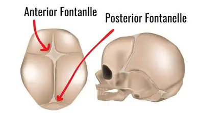
fontanelles
soft spots on baby’ s skull allow compression of skull during birth fuse and form sutures / joints before baby is 1-2 years old
ribs
12 pairs
true ribs (1-7): attach directly to sternum
false ribs (8-10): don’t attach to sternum directly (share 7th ribs cartilage)
floating ribs(11-12): don’t attach to sternum at all

pelvic girdle

pectoral girdle and upper extremity

lower extremity

anatomy of a long bone

osteon/haversian system
structural unit of a compact bone
haversian canal
space where blood vessels and nerves pass
osteocyte
bone cells
lacunae
spaces that house osteocytes
canaliculi
canals that connect haversian canals to osteocytes
volkman’s canals
connect haversian canals to periosteum horizontally
synarthrosis joints
no movement (immovable)
bones are tightly joined by fibrous connective tissue
provide strength and protection
little to no movement
ex: cranial sutures
coronal suture
between frontal and parietal bones
sagittal suture
between parietal bones
lambdoidal suture
between parietal and occipital bones
amphiarthrosis joints
slight movement (limited movement)
bones are joined by cartilage (fibrocartilage or hyaline cartilage)
provide strength and stability
allow limited movement
ex: pubic symphysis and intervertebral joints
pubic symphysis
between the two pubic bones
intervertebral joints
between vertebrae
diarthrosis joints
free movement (highly moveable)
bones are held together by a synovial joint capsule with ligaments
allow a wide range of movement
provide stability while allowing mobility
ex: ball and socket joint, hinge joint, pivot joint, saddle joint, gliding joint
ball and socket joint
shoulder joint
hip joint
hinge joint
knee joint
elbow joint
pivot joint
atlantoaxial joint (neck)
saddle joint
thumb joint
gliding joint
wrist (between carpal bones)
ankle (between tarsal bones)
meniscus
C-shaped pad of fibrocartilage that cushions the joint, absorbs shock, increases stability, and helps distribute weight across the joint surface
cartilage
flexible, gel-like matrix (made of mostly collagen and water)
large cells, spaced far apart within the matrix
chondrocytes (living cartilage cells)
no blood vessels (nutrients diffuse much slower into cells)
bone
rigid, calcified matrix(made of collagen and mineral salts)
smaller cells, tightly packed (within the matrix)
osteocytes (living bone cells)
has blood vessels (nutrients diffuse quickly into cells, allowing faster repair)
endochondral ossification
hyaline cartilage model (most long bones begin as a model of hyaline cartilage in the embryo)
cartilage calcifies and breaks down(cartilage in the center calcifies and chondrocytes die. blood vessels invade)
primary ossification center - diaphysis (bone tissue replaces the cartilage in the center)
secondary ossification centers - epiphyses (bone formation later occurs in the epiphysis. cartilage remains at the growth plate until growth stops)
mature bone (cartilage model is completely replaced by bone, except for articular cartilage and the growth plate until adulthood)
osteoclasts
bone cells that RESORB (break down) bone
osteoblasts
bone cells that BUILD bone
osteoid
new bone matrix
wolfe’s law
bone adapts to stress
importance of endochondral ossification
allows bone to respond to stress/injury by changing size, shape and density
stresses on bone INCREASE the rate bone is deposited
bone aging
young adult: thick trabeculae (more bone mass)
older male: thinner trabeculae (some bone less, more gaps)
older female: much thinner trabeculae (more bone loss, larger gaps)
osteoarthritis
joint paint (worse with activity)
stiffness and decreased range of motion
swelling
loss of joint function
treatment:
pain management
physical therapy and exercise
weight loss
joint injections
joint replacement in severe cases
Compact bone
is dense and solid, made of tightly packed osteons. It forms the outer layer of all bones and the thick shaft (diaphysis) of long bones. It provides strength and resists bending and twisting.
spongy bone
has a porous, honeycomb-like structure. It is found inside bones, especially at the ends of long bones (epiphyses) and in the interior of flat bones. It is lighter and houses marrow.
trabeculae
are the thin, branching struts of spongy bone. They are arranged along lines of stress, so they absorb and distribute forces while keeping the bone light.