ANPH 301 quiz 3
 Call Kai
 Learn
Learn Practice Test
 Spaced Repetition
 Match
Match Flashcards
 Knowt Play
1/84

There's no tags or description
Looks like no tags are added yet.
Name | Mastery | Learn | Test | Matching | Spaced | Call with Kai | Chat |
|---|
No analytics yet
Send a link to your students to track their progress
85 Terms
4 Major types of animal tissues
Epithelial Tissue
Connective Tissue
Muscle Tissue
Nervous Tissue
Epithelial Tissue
Barrier between inside and outside lines cavities, forms glands
sheets of polar cells (sheets of connected cells very close together)
apical surfaces faces “outside’ or into a body cavity
basal surfaces connects basement membrane, which connects to underlying connect tissue
Avascular and highly cellular (very little ECM) innervated
vascular
innervated
avascular
Protection, absorption, secretion, transport, filtration, lubrication
Apical surfaces often bears cilia (movement of fluids) or microvili (absorption)
Connective Tissue
Protection, connection, structure, support
Muscle Tissue
Contractile, excitable, allows multicellular movement
Nervous system
Excitable, allows for rapid communication and coordination of responses
Inside vs Outside
epithelium forms barrier inside and outside
Uterus, bladder, and digestive tract of body considered outside body\
Inside = w/in cells and tissue
outside = must cross epithelium
lining = covers inside of something
Vascular
contains blood vessels
innervated
contain nerves/ nerve endings
avascular
contains no blood vessels
get O2 nutrients —>from diffusion or cell-cell transport
Types of epithelial layers
number of layers
simple (=stratum)
stratified (=strata)
looks stratified but isnt = pseudostratified
cells change shape = stretchy tissue
1 layer of epithelial tissue
simple (=stratum)
more than 1 layer of epithelium tissue
stratified (=strata)
looks like a startified epithelium tissue but isnt
pseudostratified
cells that change shape of epithelium tissue
transitional
flat mature cells
squamous
boxy mature cells
cuboidal (wide as they are talk)
longer than wide mature cells
columnar
pseudostratified
looks stratified, but isnt
1 layer even if it looks weird
transitional
cells can change shape (stretchy tissue)
weird domed epical surface
can stretch laterally
when it stretches it flattens
looks cuboidla when stretched
Elastin —> stretchy protein
rebounds, bounces back after its stretched
found in urinary system, specifically bladder
Simple epithelium
1 layer of cells
delicate but allows for diffusion and absorption most important
simple cuboidal epithelium
In ducts and secretory portions of small glands and in kidney tubules
secretes and absorbs
Simple columnar epithelial
Ciliated tissues are in bronchi, uterine tubes, and uterus smooth (non-ciliated) tissues are in digestive tract and bladder
absorbs, it also secrete mucus and enzymes
Pseudostratified columnar epithelium
Cilated tissue lines the trachea and much of the upper respiratory tract
Secretes mucus, ciliated tissue moves mucus
goblet cells
Goblet cells
Secretes mucus
tend to be upon simple epithelium
Impact the cell shape around them
Simple squamous epithelium
Air sacs lungs and the lining of the heart, blood vessels, and lymphatic vessels
allows materials to pass through by diffusion and filtration, and secretes lubrication substances
alveoli of lungs
Lining of blood and lymphatic vessels vessels
Lines inside of hear
—> layer that never contacts outside, also called endothelium
Stratified Epithelium
more layers = can w/stand more wear and tear friction
more durable, more protection
Multiple layer = can w/stand friction → more protective functions
Protective, as well as absorption/ secretion
Lining of cheeks and esophagus, lining of some glands and ducts, skin
skin, outer layers are keratinized
protein that makes fingernails hard
Allows skin to be water proof because those cells are dead
Stratified squamous epithelium
Lines the esophagus, mouth, and vagina
protects against abrasion
Stratified cuboidal epithelium
Sweat glands, salivary glands, and the mammary glands
secretes and protects
Stratified columnar epithelium
The male urethra and the ducts of some glands
secretes and protects
Transitional epithelium
only found in urinary system (ureters, bladder, urethra)
allows the urinary organs to expand and stretch
Glands (Glandular Epithelium)
specialized for secretion
Endocrine glands
Exocrine glands
Endocrine glands
Secretes hormones w/in the body, directly into interstitial fluid and blood stream, never through duct
ductless, interstitial and for blood, hormones (ex. Insulin)
→ “inside” “sharing”
Exocrine glands
Secrete onto epithelium to “outside” via duct
→ “outside” sharing
unicellular
Multicellular
Classification of multicellular glands
simple
Compound
Acinar

Simple multicellular glands
One duct (branched or not)
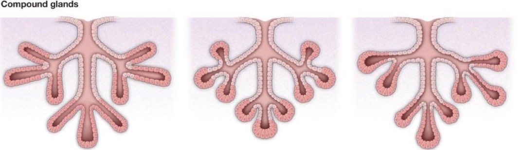
Compound multicellular glands
Multiple ducts transporting secretions
secreting parts of the gland connected by non-secreting parts
Acinar multicellular glands
Pockets (round/ spherical)
Tubular multicellular glands
Tubes/ elongated
Unicellular
No duct → goblet cells → secretes mucin → H2O → mucus
Multicellular
Ducts → tubes that conduct secretions to destinations → non secreting of gland
Exocrine secretion types
Merocrine
Apocrine
Holocrine
Merocrine
“part” “separate”
exocytosis, liquidy
Apocrine
“off/away”
apical portions of cells pinch off
nutrient rich, bacteria digest it + produce odour
Holocrine
“whole”
whole cell bursts to release secretions, very nutrient rich
lipid rich contain sebum
Connective tissue
most structurally diverse tissue type
vascular and innervated
cells widely dispersed aurrounded by large amounts of ECM/ interstitial matrix
includes amorphous ground substance that fills the space between a mesh of protein fibres
→ mesh of protein fibers (mesh = ground substances) + water + dissolved compounds that “fills”
collagen
elastin
reticular fibres
Components very depending on type
types of connective tissues
12 types, 3 categories
Supportive connective tissue
Fluid connective tissue is fluid
connective tissue proper
supportive connective tissue
provides solid, structural support
structure, movement of the body and parts, resist gravity, protection, store calcium
Cartilage:
hyaline cartilage
fibrocartilage
elastic cartilage
Bone:
spongy bone
compact bone
fluid connective tissue
Interstitial fluid connect them
not a connective tissue but acts like oen
blood
lymph
Connective tissue proper
semi-solid and flexible
most type of cell in connective tissue are fibroblasts
Fibroblasts
produce ECM fibers
collagen
elastin
reticular
Loose connective tissue proper
loose connective tissue proper: loosely packed fibres
Areolar Tissue
Adipose Tissue
Reticular Tissue

Areolar Tissue
purpose use connective tissue
found around blood/ lymphatic vessels

Adipose tissue
Fat storage → sub-cataneous layer under the skin
adults have white fat
babies have brown fat (used to generate heat)

Reticular Tissue
mesh-like frameworks for cells in non-muscular organs
liver, spleen, pancreas, lymph nodes
Dense connective tissue proper
dense connective tissue proper: densely packed fibres
regular dense tissue
irregular dense tissue

regular dense tissue
fibres are aligned (high tensible strength in one direction; tendons (muscle-bone); ligaments (bone-bone)
Irregular dense tissue
fibre orientation random (strength in all directions, not as strong as regular fibres)
Tissue membrane
epithelial membranes
synovial membrane
epithelial membranes
epithelial + connective tissue
serous membrane, mucous membrane, cutaneous membrane (skins)
Synovial membrane
connective tissue membrane encloses organ (e.g kidney) wrap around movable joints
encloses a synovial joint, w/synovial fluid
enclosed in a connective tissue
Integumentary system
skin (largest organ) and accessory structures, inducing hair, nails and glands
external barrier between body wall and environment
protects internal organs and spaces from physica; damage, radiation, pathogens, dehydration
vitamin D synthesis → calcium absorption
involved in regulation of water, temperature, ion balance
sensory reception
Lamellated corpuscle
tactile corpuscle
nocireceptor
thermoreceptor
Lamellated corpuscle
pressure sensor
tactle corpuscle
touch sensor
nocireceptor
pain sensor
thermoreceptors
heat transfer sensors
If overheated, capillary beds dilate to lose heat through skin
if cold capillary beds constrict to prevent heat transfer
less heat transfer = less blood flow
skin (integumentary)
largest organ in body
2 main layers
epidermis
dermis
closely associated underlying layer
hypodermis
deep to the hypodermis
epidermis
epithelial tisse
nocireceptor
dermis
connective tissue → receptor:thermoreceptors
hypodermis
also called subcutaneous layer
connective tissue, alot of adipose tissue
Fascia (connective tissue)
top of underlying muscle
Epidermis cell types
keratinized stratified squamous epithelium
1st line of defense
4 major cell types
keratinocytes (most common)
Langerhans cells
Melanocytes
Merkel cells
keratinocytes
produced by stem cells in deepest layer, migrate superficially
as they mature, they flatten, lose organelles, accumulate keratin and eventually die
water-impermeable and pathogen resistant surface layer that constantly sloughs off and is replenis
Langerhans cells
Immune cells (act as macrophages → cells that ‘eat’ pathogen-infected cells)
second line of defense
Melanocytes
produce pigment (protects from UV damage prevents folic acid breakdown)
found in basal layer
melanosomes stores melanin
type of melonin produce determines colour
how melanin are stored in cell important
Merkel cells
Merkel-ranvier cells/tactical epithelial cells, mechanoreceptors, detect light touch may connect to nerve endings
found in basal layer
thick skin
soles of feet, palmar surface of hands including fingertips
5 layer in epidermis
thin skin
everywhere else
4 layer in epidermis
Epidermis layers
stratum basale
stratum spinosum
stratum granulosum
stratum lucidum
stratum corneum
Stratum basale
also called stratum germinativum
single layer of mostly cuboidal basal cells overlying. Connects to basement membranes
dividing, attached via desmosomes + hemidesmosomes absorption + transport to rest of epidermis
stratum spinosum
cells migrating superficially
continual exchange of nutrient and waste
stratum granulosum
cells accumulate keratin
cells accumulate keratin and start to flatten
produce and release lamellar granules and lipids
dorm tight junctions w/ each other
Lamellar granules contain a waterproofing glycolipid
stratum lucidum
thick skin only
thin layer of tightly packed dead keratinocytes
contain eleidin → clear protein, water impermeable
stratum corneum
thick layer of dead keratinocytes
protection from physical force, pathogens, and prevention of water loss
outermost layer of dead cells constantly sloughing off
Dermis
connective tissue that underlines and supports epidermis
contain blood and lymph vessels, nerves, hair follicles, arrector pilli muscles, glands
2 layers: papillary layer and reticular layer
Papillary layer
loose areolar connective tissue
papillar between epidermis and dermis act to hold the skin together
Dermal papillae: tightly interlock the dermis and epidermis
form ridges known as fingerprints
contain tactile (meissner) corpuscles → detect light touch
deeper touch sensors = lamellar corpuscles (facinian corpuscles)
Reticular layer
thicker layer of dense irregular connective tissue deep to the papillary layer
contains hair follicles, blood vessels, nerves
pacinian corpuscles (lamellar corpuscles) → sense deep pressure