Anatomy Male and Female Perineum
 Call Kai
 Learn
Learn Practice Test
 Spaced Repetition
 Match
Match Flashcards
 Knowt Play
1/129

There's no tags or description
Looks like no tags are added yet.
Name | Mastery | Learn | Test | Matching | Spaced | Call with Kai | Chat |
|---|
No analytics yet
Send a link to your students to track their progress
130 Terms
pelvic outlet and skin between the thighs
perineum is between the
Male Perineum
the area between the anus and scrotum
Female Perineum
the area between the anus and the vagina
-pubic symphysis and body of pubic bone
-ischiopubic ramus to ischial tuberosities
-sacrotuberous ligaments
-coccyx
Pelvic Outlet is bounded by:
Sacrotuberous ligament
connects sacrum to ischial tuberosity

pelvic and urogenital diaphragms
Perineum is separated from the true pelvic cavity by the
perineum
space between the diaphragms and skin
Sacrospinous Ligament
connects sacrum to ischial spine

urogenital triangle and anal triangle
what are the two parts of the perineum?
ischial tuberosity
what divides the two trianges of the perineum
between sacrospinous and sacrotuberous ligaments
Where is the lesser sciatic foramen located?
Lesser sciatic foramen
passageway for structures entering or leaving the perineum
Pudendal nerve
innervates muscles of the perineum
Internal Pudendal Artery
supplies perineum and external genitalia
piriformis
The pudendal nerve and internal pudendal artery are located inferior to what muscle?
greater sciatic foramen
The pudendal nerve and artery pass out of
lesser sciatic foramen
The pudendal nerve and artery pass into
ischioanal fossa
after the pudendal nerve and internal pudental artery pass through the lesser sciate foramen where do they go
pubic symphysis
ischiopubic rami
ischial tuberosities
Urogenital triangle borders
Urogenital triangle

Coccyx
Sacrotuberous ligament
Ischial tuberosities
Anal triangle borders
Anal Triangle

Obturator Internis Fascia

Ischioanal Fossa

deep to anal triangle
The ischioanal fossa is located deep to what structure?
levator ani
obturator internus
The ischioanal fossa is located between what two muscles?
skin of anal triangle
floor of ischioanal fossa
junction where iliococcygeus muscle takes origin from the obturator internus muscle
which is the deeper located apex of the ischioanal fossa
obturator internus
What is the lateral side of the ischioanal fossa
levator ani
What is the medial side of the ischioanal fossa
Pudendal canal
Inferior rectal nerve, arteries and veins
Contents of ischioanal fossa
Pudendal nerve
Internal pudendal artery and vein
Contents of pudendal canal
Pudendal canal

Alcock's cana
the pudendal canal is also known as
external anal sphincter
inferior rectal nerves and arteries supply
pudendal nerve
inferior rectal nerve is a branch of the
internal pudendal artery
inferior rectal arteries and veins are branches of
Pectinate Line
line separates soma (i.e., the body wall) from viscera
Pectinate Line
coincides with anal valves

painful
ex. external hemorrhoids
Describe lesions below the pectinate line
silent unless they cause stretching of GI wall
ex. internal hemorrhoids
Describe lesions above the pectinate line
Anal columns
small longitudinal folds in the anal canal
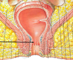
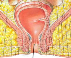
Anal valves
circular folds inferior to anal columns
Internal anal sphincter
involuntary smooth muscle

External anal sphincter
voluntary skeletal muscle

inferior rectal nerve
What nerve supplies the external anal sphincter?
perineal nerves and arteries
Pudendal nerve and Internal Pudendal artery change names to
in the urogenital triangle
Where do Pudendal nerve and Internal Pudendal artery change names to perineal nerves and arteries ?
Perineum

Perineal Membrane

deep and superficial perineal spaces
The perineal membrane separates what?
Urogenital Diaphragm
Middle layer of pelvic floor

Superficial Perineal Pouch
potential space inferior to the perineal membrane

Colles Fascia
continuous with scarpa's fascia

Glans Penis
bulging structure at the distal end of the penis (glans = acorn)
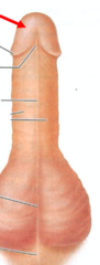
Corona of Glans
rim of glans
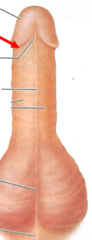
Frenulum of Prepuce
a fold of skin
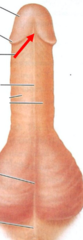
deep surface of prepuce with the glans
Frenulum of Prepuce connects what two structures?
immediately ventral to external urethral orifice
Where is the Frenulum of Prepuce located?
Penile Raphe
line on penis at fusion of urethral folds

Scrotal Raphe
division of the 2 scrotal chambers

Body of Penis

Root of Penis

Corpus Cavernosum
becomes engorged with blood during erection
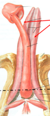
Corpus Spongiosum
surrounds the urethra
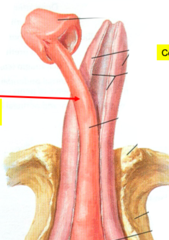
Crus of the Penis
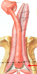
Bulb of the Penis

corpus spongiosum
The bulb of the penis is to the root, as the ______ __________ is to the body of the penis
Bulbospongious muscle
covers bulb of penis

expel contents of spongy urethra
What is the function of the bulbospongious muscle?
Ischiocavernosus muscle
covers crus of penis
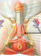
stops drainage of blood during erection
What is the function of the ischiocavernosus muscle?
Superficial Transverse Perineal Muscle

stabilizes and strengthens central tendon of perineum
What is the function of the Superficial Transverse Perineal Muscle?
Prostatic Urethra
Membranous Urethra
Spongy Urethra
3 Male Urethras
Prostatic Urethra
surrounded by prostate
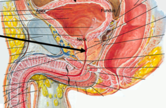
Membranous Urethra
passes through urogenital diaphragm

Spongy Urethra
runs through penis; opens at external urethral orifice

External Urethral Sphincter
voluntary skeletal muscle

urogenital diaphragm
Where is the external urethral sphincter located?
controls urination
external urethral sphincter function
Internal Urethral Sphincter
involuntary muscle

in neck of urinary bladder
Where is the internal urethral sphincter located?
prevents semen from entering the urinary bladder
What is the function of the internal urethral sphincter?
Internal Urethral Sphincter
What urethral sphincter is found only in males?
Superficial Dorsal Vein of the Penis
within the skin

Deep Dorsal Vein of the Penis
unpaired
lies along dorsal midline

Dorsal Artery of the Penis
paired arteries on the dorsum of the penis
deep to Buck's fascia
lateral to the deep dorsal vein

Source of blood that engorges Corpus Cavernosum to form erection
Deep Artery of the Penis
Buck's Fascia
deep fascia of penis

Tunica Albuginea
white fibrous capsule on testes

vein, artery, nerve
order of Deep Dorsal Vein of the Penis, Dorsal Artery of the Penis, Dorsal Nerve of the Penis
Peyronie's Disease
Idiopathic formation of fibrous plaques within the Tunica Albuginea, usually on the dorsal surface.
Presents with palpable subcutaneous mass and often abnormal curvature of the penis.
how does Peyronie's Disease present
pain during intercourse
erectile dysfunction
stenosis of spongy urethra
Peyronie's Disease (Penile Fibrosis) is associated with
cutaneous to femoral triangle, anterior scrotum/labia majora
Genitofemoral nerve innervates
anterior surface of psoas major muscle
Genitofemoral nerve descends on
Lateral cutaneous nerve of thigh
cutaneous to lateral side of the thigh
(passes medial to ASIS)
Cremaster muscle
Muscle that pulls the scrotum closer to the body in cold temperatures and relaxes to let the testicles be farther away from the body in warmer weather
Cremasteric Reflex
stroking superior part of medial thigh causes contraction of cremaster muscle which leads to retraction of spermatic cord and elevation of the testes