Topic 4: Investigating the brain
 Call Kai
 Learn
 Practice Test
 Spaced Repetition
 Match
 Flashcards
 Knowt Play
1/32

There's no tags or description
Looks like no tags are added yet.
33 Terms
CT (computed tomography) is..
X-rays pass through the head from many angles. The detector measures what gets through. A computer combines this data to create cross-sectional images. GIve crose sectional slices, viewed at any plane
CT advantages
Very fast.
Excellent for detecting bleeding, fractures and bone.
CT disadvantagesUses ionizing radiation.
Lower soft tissue contrast than MRI.
Not ideal for detailed brain structure.
CT clinical use
Detecting bleeding after head injury.
Finding skull fractures.
Identifying strokes.
MRI (Magnetic Resonance Imagining)
uses a magnetic field, hydrogen atoms align. Radio frequency waves then disturb the aligned atoms, wave stops and atomes return to orginal postion —> 3D image of brain created
MRI
Gives very good contrast between soft tissues, making it ideal for studying brain structure.
MRi disagvantge
loud, patient must be still, patient is in an enclosed space, those w metal implants (eg, pacemakers), cannot enter machine.
MRI use
Diagnose: tumours stoke, MS; follow progression of disease, plan surgery by seeing brain map. Sructure mRI can be combined w PET or fMRI
DTI (diffusion tensor imaging)
Water moves freely in all direction in grey matter, In white matter water diffused roe easily ALONG axone, measure directional movement of water, hence mapping white matter pathways
DTI advantage
Maps the brain’s white matter pathways
✅ Non-invasive
✅ Helps us understand connectivity
✅ Useful in many neurological and psychiatric conditions
✅ Can be combined with MRI
DTI disadvantage
Indirect
❌ Less accurate where fibres cross
❌ Lower spatial resolution than standard MRI
❌ Can be affected by movement
DTI Image

MRI image
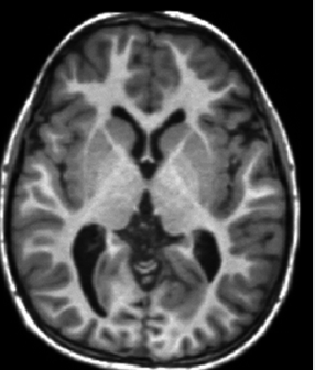
CT scan

Direct Measurement
measure activity of neurons direction: EEG MEG
Faster but less precise
Indirect measurement
measure changes in blood when nuerons are active. EG: fMRI, PET Slower but more precise
Spatial resoultion is..
How clearly can we see the detail and pinpoint the exact location? we can see WHERE things are
Temporal Resolution..
How precisely can we see timing and how quickly things change?
we know WHEN things happen
High spatial, low temporal
fMRI, PET, MRi
High temporal low spatial
EEG, MEG
EEG mesures..
electrodes placed on scalp detects electrical activity (generated by neurons) that passes through the skull.
EGG
EEG signal produces a waveform for each electrode.
Event-related potentials can be recorded by looking at the EEG signal immediately after a stimulus or event.
This can be used to examine the brains response and sometimes sequence activity to a complex stimulus.
EEG Advantage
✅ Measures brain activity almost instantly,✅ Excellent temporal resolution✅ Excellent temporal resolution
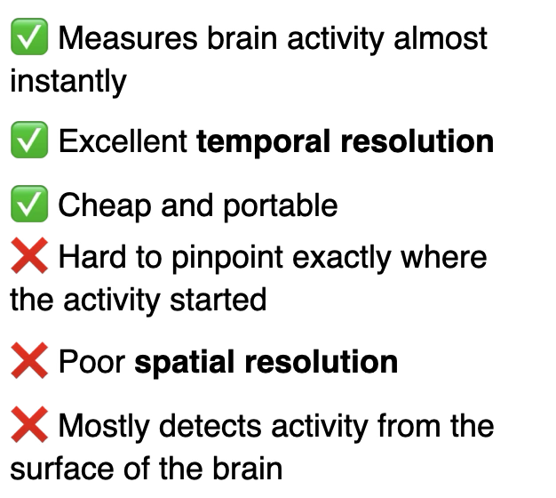
EEG disdvantage
Hard to pinpoint where exactly activity started, poor spatial resolution, detects activity from the surface of the brain
EEG use
How quickly do we recognise a face?
What happens during sleep?
Where do epileptic seizures begin?
How fast does the brain respond to a sound?
good for measuring timing of brain acitvity
MEG
meausres magnetic field that passes through the skull. Mganetic field passe through w less distortion, Hence this method offers better locaisation than EEG
MEG advantages
good temporal resultion, better spatial resolution than EEG.
MEG disadvantages
Expsnsive, nor portable, best for measuring brain acitvity near the surface of the brain
PET positron Emission Tomogrpahy)
Inject radioactive tracer, tracer travels in the bloodstream, binds to a specfci target, Scanner detects where teh tracer is using gamma ray detectors