Appendicular Skeleton Set
 Call Kai
 Learn
 Practice Test
 Spaced Repetition
 Match
 Flashcards
 Knowt Play
1/87

There's no tags or description
Looks like no tags are added yet.
88 Terms

Clavicle: collar bone

Sternal (medial) end: rounded end
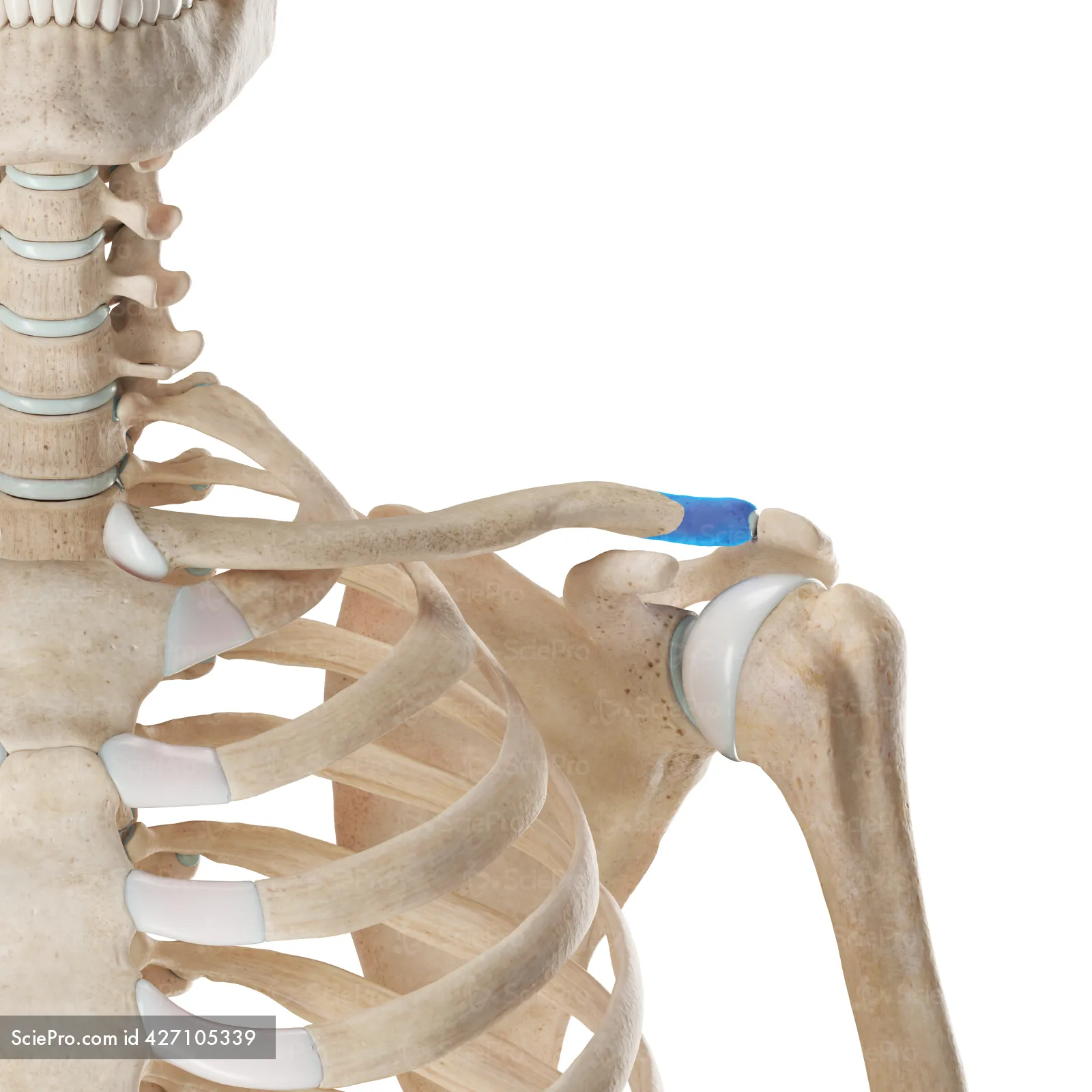
Acromial (lateral) head: flat end

Scapula: shoulder blade

Acromion: flat bone- follow it up from the spine of scapula
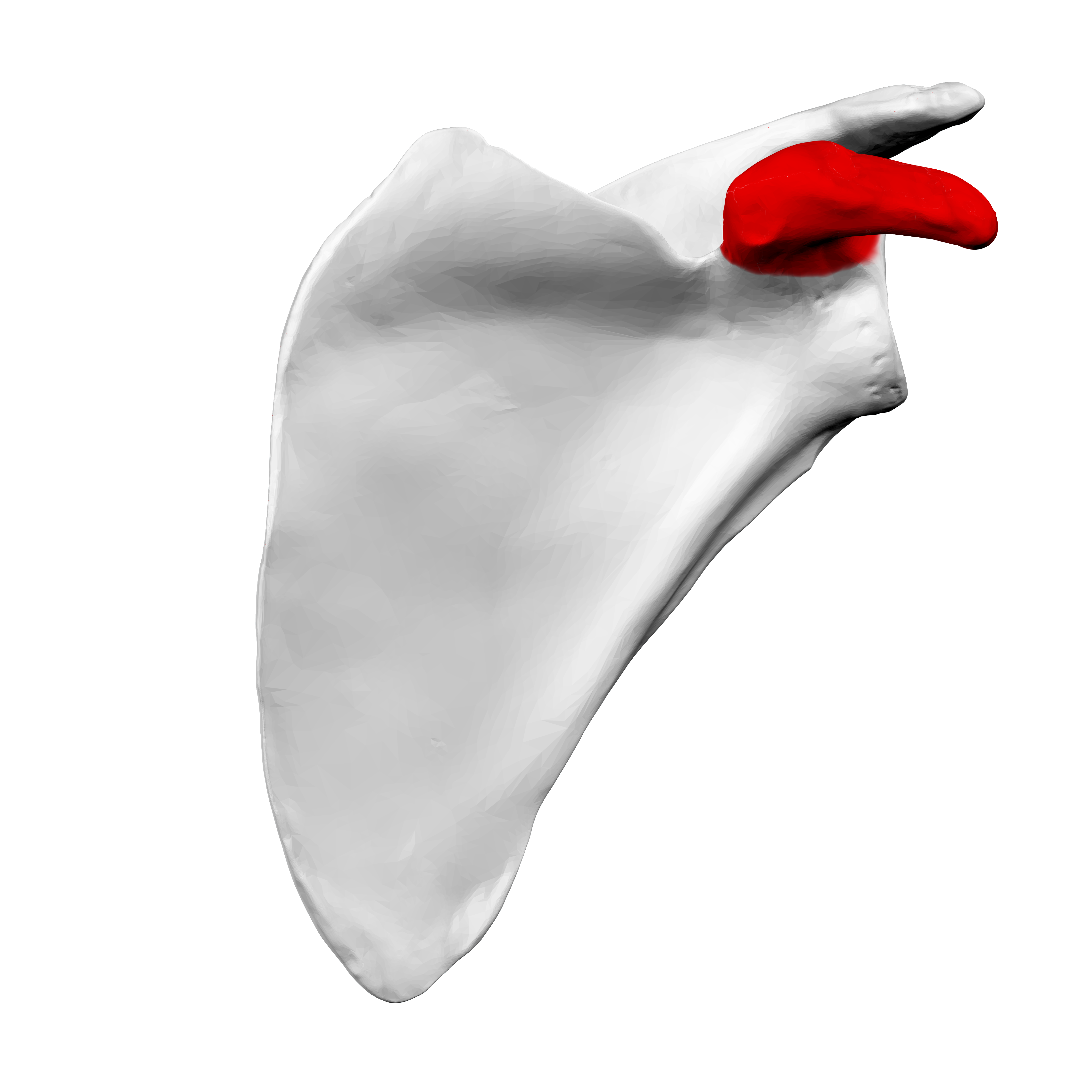
Coracoid process of scapula : facing anterior- flat bone

Glenoid cavity: where the arm bone meets the scapula: fossa [Determining whether it is the left or right scapula depends on the location of this bone]

Lateral (axillary) margin: lateral part of the scapula- would be closest to the humerus

Subscapular fossa: front of scapula

Medial (vertebral) margin: medial part closest to vertebrae

Supraspinous fossa: indentation above spine of scapula

Spine of scapula: located on the posterior portion

Infraspinous fossa: indentation below spine of scapula

Inferior angle: most inferior part of scapula-bottom part

Humerus: upper arms bone

Greater tubercle of Humerus: more proximal “bump” on head of humerus
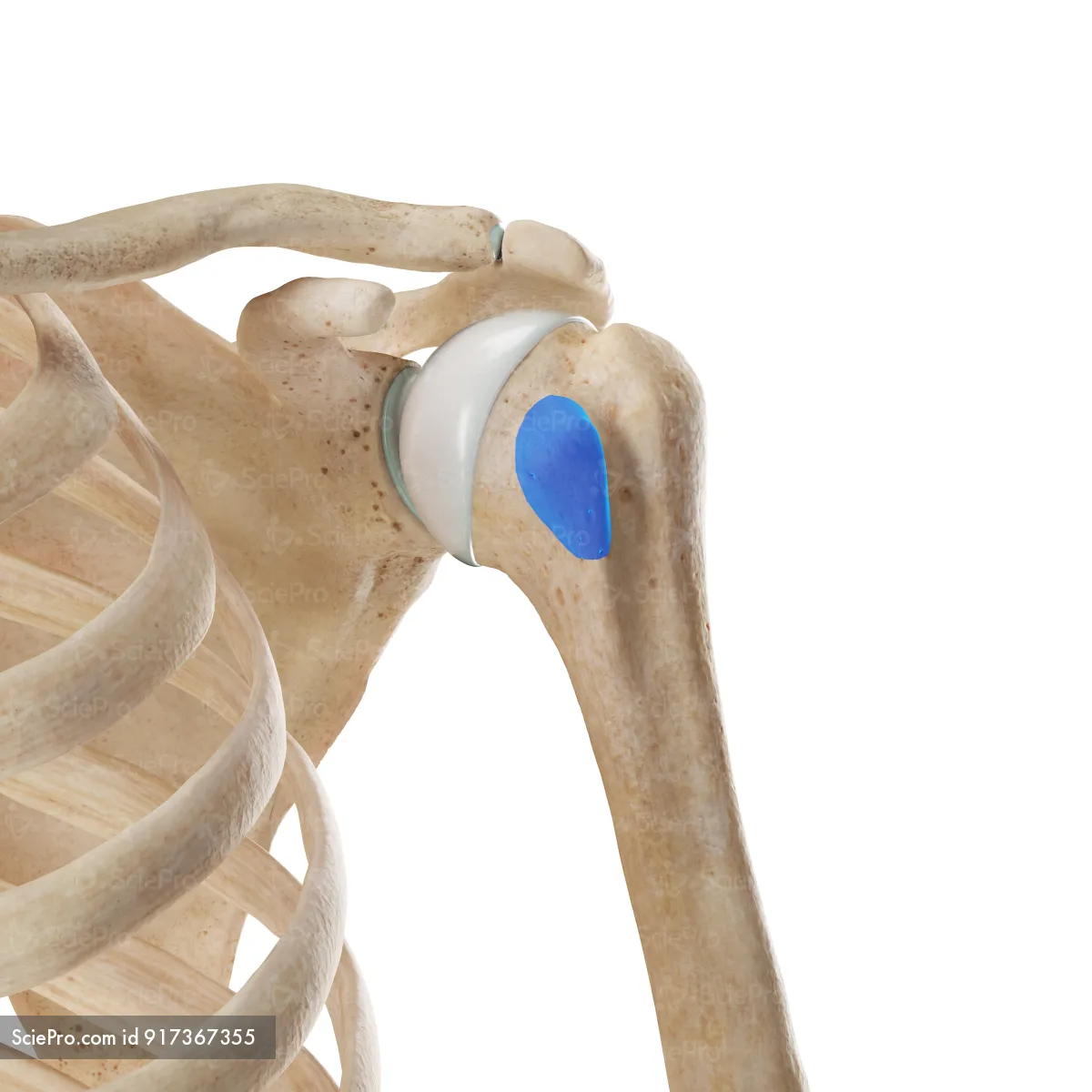
Lesser tubercle of humerus: more distal “bump”- still near head of humerus

Head of humerus: round part articulating with the glenoid cavity of scapula

Anatomical neck of humerus: where the head of humerus meets the rest of the bone [think of saturn’s ring]

Surgical Neck of humerus: where the head meets diaphysis of bone
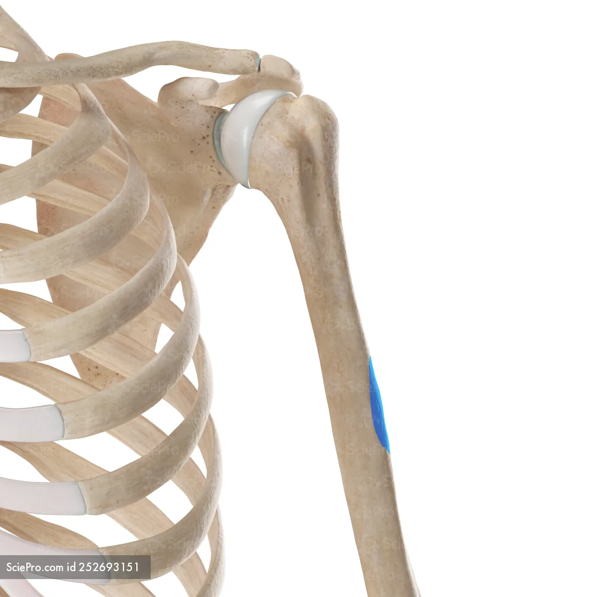
Deltoid tuberosity: roughening on diaphysis

Capitulum: on lateral part of the inferior humerus [can only be seen on the anterior view]

Trochelea: on flattened middle part of the inferior part of the humerus [can be seen on both the anterior and posterior view]

Coronoid fossa of humerus: forward facing indentation “small”

Olecranon fossa: “elbow” indentation

Radius: only bone with a nice rounded hear thats flat*this is the bone that twist in the lower part of your arm*
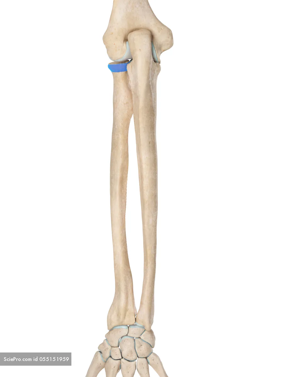
Head of radius: flat side-looks like a plate
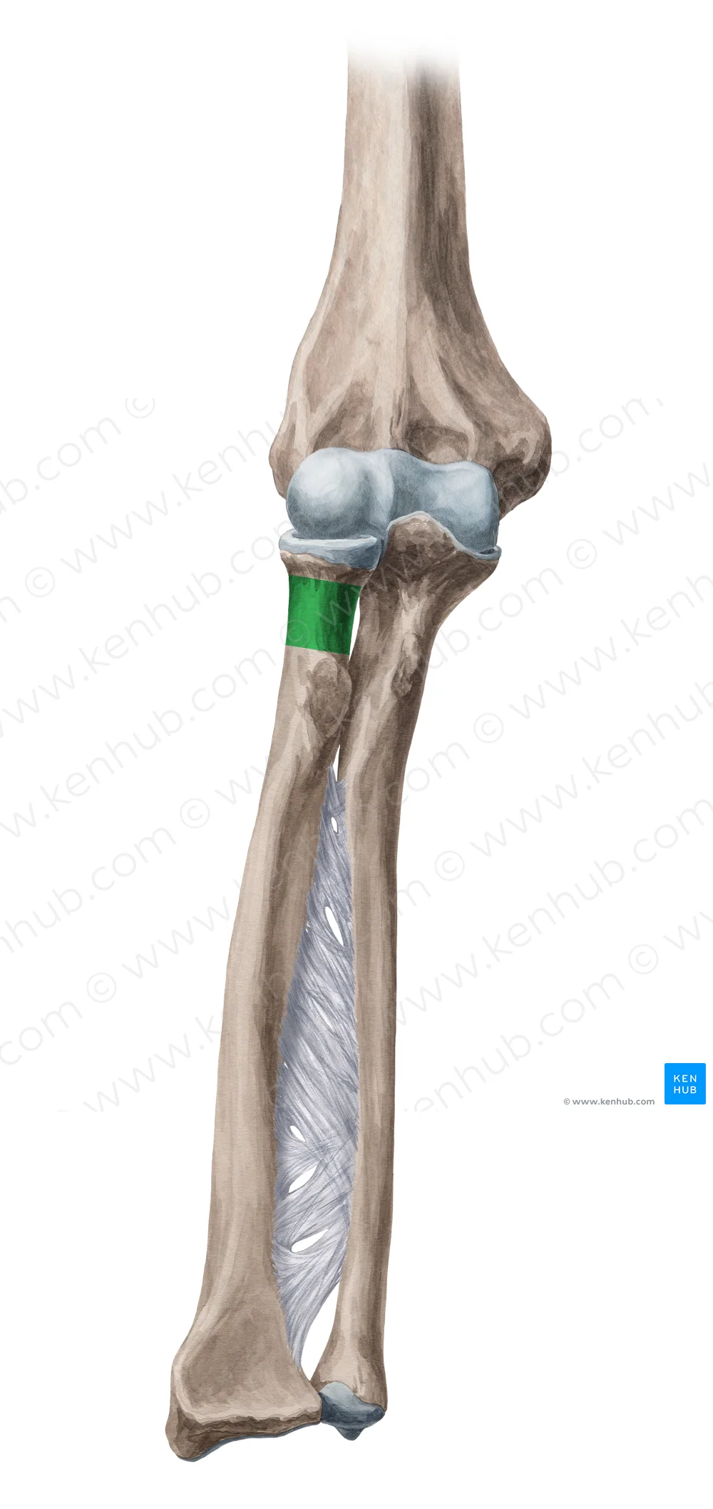
Neck of radius: distal to the head of the radius-where the head meets the diaphysis

Radial Tuberosity: roughening on radius

Styloid process of the radius bone: “point part” that's located on the most inferior part on the thumb side
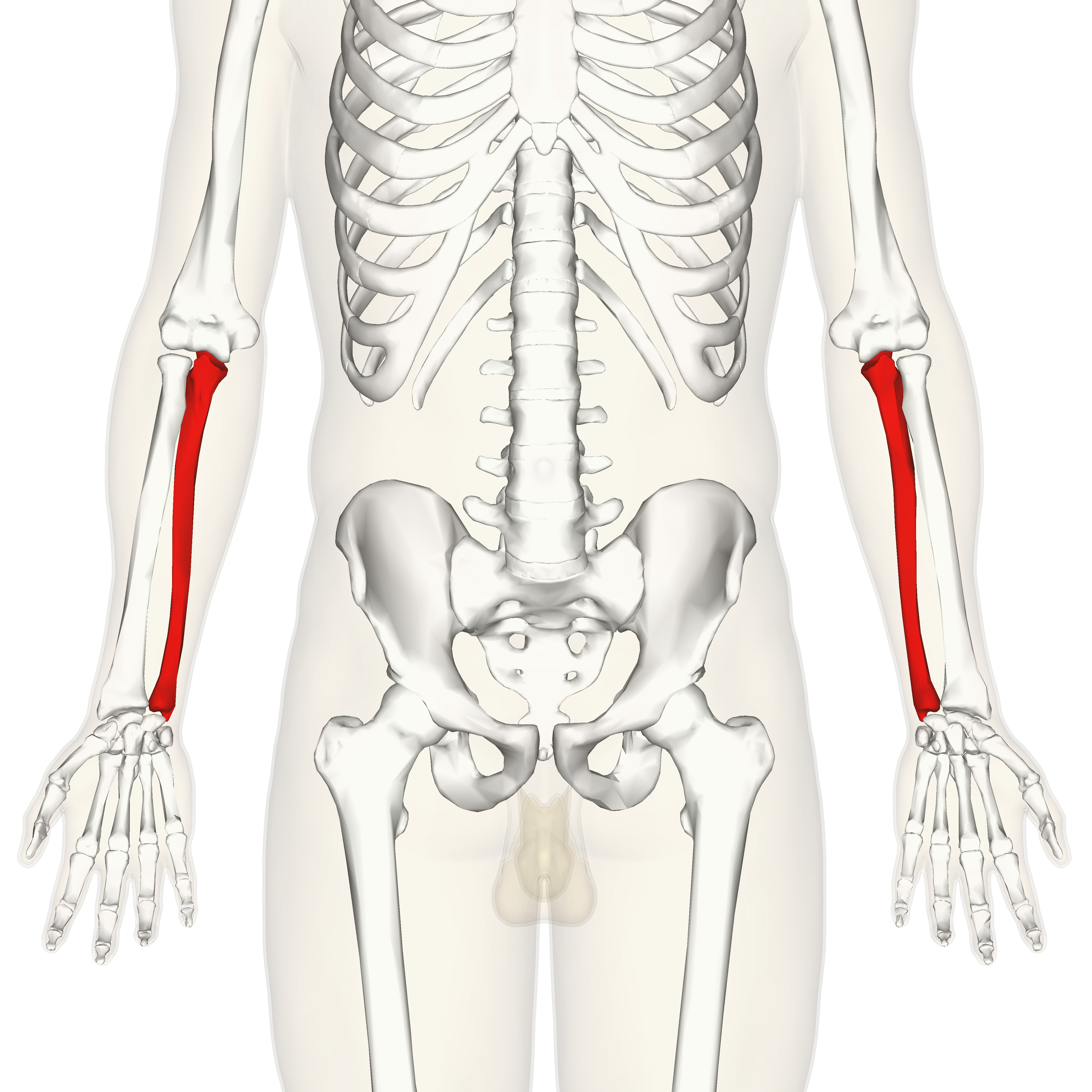
Ulna: ulna inside- medial; closest to you articulates with the trochlea of humerus

Coronoid process of the ulna bone: articulates with the coronoid fossa of humerus- superior anterior part

Olecranon process of the ulna bone: articulates with posterior fossa- the fossa located at the posterior humerus

Trochlear (semilunar) notch of the ulna: the halfmoon notch area (kinda looks claw shaped)

Radial Notch: slight dip where the head of the radius rotates

Styloid process: “pointy part” of the ulna

Carpals: wrist bones

Metacarpals: bones coming from your wrist bones
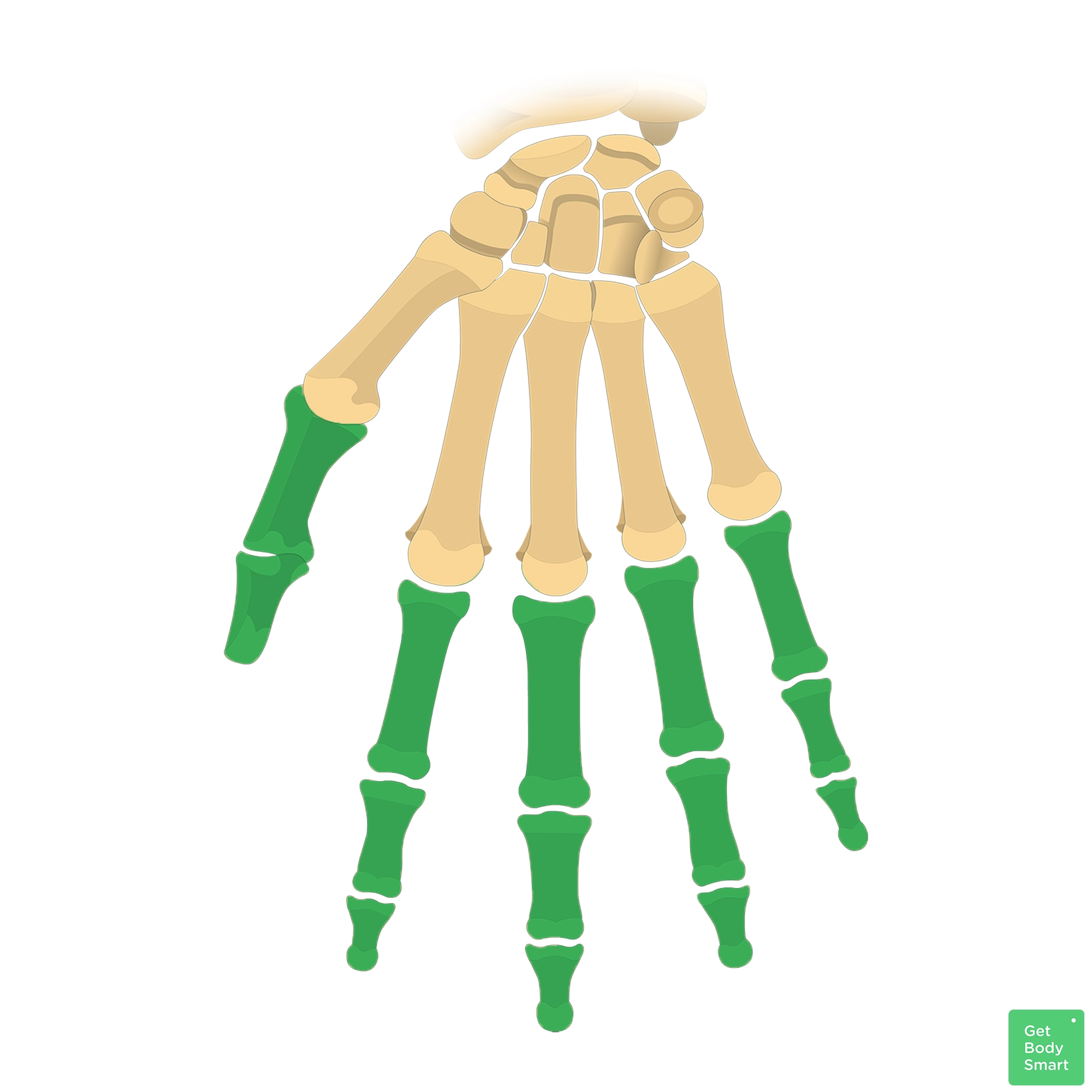
Phalanges: your fingers

Ilium: superior flat part

Iliac Crest: most superior part of the iliac crest- think the rim of it

Anterior superior iliac spine(ASIS): following along the crest the anterior area

Ischium: thicker part

Ischial tuberosity: roughening part we sit on this part
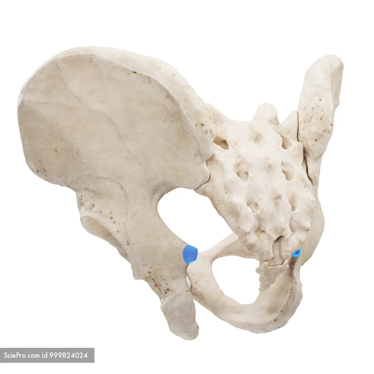
Ischial spine: little “hook” that is superior to the ischial tuberosity
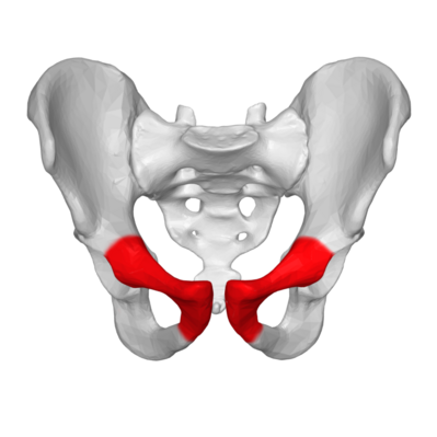
Pubis: thinner part- part that is most inferior

Symphysis pubis: thinner part where each pubic bone connects

Sacrum articulating surface( sacroiliac joint): where the sacrum and ilium meet on the medial side
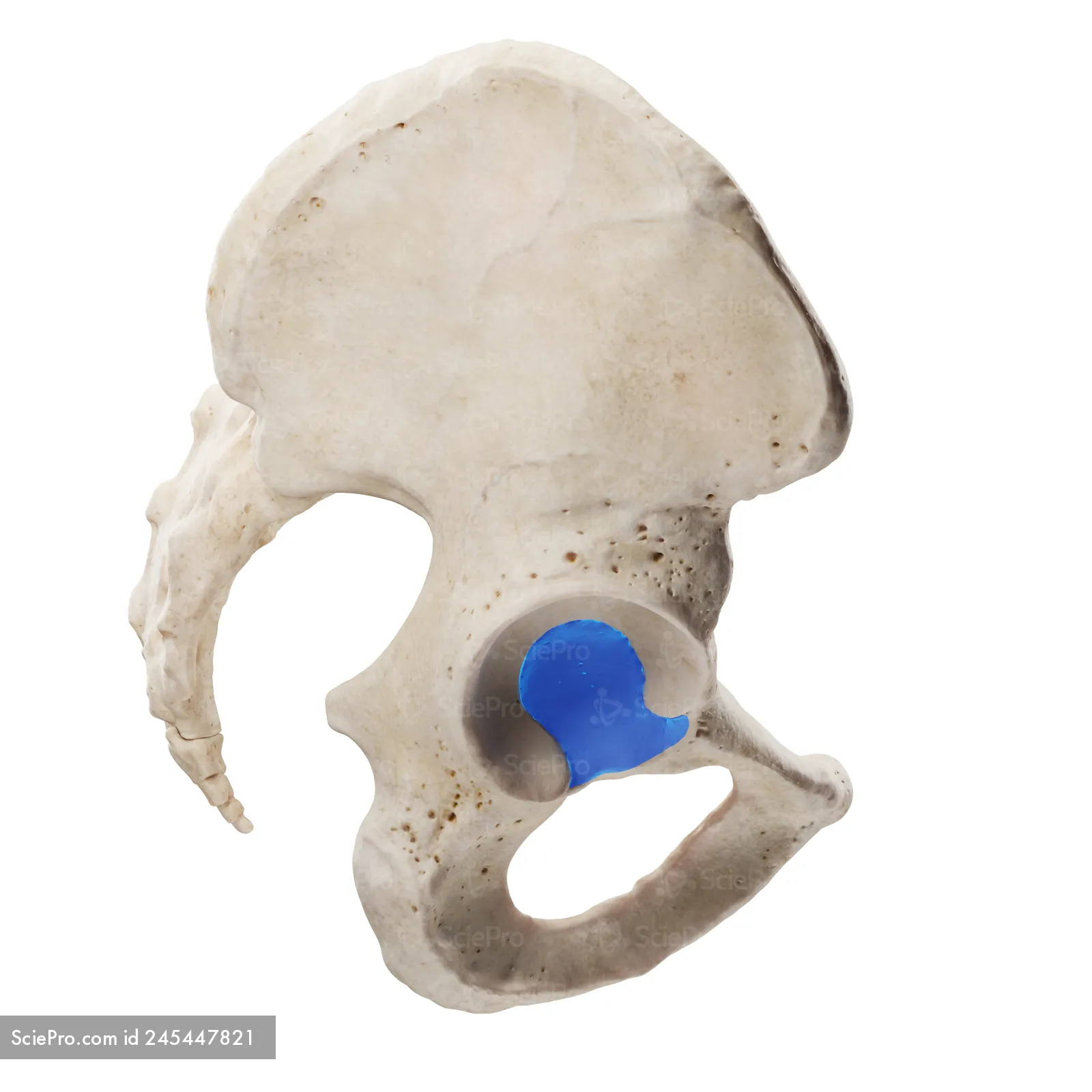
Acetabulum: where the femur articulates

Obturator foramen: big hole

Greater sciatic notch: Notch that looks kinda heart shepherd-faces posteriorly

Femur: largest bone in the body

Head of femur: round part

Neck of femur: where the “ball” of the head meets the diaphysis

Greater trochanter of femur: proximal bump seen on posterior view

Less trochanter of femur: slightly distal bump seen on posterior view

Gluteal tuberosity: roughening part

Linea aspera: line that goes down the posterior side of the femur seen on posterior view
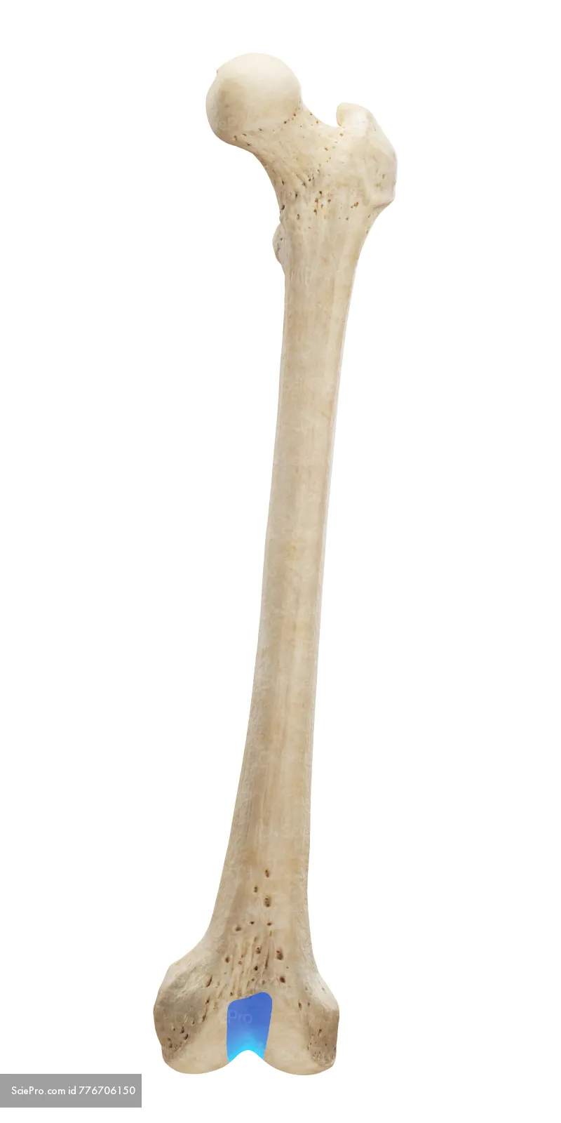
Patellar surface: “front bump” between the condyles seen on anterior view

Medial condyle of femur: bubble on medial side distal end of femur medial condyle is slightly larger than lateral seen on posterior view*

Lateral condyle of femur: bubble on lateral side on the distal end of femur seen on posterior view

Fibula: skinny bone of leg

Head of fibula: proximal end
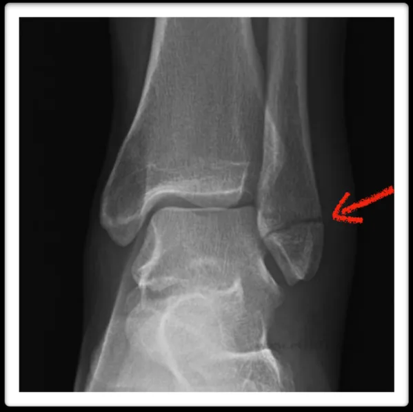
Lateral malleolus: pointy part on the outer most part of the ankle- that's the most distal

Tibia: bigger bone of the lower leg- the proximal end is club shaped & its anterior side has a bump that we all feel

Lateral condyle of tibia: lateral bump on the club part

Medial condyle of tibia: medial bump on the club part -because its medial its slightly larger

Tibial tuberosity: roughening part

Medial malleolus: bony prominence the distal end of your tibia on the inner ankle part- it articulates with the talus

Patella: tiny round bone

Tarsals: bones before your metatarsals

Talus: kind of rounded bone that

Calcaneus: the ball of your foot- your heel

Metatarsals: long bones of the foot
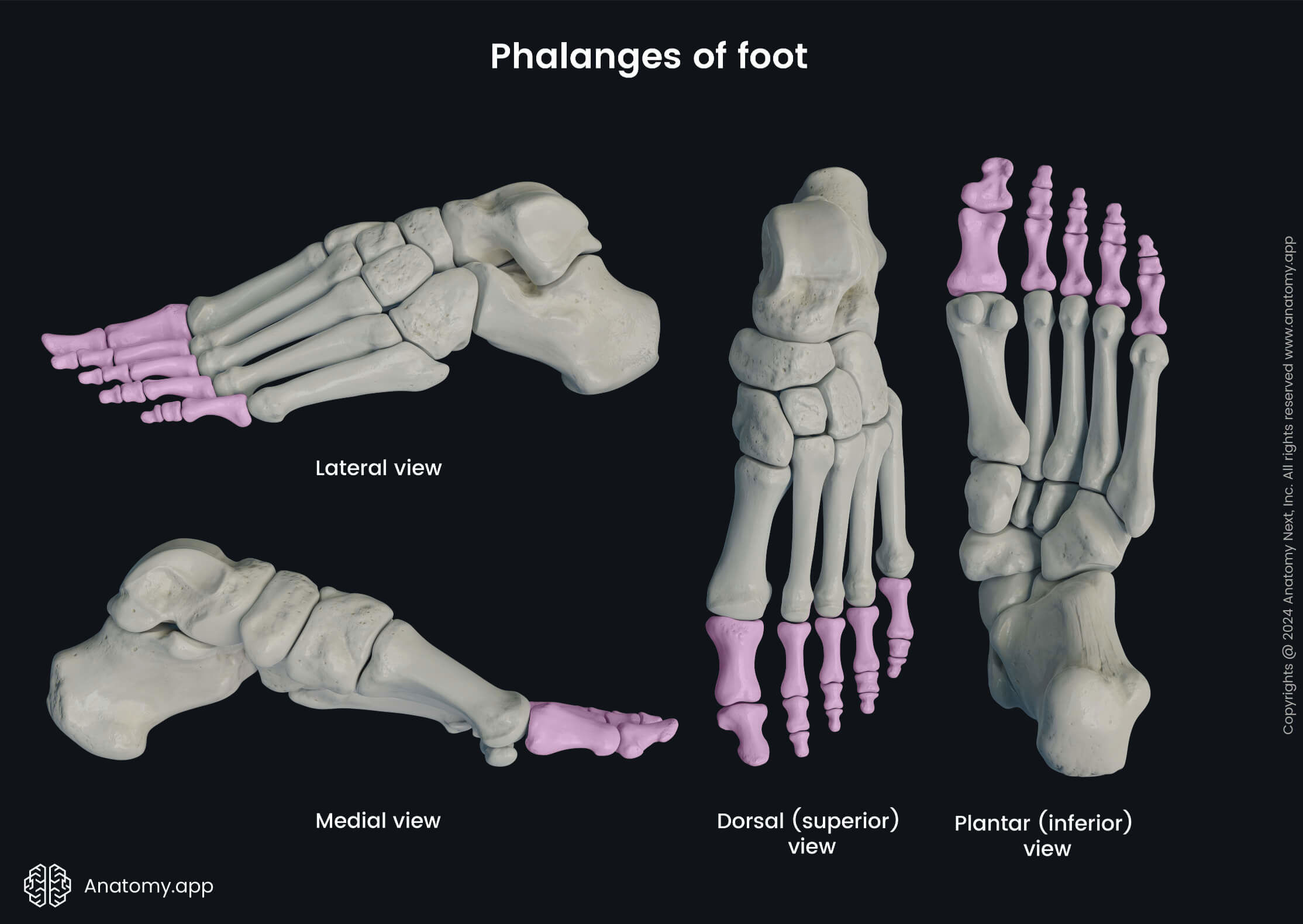
Phalanges: bones of the toes itself

Shoulder Joint: bones that make this; the scapula and humerus

Elbow Joint: bones that make this; the ulna, radius, and humerus

Hip Joint: bones that make this; head of the femur articulating with the acetabulum
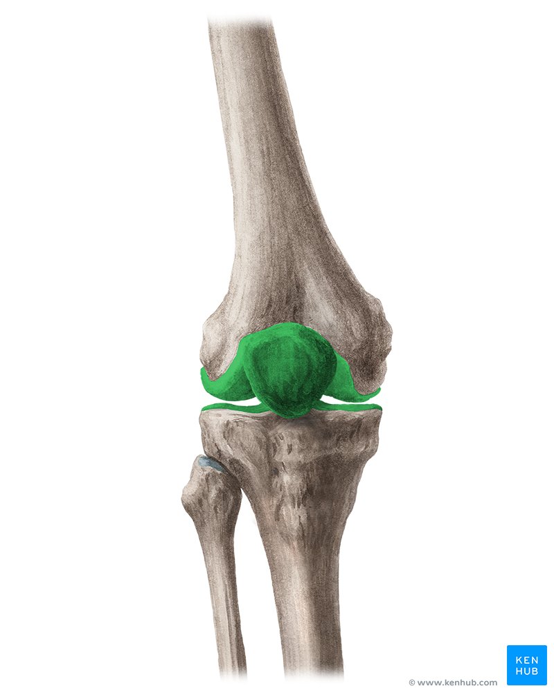
Knee Joint: bones that make this; the femur, patella, and tibia
How to tell if you are looking at the left or right Clavicle:
the inferior side of the clavicle has more bumps(so that side with the bumps would be facing towards the floor). Place the bone with the superior side facing up and the medial end facing you.
How to tell if you are looking at the left or right Scapula:
Look at the direction of the glenoid cavity is pointing to articulate with the humerus.If glenoid cavity points to the right it is a right side bone
How to tell if you are looking at the left or right Humerus:
Look at the posterior part of the humerus has a deep fossa “indentation that's one way to tell whether your looking at the anterior or posterior side of the humerus; to tell whether its your left or right humerus the head has to face medially

How to tell if you are looking at the left or right Femur:
Condyles would be facing posteriorly and look at the direction of the femoral head [head is facing right, its a left femur; head is facing left its a right femur]

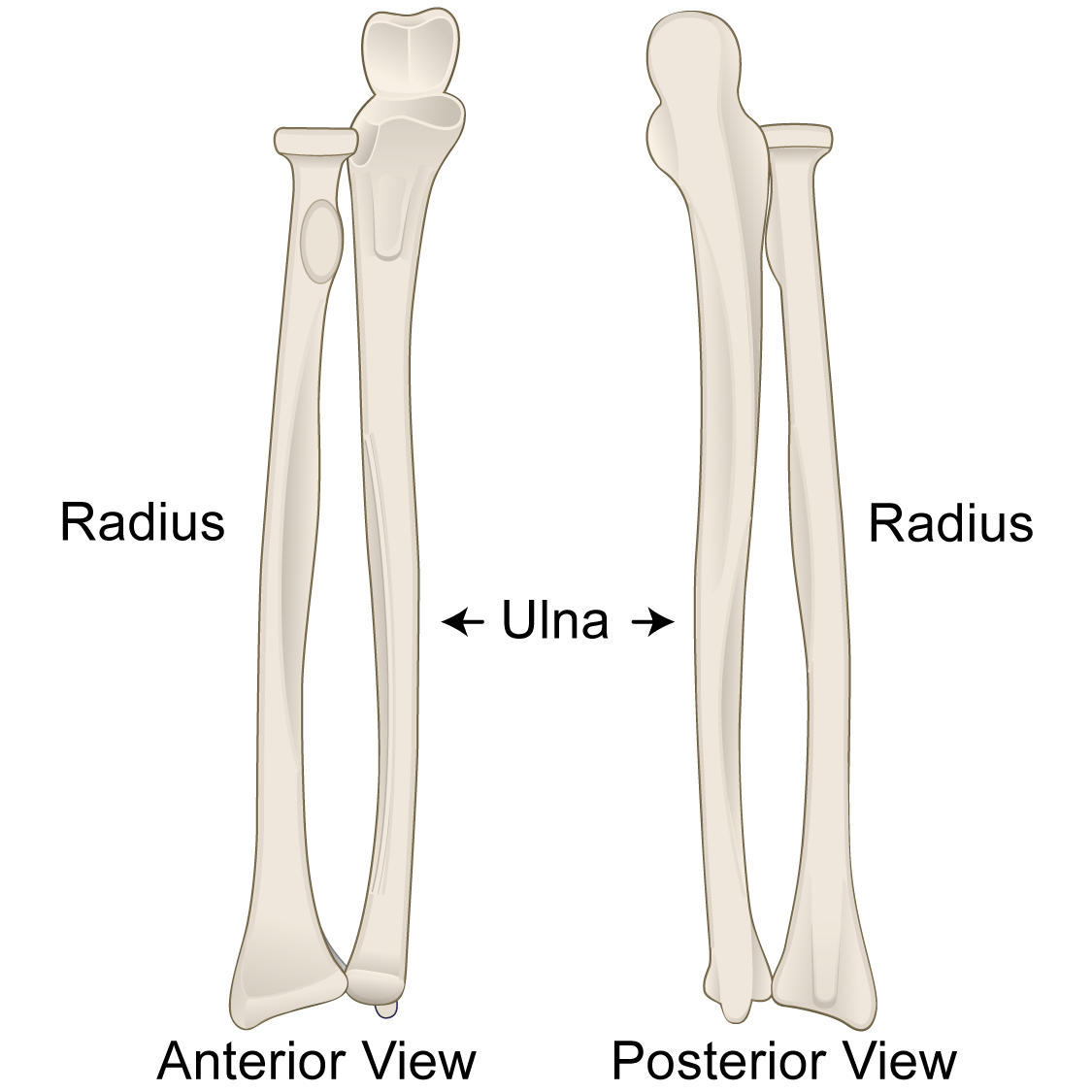
How to tell if you are looking at the anterior or posterior side of the Ulna:
If the olecranon process is prominent-the top of the hook you are looking at the posterior side

How to tell if you are looking at the left or right side of the Ulna:
If the styloid process is on the right it is a right side bone
How to tell if you are looking at the left or right Coxal bone:
Look at the direction of the acetabulum is pointing to articulate with the humeral head

How to tell if you are looking at the anterior or posterior side of the Coxal bone:
The greater sciatic notch faces posterior

How to tell if you are looking at the left or right Tibia:
If the Malleolus is on the right it is a right tibia
