labs 1-6 AP 2
 Call Kai
 Learn
Learn Practice Test
 Spaced Repetition
 Match
Match Flashcards
 Knowt Play
1/334

There's no tags or description
Looks like no tags are added yet.
Name | Mastery | Learn | Test | Matching | Spaced | Call with Kai | Chat |
|---|
No analytics yet
Send a link to your students to track their progress
335 Terms
Relaxed vs. contracted diaphragm position
Relaxed: dome-shaped, bulging up into the thoracic cavity under the ribcage. Contracted: flattens and moves down.
Diaphragm contracts: what happens to thoracic volume, pressure, and airflow?
Volume increases, pressure decreases (below atmospheric), air flows inward (inspiration).
Diaphragm relaxes: what happens to thoracic volume, pressure, and airflow?
Volume decreases, pressure increases (above atmospheric), air flows outward (expiration).
Why do volume changes move air?
Volume and pressure are inversely related. Air flows from high pressure to low pressure, so a bigger thoracic volume lowers pressure and pulls air in.
Quiet breathing: is inspiration active or passive?
Active. The diaphragm must contract, which requires ATP.
Quiet breathing: is expiration active or passive?
Passive. The diaphragm relaxes and the lungs and chest recoil, so no muscle ATP is needed.
Resting vs. forced breathing: energy requirements
Resting: only inspiration is active. Forced: both inspiration and expiration are active, extra muscles contract, and more ATP is used.
Rule for muscles that help forced breathing
A muscle that INCREASES thoracic volume lowers pressure and helps forced INSPIRATION. A muscle that DECREASES thoracic volume raises pressure and helps forced EXPIRATION.
Muscles of inspiration (I)
Diaphragm, external intercostals, sternocleidomastoid, pectoralis minor. The last two are accessory muscles for forced inspiration.
Muscles of expiration (E)
Internal intercostals, rectus abdominis, external abdominal obliques. Used in forced expiration (quiet expiration is passive).
Why does contracting the abdominal muscles help forced expiration?
They compress the abdominal organs and push the diaphragm up, and they pull the ribs down. This decreases thoracic volume and raises pressure, forcing air out.
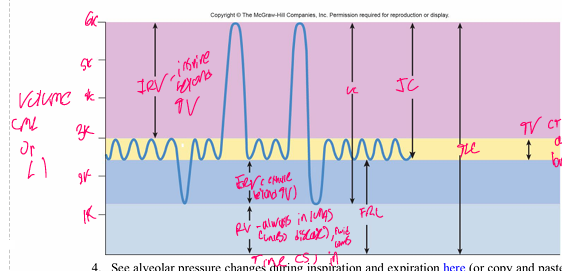
Tidal volume (TV)
Amount of air that enters and leaves the lungs during normal breathing. About 0.5 L (500 mL) in a young healthy person.
Inspiratory reserve volume (IRV)
Extra air that can be inhaled above tidal volume during a forced inspiration.
Expiratory reserve volume (ERV)
Extra air that can be forced out after a normal tidal expiration.
Residual volume (RV)
Air that remains in the lungs after a forceful expiration. About 1.2 L. It cannot be exhaled, so a spirometer cannot measure it.
Functional residual capacity (FRC)
FRC = RV + ERV. The volume of air that remains in the lungs during normal breathing.
Vital capacity (VC)
VC = TV + IRV + ERV. The maximum volume of air the lungs can exchange. About 4.5-4.8 L in a young healthy person (varies with sex, height, and age).
Total lung capacity (TLC)
TLC = TV + IRV + ERV + RV (or VC + RV). All the air the lungs can hold. About 6 L.
Inspiratory capacity (IC) and expiratory capacity (EC)
IC = TV + IRV. EC = TV + ERV.
Forced expiratory volume in 1 second (FEV1)
The amount of air forced out in the first second following a maximal inspiration.
Anatomical dead space (ADS)
Air that is not involved in gas exchange (it stays in the conducting airways). About 150 mL.
Alveolar ventilation rate (AVR)
AVR = breathing frequency x (TV - anatomical dead space). The volume of air that actually reaches the alveoli per minute.
Expired minute volume (VE)
VE = VT x breaths per minute (L/min). It does not subtract dead space, so it is larger than AVR.
Why can't a spirometer measure RV, FRC, or TLC directly?
You can't exhale the residual volume, and FRC and TLC both include it. So the lab uses a PREDICTED RV (based on sex, height, and age) to calculate them.
What does a spirometer actually record?
Airflow (L/s). Volume is calculated from the flow. Expiration appears as a downward deflection.
Why is the volume correction procedure done?
Expired air is warmer and holds more water vapor, so it has a greater volume than inspired air. Calibrating corrects for this.
Why do you zero the spirometer before each recording?
The signal drifts with temperature, so zeroing resets the baseline. The flow head must be left undisturbed during zeroing.
Why does the volunteer wear a nose clip?
So all the air they breathe goes through the mouthpiece, filter, and flow head and gets measured.
Static vs. dynamic pulmonary function tests
Static tests measure volumes and capacities, with no concern for how fast air moves. Dynamic tests depend on speed and time (forced, timed breathing).
Which measures are static?
Tidal volume, IRV, ERV, residual volume, vital capacity.
Which measures are dynamic?
FEV (forced expiratory volume), FEV1, FVC (forced vital capacity).
Forced vital capacity (FVC)
The total volume exhaled as fast and as hard as possible from full inspiration to full expiration.
How is FVC performed?
Inhale slowly until no more air fits, hold no more than 1 s, then exhale as forcefully, fully, and for as long as possible.
Vital capacity vs. FVC: why is FVC usually less than VC?
In a forced exhalation, the high pressure compresses and collapses small airways early and traps some air. So less air gets out than in a slow, relaxed exhalation.
FEV1/FVC ratio: what does it tell you?
The percentage of your FVC you can blow out in the first second, a measure of how easily air flows out. Normal is about 80%. It is lower with airway obstruction.
Restrictive lung disorder
The lungs cannot expand fully (stiff lungs or limited chest expansion). Lung volumes and capacities (VC, TLC) are DECREASED. FEV1/FVC is normal or increased. Examples: pulmonary fibrosis, scoliosis, obesity, neuromuscular disease.
Obstructive lung disorder
The airways are narrowed or blocked, so air is hard to get out. FEV1 and FEV1/FVC are DECREASED. Air gets trapped, so RV and TLC can increase. Examples: asthma, COPD (emphysema, chronic bronchitis).
Which disease type gives a lower-than-normal vital capacity? Why?
Restrictive. The lungs can't expand fully, so they can't take in as much air.
Which disease type gives a lower-than-normal FEV1? Why?
Obstructive. Narrowed airways resist airflow, so less air can be blown out in the first second.
Why might measured VC, FRC, and TLC differ from predicted values?
Predictions are averages for sex, height, and age. Differences can come from effort and technique, mouth or nose leaks, fitness, smoking, lung disease, or measurement error.
How might lung volumes change in a 74-year-old woman with breathlessness?
Lung elasticity falls with age. A restrictive problem would lower VC and TLC. An obstructive one would trap air (higher RV and FRC) and lower FEV1.
How would her breathlessness affect forceful exhalation and test results?
She can't exhale forcefully or quickly. Her FEV1 and FEV1/FVC would be lower than normal, and her FVC may also be reduced.
Hyperinflated lungs: what is it and how was it done in the lab?
Lungs held partially expanded, with air trapped and not fully exhaled. The volunteer takes a large breath, exhales a little, then breathes tidally on top of that extra volume.
Hyperinflated vs. normal breathing: rate and tidal volume
Respiratory rate increases and tidal volume decreases (faster, shallower breaths). Expect this, then check it against your own data.
Hyperinflated vs. normal breathing: IRV and ERV
The breathing sits at a higher lung volume, so IRV decreases and ERV increases. VC stays about the same. Check against your own data.
Why does a higher breathing rate with hyperinflation affect blood gases?
Expired minute volume increases, so more CO2 is blown off. Blood CO2 drops and pH rises.
How does hyperinflated breathing feel? Why?
Uncomfortable and tiring, like it takes effort. Chest and neck muscles must work harder to move air at that high lung volume.
Would an inflated chest alter breathing in a 74-year-old woman?
Yes, it would be uncomfortable. She would likely breathe faster and take smaller breaths.
Effect of hyperinflated breathing on FEV1
FEV1 decreases compared to normal breathing, which mimics what happens in obstructive disease. Check your own data.
Why do obstructive patients get hyperinflated lungs?
Air becomes trapped because they can't exhale it fully. The chest and neck muscles must work harder to assist breathing.

Label the spirogram: all volumes and capacities (image card)
IRV (top), TV, ERV, RV (bottom). Capacities: IC = TV + IRV, EC = TV + ERV, VC = IRV + TV + ERV, FRC = ERV + RV, TLC = everything. Axes: time (x) and volume in L or mL (y).
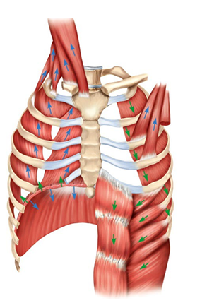
Label the respiratory muscles
Inspiration (I): diaphragm, external intercostals, sternocleidomastoid, pectoralis minor. Expiration (E): internal intercostals, rectus abdominis, external abdominal obliques.


Label the volumes on the colored lung diagram
Top blue: IRV.
Light blue: TV.
Orange: ERV.
Bottom red-orange: RV.


Label the FVC recording
Flow graph: where the sharp downward drop begins = forced expiration starts, and the lowest point = maximal expiration. Volume graph: volume 1 s into the exhale = FEV1, and the final plateau = FVC.


What is this? Label.
This is a vein.
Valve is Venous Valve


What is this? Label.
Capillary


What is this? Label.
Artery
Tunica interna is made up of basement membrane and endothelium


Label and explain
A. Arterial anastomosis
Provides collateral (alternative) routes of blood supply to organs.
Occurs in: Around joints, heart, brain.
B. Arteriovenous anastomosis
Allows blood to bypass capillary bed; good for preventing heat loss.
Occurs in: Extremities (fingers, toes, ears).
C. Portal system
Has two sequential capillary beds so items put in bloodstream by one organ are delivered directly to a second organ.
Occurs in: Liver (hepatic) and pituitary (hypophyseal).
D. Venous anastomosis
Allows for alternative drainage routes from organs.
Occurs in: Limbs (veins of hand/foot) and head/neck.
E. Typical (simple)
Designed for most efficient exchange of gases, fluids, and nutrients.
Occurs in: Most tissues/organs (muscles, skin, lungs).


What is this? And Label.


Label
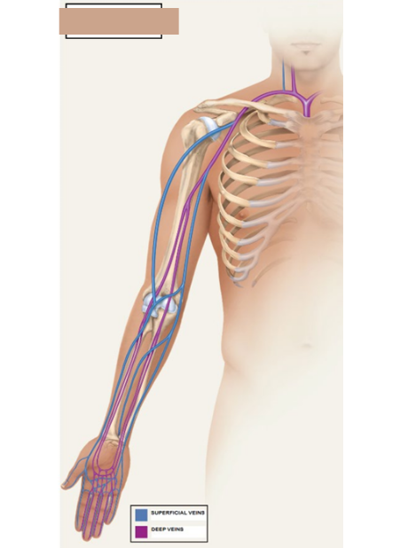
What? and Label
Veins


What? and label
Arteries


label
arteries

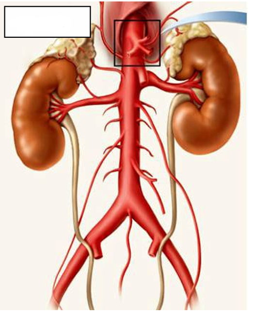
Label
Arteries


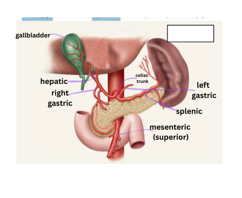
What is this? Label
Arteries, branches of the celiac trunk

Label
Veins


Label
Arteries


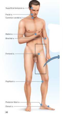
Label.


label
Tunica interna (intima)
Innermost layer of a vessel. Endothelium on a basement membrane. Its smooth surface reduces friction for blood flow.
Tunica media
Middle layer of smooth muscle and elastic fibers. Thickest in arteries. Controls vessel diameter (vasoconstriction and vasodilation).
Tunica externa (adventitia)
Outer layer of connective tissue (collagen). Protects, anchors, and supports the vessel.
Capillary wall - structure and function
Only endothelium on a basement membrane (no tunica media or externa). The very thin wall gives a short diffusion distance for exchange of gases, nutrients, wastes, and fluids.
Artery wall - structure and function
Thick tunica media (muscle and elastic fibers) and a small lumen. Withstands high pressure, recoils to keep blood moving, and vasoconstricts or vasodilates to regulate flow.
Vein wall - structure and function
Thinner wall, thin tunica media, large lumen, and valves. The large lumen holds a lot of low-pressure blood. Valves prevent backflow and help return blood to the heart against gravity.
Direction of blood flow in arteries vs. veins
Arteries carry blood AWAY from the heart. Veins carry blood TO the heart. This is true regardless of oxygen content.
Which vessels carry oxygenated vs. deoxygenated blood?
Oxygenated: systemic arteries and pulmonary veins. Deoxygenated: systemic veins and pulmonary arteries. Capillaries are where blood gains or loses oxygen.
How to identify an artery on a slide
Thick wall with a thick tunica media, round shape, and a small, regular lumen.
How to identify a vein on a slide
Thinner wall with a thin tunica media, and a larger, irregular lumen that is often flattened.
Why is the vein flat while the artery is round on the slide?
The vein has a thin, less muscular, less elastic wall and carries low pressure, so it collapses. The thick, elastic artery wall holds its round shape.
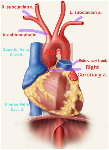
Aorta and vena cava (superior and inferior)
The aorta delivers blood to the entire systemic circuit. The vena cava receives blood from the entire systemic circuit and delivers it to the right atrium.
Coronary arteries and veins
Arteries deliver blood to the myocardium. Veins receive blood from the myocardium.
Brachiocephalic artery and vein
The artery delivers blood to the right arm and right side of the head and neck. The vein receives blood from the head, neck, and arm and delivers it to the superior vena cava.
Subclavian artery and vein
The artery delivers blood to the shoulder and arm (next: axillary artery). The vein receives blood from the arm (from the axillary vein) and delivers it to the brachiocephalic vein.
Intercostal arteries and azygos vein
Arteries deliver blood to the intercostal muscles and chest wall. The azygos vein receives blood from the same area and delivers it to the superior vena cava.
Pulmonary arteries and veins
Arteries carry deoxygenated blood from the right ventricle to the lungs. Veins carry oxygenated blood from the lungs to the left atrium.
Celiac trunk
Branch of the abdominal aorta that delivers blood to the stomach, liver, and spleen (via the gastric, hepatic, and splenic arteries).
Gastric artery and vein
The artery delivers blood to the stomach. The vein receives blood from the stomach and delivers it to the hepatic portal vein.
Splenic artery and vein
The artery delivers blood to the spleen. The vein receives blood from the spleen and delivers it to the hepatic portal vein.
Hepatic artery and hepatic veins
The hepatic artery delivers oxygenated blood to the liver. The hepatic veins receive blood from the liver and deliver it to the inferior vena cava.
Hepatic portal vein
Receives blood from the stomach, intestines, and spleen (via the gastric, mesenteric, and splenic veins) and delivers it to the liver. It has no matching artery.
Mesenteric arteries and veins (superior and inferior)
Arteries deliver blood to the intestines. Veins receive blood from the intestines and deliver it to the hepatic portal vein.
Renal artery and vein
The artery delivers blood to the kidney. The vein receives blood from the kidney and delivers it to the inferior vena cava.
Gonadal arteries and veins
Arteries deliver blood to the gonads (ovaries or testes). Veins receive blood from the gonads and return it toward the inferior vena cava.
Lumbar arteries and veins
Arteries deliver blood to the lower back and posterior abdominal wall. Veins receive blood from the same area and deliver it to the inferior vena cava.
Iliac arteries (common, internal, external)
The common iliac delivers blood to the pelvis and legs. It splits into the internal iliac (pelvic organs) and the external iliac (leg, next: femoral artery).
Iliac veins (common, internal, external)
Receive blood from the pelvis and leg and deliver it to the inferior vena cava.
Axillary artery and vein
The artery delivers blood to the armpit and shoulder (next: brachial artery). The vein receives blood from the arm (from the brachial vein) and delivers it to the subclavian vein.
Brachial artery and vein
The artery delivers blood to the upper arm (next: radial and ulnar arteries). The vein receives blood from the forearm (from the radial and ulnar veins) and delivers it to the axillary vein.