PATH 201 - LECTURE 12
 Call Kai
 Learn
 Practice Test
 Spaced Repetition
 Match
 Flashcards
 Knowt Play
1/69

There's no tags or description
Looks like no tags are added yet.
70 Terms
what is the pulmonary defence mechanism around larger microrganisms
trapped in the mucocilliary blanket that lines the nose and the upper respiratory tract and transported by cilliary action to the back of the throat where it is swaller
what is the pulmonary defence mechanism around smaller microrganisms
smaller than 5 micrometres are carried in to the alveoli where they are phagoyctosed by leukocytes
what are the mechanisms by which microorganisms evade pulmonary defenses
attachment and entry into epithelial cells in the lower respiratory tract
release of toxins that impair cilia
resistance to killing following phagocytosis
what are some examples of upper respiratory tract infections
sore throat, tonsilitis, pharyngitis, laryngitis, sinusitis and the common cold
are upper respiratory infections caused by virus or bacteria
mostly virus
what is pneumonia
infection of the lung tissue
when does pneumonia occur
when hosts immune system is compromised
when local defence mechanism of the lung is compromised
what are some examples of compromised local defences of the lungs
loss or suppression of the cough reflex
impaired ciliary function
thick mucus
impaired phagocytic activity
pulmonary congestion the edema
upper respiratory infection
what are the anatomical classifications of pneumonia
lobar
bronchopneumonia
describe lobar pneumonia
when an entire lobe is infected
describe bronchopneumonia
when infection starts in bronchi and bronchioles and spreads to adjacent lung tissue in a patchy way
not confined to one lob or lung
when is it useful to classify pneumonia by etiological agent
it can determine treatment
what are some examples of pneumonia classified by clincal setting
community acquired pneumonia
hospital acquired
aspiration pneumonia
what is the most common causative organism of community acquired bacterial pneumonia
streptococcus pneumoniae
does bacterial pneumonia present as lobar or bronchi
mostly lobar
but can be both
what are the 4 stages of lobar pneumonia
congestion
red hepatizatin
grey hepatization
resolution
over what duration does the first stage of lobar pneumonia occur
day 1
describe the congestion phase of pneumonia
dilated and congested capillaries in the alveoli wall
fibrin rich fluid exudate in the alveolar spaces
over what duration does the second stage of pneumonia occur
days 2-4
describe the red hepatization phase of pneumonia
fibrinous exudate, neutrophils and some RBCs in the alveolar spaces
capillaries in the alveolar wall still congested
over what druation does the 3rd stage of lobar pneumonia occur
day 5-8
describe the grey hepatization phase of lobar pneumonia
Fibrinous exudate in the alveolar space now has macrophages and few neutrophils
macrophages contain hemosiderin
alveolar wall congestion has subsided
what is hemosiderin
brown haemoglobin derived pigment from phagocytosis of red blood cells
Describe the fourth stage of lobar pneumonia
resolution - enzymatic digestion of the exudate occurs, followed by expectoration and phagocytosis by macrophages

what is occuring here
stage 1 of lobar pneumonia - congestion

what is occuring here
stage 2 red hepatization
nuetrophuls are the cells with the dark purple stained nuclei

what is occuring here
stage 3 of pneumonia - grey hepatization

what is occuring here
bronchopneumonia
inflammation is patchy and surrounding the bronchi
what are common complications of bacterial pneumonia
pleural effusion
emphysema
lung abscess
respiratory and circulatory failure
what is pleural effusion
fluid accumulation between the lung and ribcage in the pleural space
what is lung abscess
necrotic area in the lung
what kind of pneumonia do viruses produce
atypical pneumonia
what is atypical pneumonia
inflammaotry exudate is in the alveolar walls rather than inside the alveolar spaces
composed primarily of mononuclear cells such as lymphocutes, macrophages and plasma cells (instead of neutrophils)
what bacteria can lead to atypical pneumoia
mycoplasma pneumoniae
chlamydophila pneumoniae

whats this
viral/atypical pneumoniae

whats this
complication of viral pneumoniae where hylaine membranes are lining the alvoeli and causing acute resperitaoty distress syndrome
gas exchange cannot occur
what causes tubercolosis
myobacterium tubercolosis
how is tb transmitted
cough sneeze laugh sing
can people wiht latent TB or extrapulmonary TB transmit the infection
no
describe mycobacterium tubercolosis
aerobic bacillus with a lipid capsule
describe how myobacterium tubercolosis can be visualised
not visible under gram stain
visualised by ziehl-neelsen stain - pink
what happens after the initial tubercolosis infection
mycobaterial antigen enter the hilar lymph nodes and presented to T cells which differentiate into TH1 cells
TH1 cells produce IFN-y which activates macrophages
macrophages secrete TNF which leads to monocyte and macrophage recruitment
activated macrophages become epitheliod cells
epitheliod cells, monocytes and lymphocyte form TB granulomas
what is a TB granuloma
infection cant be cleared and so they develop a granuloma to contain it
what are giant cells
fusion of mutliple macrophages creates giant cells
what is caseous necrotic core of TB granuloma
centre is cheesy and necrotic
collateral tissue damage as a result of the immuen response and cytokine release
what is granuloma surroudned by
fibrosis
which is an attempt at healing

what r all the arrows


what are all the arrows

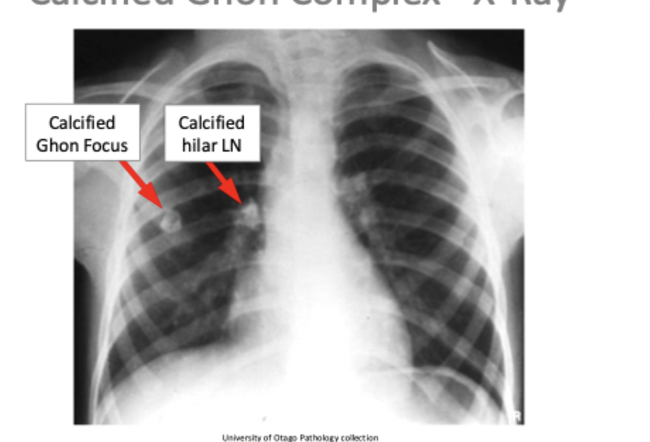
what is the gohn focus
area of primary infection in the lung that develops granulomas
what is the ghon complex
hilar lymph nodes that were involved in the first infection that develop granulomas + ghons focus
together make the ghon complex
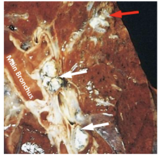
what are the red and white arrows
red arrow is the ghon focus
white arrows are the hilar lymph nodes involved in the intial infection that also develop granulomas
what are the possible outcomes after primary infection
immunity against TB
latent TB
progressive TB
describe immunity against TB as a possible outcome of the primary infection
TH1 response leads to killing of the organism
ghon complex heals by fibrosis
fibrosis followed by calcification
the pateint will then have acquired immunity against TB
describe latent TB as a possible outcome of TB
the organism may remain dormant within the intial ghon focus or in the extrapulmonary sites
it can get reactivated when the hosts immunity drops
describe progressive primary TB as a possible outcome of the primary infection
in immunosurpressed people the infection progresses and the immune resposne results in extemsive necrosis and damage
what is miliary TB
during progressive primary TB
spread of the organism can lead to TB granulmoas in multiple organs


scattered millet seed like granulomas
milliary tb
what is the result of reactivation of TB
reactivate or reinfection results in rapid mobilisation of a vigorous immune reaction
large necrotic granulomas with lots of fibrosis around them form
where do secondary lesions due to secondary TB form
in the apical parts of the lungs where oxygen tension is the highest bc mycobacterium tubercolosis is an aerobic organism
what is chronic fibrocaseous TB
secondary TB
called that due to the massive areas of central caseous necrosis surrounded by fibrsos
what is cavitary TB
secondary TB - extensive tissue destruction destroys adjacent bronchial walls
patient will then cough up bacteria laden caseous material which leaves cavities - therefore cavitary TB
does secondary TB lead to miliary TB
yes it can but not as common as it is with primary TB

what are the arrows pointing to
The left arrow is TB lesions with extensive caseation and fibrosis - fibrocaseous TB
The right arrow is the apical cavity created after coughing up caseous necrotic matieral - cavitary TB
how is TB defintively diagnosed
identifying M. tubercolosis in a clincal sample using Z-N stain or PCR
what are some observations that are used in the diagnosis of TB
cough, sputum, night fever sweat
granulomas
chest x ray
mantoux test/interferon gamma release assay
what is the treatment for active TB
2-phase treatment
intensive phase: bacteriocidal
confirmation phase: sterilization
describe intensive phase treatment
4 drugs given for 2 months
describe confirmation phase
2 drugs given for 4 months
Describe the treatment against latent disease
patient under 40 - 1 drug 9 months or 2 drugs 4 months