Lecture 12
 Call Kai
 Learn
 Practice Test
 Spaced Repetition
 Match
 Flashcards
 Knowt Play
1/41

There's no tags or description
Looks like no tags are added yet.
42 Terms
Oxyanion hole (review)
Two hydrogens not part of reaction when catalyzed on benchtop. Rate-limiting step was tetrahedral intermediate with full negative charge sitting out in air/solution. Worst thing to do with a charge is have it uncompensated. Activation energy really high for uncatalyzed reaction. 3,000 years half-life for peptide cleavage. Delta delta G of 40 kilojoules per mole. Catalyzed reaction has this. Stabilizes by a lot. Makes it 100 million times faster. Physical basis for catalysis: hydrogen bonds facilitated by oxyanion hole stabilize high-energy state, goes faster.
Michaelis-Menten summary (spatial separation of Km and Kcat)
affect Km or Kcat
Enzyme plus substrate goes to enzyme-substrate complex, times K2 gives enzyme plus product.
Two processes: binding (has Km associated) and kinetic process (velocity equals K2 times ES).
Binding part is responsible for Km—anything to do with recognition (complementary size, hydrophobic, etc.). Changes in specificity pocket affect Km (affecting affinity).
Anything to do with catalysis—fixes or changes chemistry in active site—affects K2, Kcat, or Vmax.
Spatially restricted: active site where chemistry occurs—changes affect K2/Kcat.
Anything affecting binding affects Km. Completely spatially separated. Having that mapped in your head allows answering any question about mutations.
Mutation in specificity pocket (clicker)
Correct answer: C (specificity pocket). Can tell right away because Km increased. If Km increased, you did something to binding affinity—probably happened in specificity pocket.
Two mutations affect inhibitor binding (clicker)
Correct answer: C. Decreasing affinity. Increasing Ki. Ki is a Kd, just like Km is a dissociation constant. Binding getting worse.
Type of inhibition (clicker)
Correct answer: A (competitive inhibition). Because K2 isn't affected. Competitive inhibitor competes with substrate—binds same site. Changed Km by an order of magnitude. Also changed Ki. Both affected by mutations the same way. Probably binding in same place as substrate—competitive inhibitor.
Serine proteases (comparison)
what does each bind to
Chymotrypsin recognizes large apolar molecules—cuts after large apolar molecules.
aromatic rings and methionine
Trypsin has aspartic acid in bottom of recognition pocket (as opposed to alanine in chymotrypsin). Charged amino acid recognizes side chains of lysine and arginine.
Elastase: two valines stuck in there (big methyl-containing side chains not present in trypsin and chymotrypsin)—recognizes small amino acids.
nonpolar
• Alanine (Ala) (most preferred)
• Valine (Val)
• Glycine (Gly)
• Serine (Ser)
• Leucine (Leu)
• Isoleucine (Ile)
Not entirely: putting these two amino acids in chymotrypsin doesn't make it as good as elastase. Making aspartate in bottom doesn't make it a good trypsin—get some activity, but other things are happening too, don't know what.
New serine protease "blue jays" (clicker)
Isolate new serine protease named blue jays. Has lysine at bottom of recognition pocket. Which amino acid likely to cut after? Lysine has positive charge. Recognizes and cuts after amino acids with negative charges—D and E. Exactly opposite of trypsin (trypsin has negative charge, recognizes lysine or arginine). Reversing the charge. Correct answer: D and E.
Metabolic enzymes function as
oligomers
Very few have one subunit. If functions as oligomer, take it as a clue: probably cooperative.
ATCase
full name
what does it do
substrates
structure
Aspartate transcarbamoylase
First step in pyrimidine biosynthesis. Make DNA—has purines and pyrimidines, made in roughly equal amounts. First step in making CTP. Starts with carbamoyl phosphate and aspartate.
Has 12 subunits: six catalytic subunits (C6) and six regulatory subunits (R6). -
Catalytic trimer (one visible, one behind).
Six regulatory subunits (three visible, three behind).
Small r = regulatory subunit (lowercase). Not to be confused with capital R (R state).
ATCase kinetics
(sigmoidal)
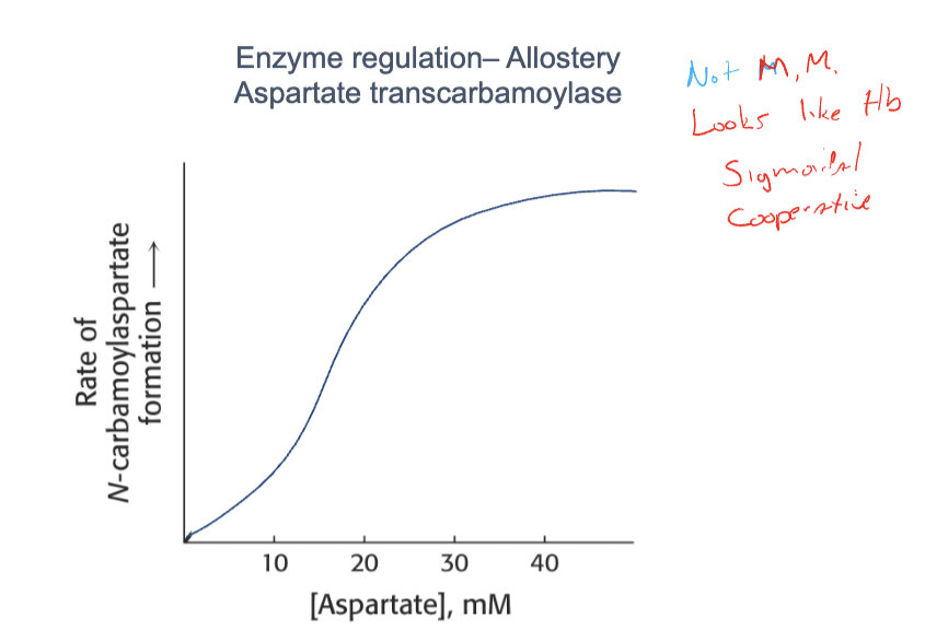
Activity as function of aspartate: not Michaelis-Menten. S-shaped, sigmoidal. Looks like hemoglobin. Means cooperative. Sigmoidal behavior because large complex has low affinity T state and high affinity R state. Like hemoglobin: low affinity state for oxygen (T) and high affinity state for oxygen (R).
Snapshot at low aspartate: all molecules in low affinity state—activity based on that state, basically none in high affinity state.
At midpoint: about half in high affinity state, half in low affinity state. Activity based on those amounts. Exactly like hemoglobin.
ATCase T and R states (Km)
Which state has higher Km? T state. Higher Km = low affinity. Read graph: 50% point gives Km of T state and Km of R state. Lower activity in T state because less substrate bound. K2 probably exactly the same in R and T states. What's different: binding ligand (aspartate) tighter in R state.
Regulatory subunit (R chain) acts
R chain acts like a ligand.
Intact protein: full protein looks like this. Remove regulatory subunit (no R chains): looks like R state. R chain removed: looks like R state. Means regulatory chain stabilizes T state.
View subunit of protein as a ligand for catalytic site—acts like ligand, stabilizes T state, pushes curve over.
r chain by itself has no activity.
r chain binds CTP (end product). CTP binds here. With CTP, decrease activity.
CTP binding (clicker)
Based on effect that CTP makes activity go down. What is true about CTP binding? Adding CTP (red) vs without (black). Correct answer: A. Looks remarkably like hemoglobin. Anything that stabilizes T state shifts curve to right. Anything that shifts curve left stabilizes R.
Feedback inhibition (definition)
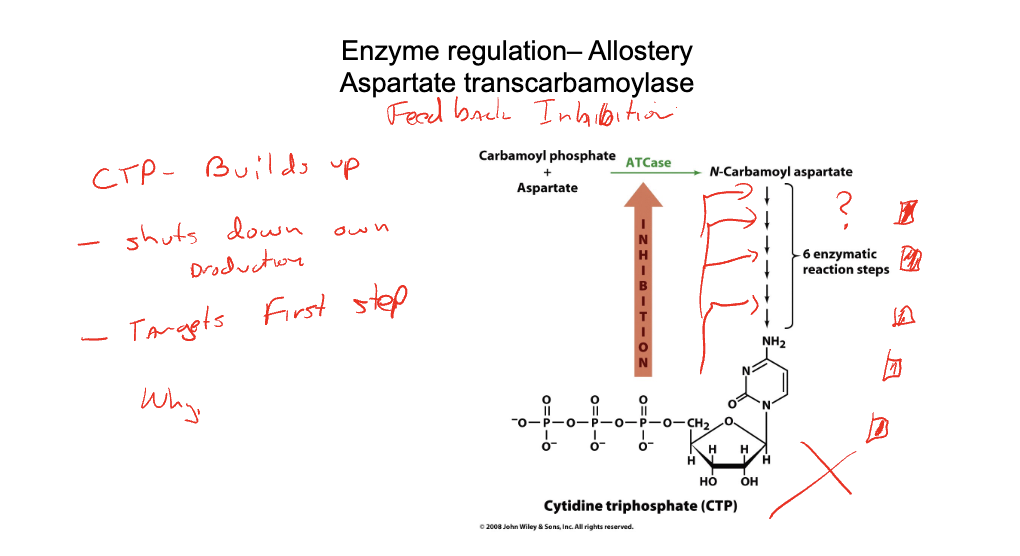
Reaction is first step in ultimate synthesis of CTP. Six more enzymatic steps.
Inhibition called feedback inhibition. CTP builds up and shuts down its own production. Makes sense—don't want a machine constantly making stuff. Find this everywhere in metabolism.
Why feedback inhibition targets the first step
Why does it go back to the very beginning? Shuts down right here, targets first step. If making piles of stuff, accumulating intermediates not used for anything, then decide at the end you don't need anymore—you have a bunch of reagents you don't need and have to break them down again. Most efficient: don't make all intermediates, go back to original stuff and say we don't need any more DNA, stop right away. Don't make even first parts.
Purine/pyrimidine coordination (clicker)
DNA purine base pairs with pyrimidine. Relative amounts of purines and pyrimidines must be tightly coordinated—don't want too much of one or the other, relative amounts precisely maintained. CTP (pyrimidine) decreases activity of ATCase, slowing pyrimidine synthesis.
What other molecule might bind ATCase, and how would activity be affected? Correct answer: B (ATP, a purine, would increase activity). If you have a lot of one, it means you have a lot of purines and need to make more pyrimidines. Putting you in position to make more. Feedback inhibition slows process when making too much of your own product. But if you have a partner thing needed alongside another thing like DNA, if you have purines, need to increase pyrimidines to bring up to that amount. That's exactly what happens.
ATCase allosteric regulation
(ATP and CTP)
r chain can bind CTP (pyrimidine) or ATP (purine).
CTP decreases activity.
ATP increases activity.
Binds same site in r chain, different effects. ATP stabilizes R state (pulls curve this way). CTP stabilizes T state (pushes curve this way). Interpret any enzyme reaction this way.
Allosteric effector table
Make table: effector, binds more tightly to T or R. Only thing different in any enzymatic system: what are the nouns—allosteric effectors, ligands that bind. Here: aspartate, CTP, ATP. Aspartate stabilizes R state (catalyzes transition to that).
CTP stabilizes T state.
ATP stabilizes R state.
Every enzyme that is an oligomer will look exactly like that. Only thing different: nouns and verbs. Behavior: things that stabilize T or R—approximate as T to R state transition, two states moved back and forth by ligands stabilizing T or R. Only thing different between systems: nouns and verbs.
Covalent modification (definition)
common type
energy
Attachment of a chemical group. Performed with enzymes.
Why do that?
Non-covalent binding allows dialing activity in (need four orders of magnitude of ligand to go from 1% to 99%).
Covalent modification gives switch-like behavior. Generally very stable. When enzyme phosphorylates something, phosphorylated form usually turns enzyme completely into T or completely into R. Locked in—go from 1% to 99%. Generally stable, tight, switch-like. Most common: phosphorylation. 30% of all enzymes/proteins are phosphorylated. Also acetylation, methylation, carboxylation, ubiquitination—slew of things.
Phosphorylation mechanism
what gets phosphorylated
characteristics after phosphorylation
Enzyme has many side chains that are serine, tyrosine, or threonine.
Distinguishing feature: hydroxyl groups (OH groups).
Kinases (some more general than others) take ATP, take end phosphate group, put it on protein, produce ADP. Phosphorylated version of enzyme. Most of the time phosphorylation makes enzyme active—but doesn't have to; can be anything. Adds a minus-two charge—imagine how that might change stability of a particular state. Introduce Coulomb interactions.
Phosphorylation reversed by phosphatase—removes phosphate group to make ortho phosphate (free phosphate). Doesn't make ATP out of ADP—decoupled that process.
Phospho bond energy
Phospho bond energy about 25 kilojoules per mole. Good amount. If bond is 100 kilocalories per mole and you want to use it going on and off, if really stable you have to have an enzyme to do that work. Want something just strong enough to keep equilibrium where you want. 25 kilojoules per mole allows on/off: unphosphorylated enzyme, R less than 1%, T greater than 99%. Phosphorylated: R greater than 99%, T less than 1%. Switch-like behavior. Don't have to have huge amount of energy expended to break interaction.
Glycogen phosphorylase (example)
Enzyme catalyzes reaction: glycogen in muscles (intermediate energy storage vehicle in cell—when you have a lot of glucose, put it together in glycogen).
Phosphorylase A (the active, phosphorylated form of the enzyme glycogen phosphorylase) catalyzes cleavage of glycogen to produce shorter chain of glycogen plus one glucose molecule. Glucose is source of sugar—need to do metabolic things to run a race. Works like classic enzyme.
Dimer—functions as dimer, which means cooperative, behaves just like ATCase.
Two states.
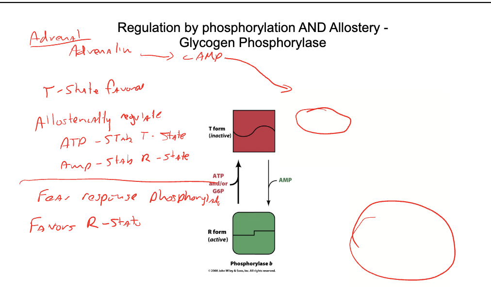
T state favored under ordinary circumstances (when not phosphorylated). Allosterically regulated. ATP stabilizes T state.
AMP stabilizes R state.
Makes sense: a lot of ATP means a lot of energy—don't need to break glycogen down, shut it off, make it T state. AMP is signal for not enough energy—turn it on. Allosteric chart: exactly same cooperative curves. Add ATP pushes to the right. Add AMP pushes to the left.
Glycogen phosphorylase what is it
Enzyme catalyzes reaction: glycogen in muscles (intermediate energy storage vehicle in cell—when you have a lot of glucose, put it together in glycogen).
catalyzes the breakdown of stored glycogen into glucose-1-phosphate by breaking α-1,4-glycosidic bonds
Glycogen phosphorylase 2
Phosphorylase A (the active, phosphorylated form of the enzyme glycogen phosphorylase) catalyzes cleavage of glycogen to produce shorter chain of glycogen plus one glucose molecule.
Glucose is source of sugar—need to do metabolic things to run a race. Works like classic enzyme.
glycogen phosphorylate structure
Dimer—functions as dimer, which means cooperative, behaves just like ATCase.
Two states.
T state favored under ordinary circumstances (when not phosphorylated). Allosterically regulated. ATP stabilizes T state.
AMP stabilizes R state.
Makes sense: a lot of ATP means a lot of energy—don't need to break glycogen down, shut it off, make it T state. AMP is signal for not enough energy—turn it on. Allosteric chart: exactly same cooperative curves. Add ATP pushes to the right. Add AMP pushes to the left.
Glycogen phosphorylase other regulation
Can also be phosphorylated. Why? If walking in woods and see a bear, you don't want to be subject to whether you've eaten enough to get energy. Want to convert all glycogen phosphorylase into active form to get away from bear. Fear response: adrenal gland produces epinephrine (adrenaline).
Glycogen phosphorylase other regulation mechanism
Cascade: adrenaline goes to cyclic AMP, goes to phosphorylase kinase.
Phosphorylates this glycogen phosphorylase.
When phosphorylated, favors R state (active state). Also turns off all regulation by ATP. If trying to get away, trying to produce ATP—don't want ATP shutting it down. Want to keep going, producing as much ATP.
Glucose can be toxic, so that allows turning it off the other way. Generally stuck on. Provides another mechanism. Normal titrate based on what you need.
If really need something, need switch-like behavior—phosphorylation turns it on immediately.
Most enzymes have two modes: normal everyday mode (dial it in precisely) and "oh my God, I got to get out of here" mode (tied to hormonal systems).
Oxyanion hole is
basis of catalysis

regulatory strategies

Enzyme regulation allostery example
Aspartate transcarbamoylase (ATCase)

Enzyme regulation– Allostery graph
ATCase

The sigmoidal behavior can be modeled as
two states in equilbrium; each state obeys M-M

r chain stabilizes T state
Enzyme regulation– Allostery 3

CTP decreases activity and targets first step
Enzyme regulation– Structural basis for cooperativity.
Aspartate transcarbamoylase

ATP stabilize R
CTP stabilize T
ATCase table

Regulation by phosphorylation kinase

serine, threonine, or tyrosine
adds -2 charge
Regulation by phosphorylation phosphotase

phosphorylation graph

phosphorylation energy and shift

switch like behavior
Regulation by phosphorylation AND allostery
Glycogen Phosphorylase

Enzyme regulation– Covalent Modification

regulation by phosphorylation AND Allostery -
Glycogen Phosphorylase