helminths: intestinal nematodes
 Call Kai
 Learn
 Practice Test
 Spaced Repetition
 Match
 Flashcards
 Knowt Play
1/29

There's no tags or description
Looks like no tags are added yet.
30 Terms
intestinal species
enterobius vermicularis (pinworm), trichuris trichiura (whipworm), ascaris lumbricoides (large intestinal roundworm), necator americanus (new world hookworm), ancylostoma duodenale (old world hookworm), strongyloides stercoralis (threadworm)
intestinal-tissue species
trichinella spiralis (trichina worm), dracunculus medinensis (guinea worm)
buccal capsule
long oral cavity aka buccal cavity
chitin
shell made of thick nitrogen-containing polysaccharide coating
copepod
freshwater fleas
copulation
mating of select worms
corticated
eggs containing a mammillated albuminous material
cuticle
surface covering present on adult nematodes
decorticated
eggs lacking an outer mammillated albuminous coating
egg
female sex cells after fertilization. vary in size & shape
embryonated
fertilized egg
filariform larva
infective, non-feeding stage that occurs after the rhabditiform larvae completes their second molt
gravid
pregnant
larva
juvenile worms. located inside fertilized eggs, emerge & continue to mature
nematode
multicellular parasites that appear round in cross section
retroinfection
infective pinworm eggs that migrate back into host body, develop, and reproduce rather than becoming dislodged
rhabditiform larva
average immature newly hatched hookworm characterized by the presence of a buccal cavity/capsule and a small genital primordium
unembryonated
unfertilized egg
zoonosis
parasite whose normal host is an animal
3 basic morphologic forms nematoda assume
eggs
larvae
adult worms
develop from maturing larvae
sexes separate (females larger than males)
have digestive & reproductive system
intestinal nematodes life cycle
individuals are similar but organism specific. fertilized adult female nematodes lay eggs in intestine and are shed in stool. mature in soil & infect new host in 2-4 weeks. most members have ability to exist independent of a host (free living)
lab diagnosis of intestinal nematodes
specimen of choice depends on organism present: cellophane tape prep, stool, tissue, infected skin ulcers. recovery of eggs, larvae, or occasional worm is necessary for diagnosis. serologic tests available
pathogenesis & clinical symptoms of nematodes
severity factors: # worms present, length of time infection persists, overall health of host. reinfection & auto-infection can also determine severity.
some asymptomatic, others have symptoms. most cause intestinal infection symptoms: abdominal pain, N/V, diarrhea, fever, eosinophilia. other symptoms: skin irritation, skin blisters, muscle involvement.
enterobius vermicularis (pinworm) egg
shape: oval, one-side flattened.
embryo: stage of development varies, may be unembryonated, embryonated, mature.
shell: double-layered, thick, colorless.

enterobius vermicularis (pinworm)
specimen is cellophane tape prep from perianal region. humans only known host, initiated following ingestion of infected eggs. pregnant female worm migrates outside body & deposits eggs. eggs can survive days to weeks outside host. retroinfection & autoreinfection can occur. found worldwide, particularly in temperate areas.

enterobiasis
pinworm infection. most common helminth to cause infection in US. infections more frequent in school or preschool children. may be responsible for transmission of Dientamoeba fragilis. intense itching, inflammation of anal or vaginal areas, difficulty sleeping, intestinal irritation, N/V

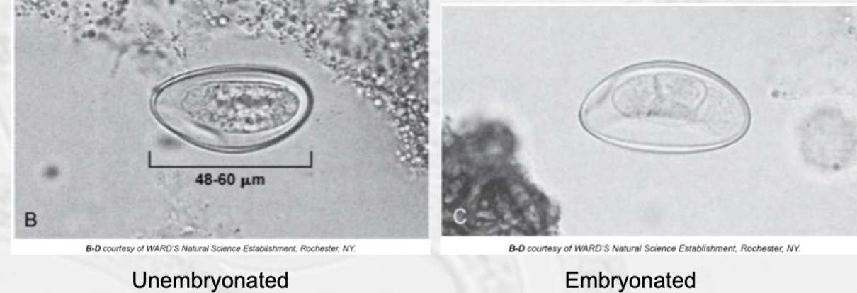
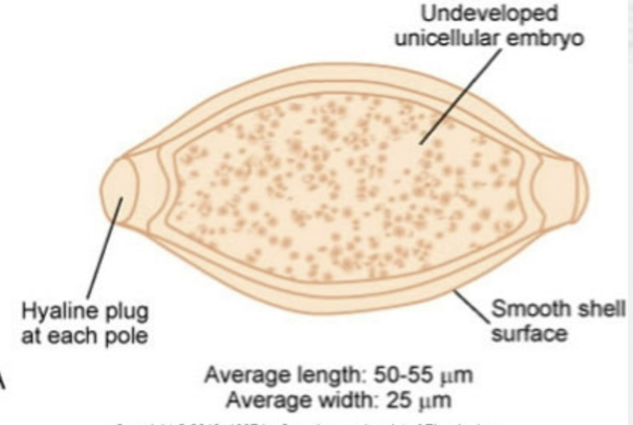
trichuris trichiura (whipworm) egg
shape: barrel, football; hyaline polar plug at each end.
embryo: unicellular, undeveloped.
shell: smooth, yellow-brown color b/c of bile contact

trichuris trichiura (whipworm) adults
size: males usually smaller than females
anterior end: colorless, ressembles a whip handle, contains slender esophagus.
posterior end: pinkish-gray, resembles whip itself, contains digestive & reproductive systems; males possess prominent curled tail.

trichuris trichiura (whipworm)
specimen: stool. eggs prominent using zinc sulfate flotation method. adults worms may be visible on macroscopic exam. ingestion of infective eggs causes human infection, larvae emerge in small intestine, adults take up residence in colon, female lays eggs following copulation which pass thru stool. 3rd most common helminth. found primarily in warm climates where poor sanitation common. children & those in psychiatric hospitals at risk. can be asymptomatic.

trichuriasis
whipworm infection.
heavy infections (500-5000 worms) have various symptoms: abdominal tenderness & pain, weight loss, weakness, mucoid or bloody diarrhea.
children who are infected present w/ symptoms similar to ulcerative colitis. as few as 200 worms can cause chronic dysentery, severe anemia, possible growth retardation. in treated children, catch-up growth is noted. increased rectal prolapse & peristalsis are common.