anatomy exam 4
 Call Kai
 Learn
 Practice Test
 Spaced Repetition
 Match
 Flashcards
 Knowt Play
1/87

Earn XP
Description and Tags
ch 19: 1-34; ch 20: 35-
88 Terms
heart location and orientation
in mediastinum
apex: left of midline, 5th intercostal space
base: 2nd intercostal space
4 corners of the heart
superior right: costal cartilage of 3rd rib and sternum
inferior right: costal cartilage of 6th rib, lateral to sternum
superior left: costal cartilage of 2nd rib, lateral to sternum
inferior left: 5th intercostal space, at midclavicular line

layers of pericardium
pericardium: triple layered sac enclosing the heart
fibrous pericardium: outermost; dense connective tissue; holds heart in place + keeps it from overfilling
serous pericardium: parietal layer (attached to inner layer of fibrous pericardium), visceral layer (epicardium, part of heart wall)
parietal cavity: contains serous fluid in between

layers of heart wall
epicardium: visceral layer of serous pericardium
myocardium: cardiac muscle, arranged in circular and spiral patterns
endocardium: endothelium resting on a layer of connective tissue, lines internal walls of heart and covers valves

external landmarks of the heart
coronary sulcus
anterior interventricular sulcus
posterior interventricular sulcus
blood vessels feed heart, deliver nutrients and oxygen, and take away co2 and wastes
right atrium
receives deoxygenated blood from body through: superior vena cava, inferior vena cava, coronary sinus
pectinate muscles: increase contraction power
crista terminalis: landmark used to locate veins entering right atrium
fossa ovalis: remnant of foramen ovale in fetal heart
right ventricle
receives blood from right atrium, pumps blood to pulmonary trunk to right and left pulmonary arteries
chordae tendineae: prevents inversion of tricuspid (and biscupid) valves
papillary muscles: pulls on chordae tendineae
trabeculae carneae: network of muscles attached to papillary muscles
left atrium
makes up heart’s posterior surface
receives oxygenated blood from lungs through right and left pulmonary veins
pumps blood to left ventricle
left ventricle
forms apex of heart; most muscular (pumps blood to entire body)
internal walls are same as right ventricle (trabeculae carneae, chordae tendineae, papillary muscles)

heart valve structure
composed of endocardium; connective tissue core
atrioventricular valves: prevents backflow of blood from ventricles into atrium; closing produces first sound from heart (“lub”)
semilunar valves: prevents backflow of blood from pulmonary trunk to ventricles; closing produces second sound from heart (“dub”)

right atrioventricular valve
tricuspid valve; best heard at inferior right corner (5th intercostal space, right sternal margin)

left atrioventricular valve
bicuspid (mitral) valve; best heard at apex
in 5th intercostal space + midclavicular line

aortic valve
semilunar valve; best heard at superior right corner (2nd intercostal space, right sternal margin)

pulmonary valve
semilunar valve; best heard at superior left corner (2nd intercostal space, left sternal margin)
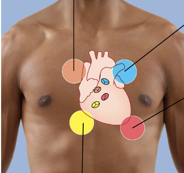
cardiac skeleton
surrounds all 4 valves; composed of dense connective tissue
anchors valve cusps
prevents overdilation of valve openings
main point of insertion for cardiac muscle
blocks direct spread of electrical impulses

blood flow pathway
deoxygenated blood enters superior/inferior venae cavae
enters right atrium
passes tricuspid valve into right ventricle
pumped to pulmonary trunk, enters right/left pulmonary arteries
deoxygenated blood enters lungs, becomes oxygenated
oxygenated blood enters left atrium through right/left pulmonary veins
passes bicuspid valve into left ventricle
pumped to aorta, into systemic circuit

heartbeat
70-80 bpm at rest
systole: contraction of heart chamber
diastole: relaxation of heart chamber
systole and diastole can also refer to: contraction and relaxation of ventricles, blood pressures
heart wall structure
atria - thin walls; ventricles - thick walls
left ventricle: has to pump to systemic circuit (entire body); 3x thicker muscle than right ventricle (more pumping force)

cardiac muscle tissue
forms myocardium
striated, short, branching, uni- or bi-nucleate, intercalated discs
contracts by sliding filament mechanism
cells separated by endomysium; binds adjacent cardiac fibers and contains blood vessels and nerves
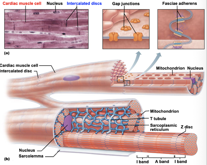
intercalated discs
interlock adjacent sarcolemmas; 2 distinct regions
fasciae adherens: long desmosome-like junctions (interlock)
gap junctions: allow ions to pass between cells and transmit contractile signal

conducting system of the heart
specialized cardiac muscle cells conduct electrical impulses through myocardium
pace of inherent rate of contraction set by sinoatrial (SA) node
conducting system of the heart - pathway
sinoatrial (SA) node → through internodal fibers → atrioventricular (AV) node
atrioventricular (AV) node → through cardiac bundle → atrioventricular (AV) bundle
atrioventricular (AV) bundle→ bundle branches aka Branches of His → apex
bundle branches → Purkinje fibers (subendocardial network) → back up though ventricles
innervation of conduction system
visceral sensory, parasympathetic, sympathetic (all pass through cardiac plexus)
parasympathetic: vagus n. branches; decrease heart rate, increase vasodilation; restricted to SA node, AV node, coronary arteries
sympathetic: to heart from cervical and upper thoracic chain ganglia; innervate same areas as parasympathetic fibers + cardiac musculature throughout heart; increase heart rate, strength of contraction, vasoconstriction
autonomic input of conduction system
controlled by cardiac centers on reticular formations of medulla
cardioinhibitory center: influences parasympathetic neurons
cardioaccelatory center: influences sympathetic neurons

coronary arteries
blood supply to muscular walls and tissues of the heart itself; 2 branches from aorta
right coronary artery, left coronary artery
both run in coronary sulcus
left coronary artery (LCA)
arises from left side of aorta, passes posterior to pulmonary trunk and divides into 2 branches
circumflex artery: follows coronary sulcus posteriorly; supplies left atrium + posterior part of left ventricle
anterior interventricular artery: aka left anterior descending artery; descends towards apex of heart; supplies interventricular septum + anterior walls of both ventricles

right coronary artery (RCA)
arises from right side of aorta, descends in coronary sulcus on anterior surface (between right atrium and right ventricle)
marginal artery: branches from inferior border of RCA
posterior interventricular artery: aka posterior descending artery; emerges from posterior part of coronary sulcus

cardiac veins
carry deoxygenated blood from heart wall to right atrium; occupy sulci on heart’s surface

coronary sinus
largest cardiac vein; runs in posterior part of coronary sulcus
returns majority of venous blood from heart to right atrium
3 tributaries: great cardiac v., middle cardiac v., small cardiac v.
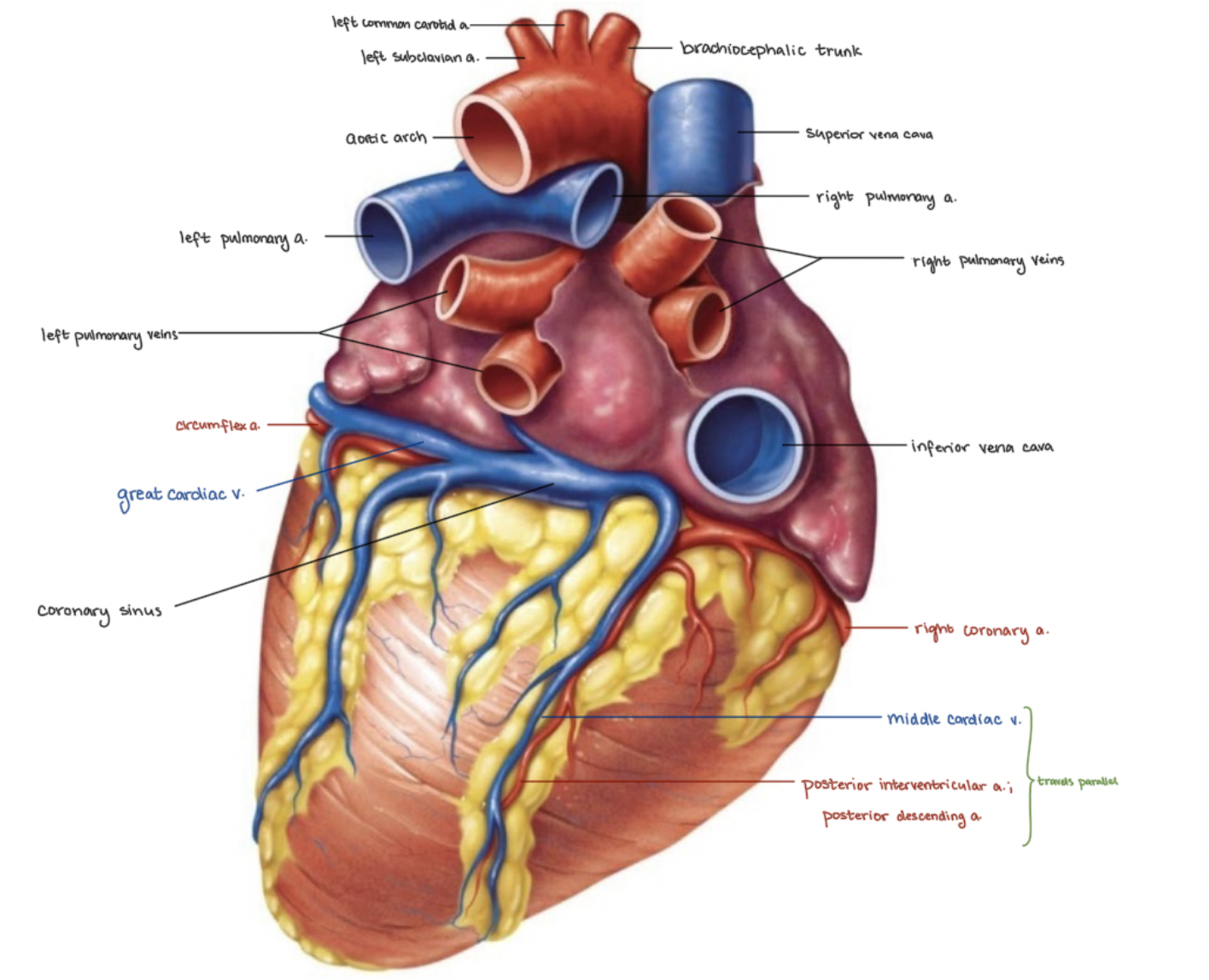
coronary artery disease (CAD)
caused by atherosclerosis: accumulation of fatty deposits in inner lining of body’s arteries
common symptom: thoracic pain; caused by inadequate oxygenation of heart muscle cells which weaken but don’t die
myocardial infarction: aka heart attack; prolonged or complete blocked coronary artery
silent ischemia: silent but fatal heart attack; 1st and only clear symptom of coronary artery disease
heart failure
progressive weakening of heart
cannot meet boy’s demands for oxygenated blood
arrhythmias
variation from normal heart rhythm; called fibrillation
2 main types: ventricular fibrillation, atrial fibrillation
ventricular fibrillation
rapid, random firing of electrical impulses in ventricles → ventricles are unable to pump blood into arteries
results from crippled conducting system
most common cause of cardiac arrest and sudden death in patients with hearts damaged by CAD
atrial fibrillation (A-fib)
multiple waves of electrical impulses circle within atrial myocardium → random stimulation of AV node → signals ventricles to contract quickly and irregularly
promotes formation of clots due to lack of smooth blood flow through heart → leads to strokes
occurs in episodes; characterized by anxiety, fatigue, shortness of breath, palpitations
often associated with CAD or heart-valve disease
types of blood vessels
arteries: carry blood away from heart
arterioles: smallest arteries
capillaries: smallest blood vessels
venules: receive blood from capillaries
veins: receive blood from venules
tunics that form arterial or vein walls
3 layers
tunica intima
tunica media
tunica externa
lumen: central blood-filled space of a vessel

tunica intima
innermost tunic
contains the endothelium (composed of simple squamous epithelium)
vessels that are less than 1mm also habe a subendothelial layer
tunica media
sheets of smooth muscle
contraction = vasoconstriction
relaxation = vasodilation
tunica externa
composed of connective tissue
mostly collagen and elastic fibers

elastic arteries
largest arteries (1cm-2.5cm diameter)
includes aorta and its major branches
sometimes called conducting arteries
high elastic content dampens surge of blood pressure
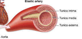

muscular arteries
lie distal to elastic arteries; diameter ranges from 0.3cm-1mm
includes most named arteries
tunica media (smooth muscle) is thick
internal and external elastic membrane


arterioles
smallest arteries; diameter from 10micrometers-0.3mm
tunica media contains 1 or 2 layers of smooth muscle cells
larger arterioles posses all 3 tunics
diameter controlled by local factors in tissues, sympathetic nervous system

capillaries
smallest blood vessels; diameter from 8-10 micrometers
red blood cells pass through in a single line
composed of 1 layer f endothelial cells (simple squamous) surrounded by basement membrane
renew and refresh the surrounding tissue fluid of cells
deliver o2 and nutrients; remove co2 and wastes
site-specific functions of capillaries
lungs: o2 enters blood, co2 leaves
small intestines: receive digested nutrients
endocrine glands: pick up hormones
kidneys: remove nitrogenous wastes
3 types: continuous, fenestrated, sinusoids

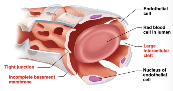
continuous capillaries
most common types; occurs in most organs
least permeable- tight junctions and desmosomes join epithelial cells; block passage of small molecules but don’t surround whole perimeter of endothelial cells
intercellular clefts: gaps of unjoined membranes; allow small molecules in and out of capillaries
strengthened and stabilized by scattered pericytes external to endothelial cells

fenestrated capillaries
joined by tight junctions and desmosomes; contain intercellular cells
pores (fenstrations) in endothelium
occur only where high rates of exchange occur- small intestine, glomeruli of kidneys, endocrine glands

sinusoid capillaries (sinusoids)
follow a twisted path; expanded large diameter and narrowed regions
most permeable (wide, leaky) because: usually fenestrated, fewer cell junctions in endothelial cells, open intercellular clefts
occurs where there is an extensive exchange of large materials (proteins, cells) between blood and surrounding tissue (e.g. bone marrow, spleen)
capillary permeability
molecules pass in and out through 4 routes
direct diffusion across endothelial cell membranes
intercellular clefts
fenestrations
pinocytotic vesicles


low permeability capillaries - blood brain barrier
continuous capillaries with complete tight junctions
no intercellular clefts
vital molecules (e.g. glucose) brought through by highly selective transport membranes in plasma membranes of endothelial cells
not a barrier against uncharged and lipid-soluble molecules (o2, co2, some anesthetics)

capillary beds
network of capillaries running through tissues
precapillary sphincters: regulate flow of blood to tissues
tendons and ligaments are poorly vascularized
epithelia and cartilage are avascular (receive nutrients from nearby connective tissue)

venous vessels (veins)
conduct blood from capillaries toward heart
blood pressure is much lower than in arteries
venules: smallest veins; diameter from 8-100 micrometer; smallest venules called postcapillary venules
venules join to form veins
structural differences of veins from arteries
lumens are larger (65% of body’s blood in veins at any given time)
tunica externa is thicker
less elastin in walls
walls are thinner than comparable arteries
mechanisms to counteract low venous pressure
valves in some veins
particularly in blood
prevent backflow
not located in veins of thoracic and abdominal cavities
skeletal muscle pump
muscles press against thin-walled veins
vascular anastomoeses
interconnection of vessels
organs receive blood from more than 1 arterial source
neighboring arteries form arterial anastomoses
provide collateral channels
many along smaller branches of coronary artery
poorly developed in kidneys, spleen, part of bone diaphyses closest to epiphyses, central artery of retine
veins anastomose more freely than arteries
pulmonary circuit - pulmonary trunk
brings blood to and from lungs
pulmonary trunk: large artery that leaves the right ventricle
divides into right and left pulmonary arteries
divides into several lobar arteries- 3 in right lung, 2 in left
becomes pulmonary arterioles
then becomes pulmonary capillaries that surround lung alveoli (small air sacs)
pulmonary circuit
newly oxygenated blood enters venules and then progressively larger veins
superior/inferior pulmonary veins exit medial aspect of each lung; carry oxygenated blood into left atrium
overall vessels of pulmonary circuit- thinner walls than systemic, max arterial pressure is lower in pulmonary circuit
systemic circulation - pulse points
arterial pulses can be palpated in muscular arteries
can be used to determine heart rate + assess blood flow to a body region after trauma, surgery, or disease
aortic arch - vessels
aorta: largest artery in body; 3 parts
ascending aorta: from left ventricle; branches into right/left coronary a.’s
aortic arch: posterior to manubrium; branches into brachiocephalic trunk, left common carotid, left subclavian a.
descending aorta: continues from aortic arch; thoracic aorta (through diaphragm, T5-T12 region), abdominal aorta (ends at L4)
abdominal aorta divides into right/left common iliac a.’s - supply pelvis and lower limbs
arteries of head and neck
common carotid
right common carotid: originates from brachiocephalic trunk
left common carotid: originates from aortic arch
branches of subclavian arteries
vertebral a.’s
thyrocervical trunk
costocervical trunk
common carotid arteries
located in anterior triangle of neck
2 branches:
external carotid a.
internal carotid a.
external carotid artery - branches
superior thyroid a.
lingual a.
facial a.
occipital a.
posterior auricular a.
superficial temporal a.
maxillary a.
internal carotid artery - branches
ophthalmic a.
anterior cerebral a. - anastomoses with anterior communicating a.; forms part of cerebral arterial circle
middle cerebral a.
subclavian arteries
right subclavian a. - originates from brachiocephalic trunk
left subclavian a. - arises as 3rd branch from aortic arch
supplies brain (vertebral a.’s)
supply neck and thoracic wall - thyrocervical trunks, costocervical trunks, internal thoracic a.’s
vertebral arteries
supply posterior brain
right/left vertebral a.’s join to become basilar a.
basilar a. branches into 2 posterior cerebral a.’s
posterior communicating a.’s connect posterior cerebral a.’s to middle cerebral a.’s
cerebral arterial circle (circle of Willis)
forms anastomosis between internal carotid and vertebral a.’s
provides alternate routes for blood to reach brain areas if either becomes occluded (blocked)
thyrocervical trunk
sends 2 branches posteriorly over scapulae
sends 1 branch to inferior part of thyroid gland
costocervical trunk
sends a branch superiorly to deep muscles of neck
sends a branch inferiorly to intercostal spaces
arteries of the thorax
internal thoracic a.’s
branch from subclavian a.’s
supply anterior thoracic walls
anterior intercostal a/’s
branches of internal thoracic a.’s
posterior intercostal a.’s
inferior 9 pairs arise from thoracic aorta
arteries of the upper limb
arise from subclavian a.
axillary a. descends through axilla, branches into:
thoracoacromial a.
lateral thoracic a.
subscapular a.
anterior/posterior circumflex humerus a.’s
axillary a. becomes brachial a. at inferior border of teres major
used in measuring bp with sphygmomanometer
major branch - deep artery of arm (deep brachial a.); serves triceps
arteries of the forearm
axillary a. divides into radial a. and ulnar a. at cubital fossa
branches of radial and ulanr a.’s join, form 2 horizontal arches
superficial/deep palmar arches
digital a.’s - branch from superficial/deep palmar arches; supply fingers
arteries of abdominal aorta - midline branches
about half of the entire arterial flow is present in these vessels
3 midline branches bring blood to digestive tract:
celiac trunk
superior mesenteric a.
inferior mesenteric a.
arteries of the abdominal aorta - all branches
(from superior to inferior)
inferior phrenic a.’s
celiac trunk
superior mesenteric a.
suprarenal a.’s
renal a.’s
gonadal (testicular or ovarian) a.’s
inferior mesenteric a.
lumbar a.’s
median sacral a.
common iliac a.’s
arteries of the pelvis and lower limbs
at sacroiliac joint level (on pelvic brim); common iliac a.’s branch into 2
internal iliac a. - mainly supplies pelvic organs
external iliac a. - supplies the lower limb
external iliac artery - branches
(superior to inferior)
femoral a.
popliteal a. - branches into:
anterior tibial a.
posterior tibial a.
fibular a. - branches from posterior tibial a.sy
systemic veins
important differences in a. and v. distributions
1) 3 major veins enter right atrium: superior/inferior venae cavae, coronary sinus
2) all medium and large arteries hav deep location accompanied by deep veins (with similar names, e.g. radial a. and radial v.)
superficial v.’s lie beneath skin unaccompanied by any arteries in some regions (e.g. median cubital v.)
3) 2+ parallel veins drain a body region rather than 1 larger vein
multiple veins anastomoses tp form a venous plexus in some regions (e.g. dorsum of hand)
4) unusual patterns of venous drainage
brain: dural venous sinuses (dura mater endothelium-lined channels)
liver: hepatic postal system (digestive sub-circulation)
venae cavae and tributaries - arterial system
superior vena cava
return blood to right atrium from regions superior to diaphragm
arises from union of right/left brachiocephalic v.’s
brachiocephalic v.’s - formed by union between internal jugular v. and subclavian v.
tributaries
fill in
veins of the head and neck
most brain v.’s drain into intracranial dural venous sinuses
form an interconnected series of channels in skull
lie between 2 layers of cranial dura mater
superior/inferior sagittal sinuses
straight sinus
transverse sinuses
sigmoid sinus - becomes internal jugular v. as it leaves skull through jugular foramen
internal/external jugular v.’s
internal jugular veins
drains blood from brain
lies lateral to internal carotid a. (superiorly)
lies lateral to common carotid a. (inferiorly)
joins subclavian v. to create brachiocephalic v.
external jugular veins
tributaries that drain posterior scalp, lateral scalp, and some of face
not accompanied by corresponding a.
empty into subclavian a.
veins of the thorax
brachiocephalic v.’s - receives blood draining from 1st few intercostal spaces
azygos system - group of v.’s; receives blood from other intercostal spaces + some thoracic viscera
azygos v. - joins superior vena cava
hemiazygos v.
accessory hemiazygos v.
veins of the upper limb - deep veins
follow paths of companion a.’s
have same names
all v.’s (except the largest) are 2 parallel v.’s that flank their a. on both sides
veins of the upper limb - superficial veins
visible beneath skin; form anastomoses frequently along course
cephalic v.
basilic v.
medial cubital v. - used to obtain blood or administer IV fluids
median v. of forearm
venae cavae and tributaries - venous syste,
inferior vena cava
widest blood vessel in body
returns blood to right atrium from body regions inferior to diaphragm
arises from union of 2 common iliac v.’s
veins of the abdomen
v.’s from paired abdominal organs, pelvis, and abdominal wall drain directly into inferior vena cava
blood from digestive organs returns via hepatic portal system
most share names of corresponding a.’s
lumbar v.’s
gonadal (testicular or ovarian) v.’s
renal v.’s
suprerenal v.’s
hepatic v.’s