Skin, Hair, and Nails Lecture
 Call Kai
 Learn
 Practice Test
 Spaced Repetition
 Match
 Flashcards
 Knowt Play
1/84

There's no tags or description
Looks like no tags are added yet.
85 Terms
Inspection and palpation
The techniques used to simultaneously assess the skin, hair, and nails
Color awareness acknowledges that injuries may appear differently in certain skin tones, color blindness is ignoring this fact
The difference between color awareness and color blindness
Natural when available
Which is more preferable, natural or fluorescent light?
Albinism
Lack of melanin can lead to this variation in skin color
Vitiligo
Melanocytes being destroyed due to an autoimmune disorder, appears as white/lighter patches on skin
Melasma
Due to hormonal changes and worsened with exposure to sunlight, also known as chloasma or “mask of pregnancy” as
Hemosederin staining
Blood pooling in lower extremities and staining them red
Striae
Normal finding, stretch marks, start out red and fade to silver color
Cyanosis
Due to lack of oxygen, grayish blue on light skin and ashen gray on darker skin
Looking at mucous membranes
How can you better inspect cyanosis on darker skin?
Eccyhymosis
Bruise, appears dark red, purple, yellow, or green in light skin and deeper bluish or black in dark skin
Palpation and ask if tender
How can you better assess a bruise in darker skin tones?
Erythema
Redness, appears red in light skin and deep brown or purple in dark skin
Cellulitis
Infection of the skin, temperature increases
Excoriation
Abrasion or scratch, causes loss of epidermis
Pallor
Paleness, whiter skin in light skin and lighter, more yellowish brown or ashen in dark skin
In the conjunctiva of the eye
How can you better assess pallor in dark skin?
Petechiae
Pinpoint bruises from broken capillaries, reddish purple in light skin and dark skin

Oral mucosa or sclera
How can you better assess for petechiae in darker skin tones?
Jaundice
Excess bilirubin and common in liver failure, yellowish color in light skin and yellowish-green in darker skin
Sclera of the eye, palms of hands, soles of feet
How can you better assess for jaundice in darker skin tones?
Palpation, patient will present with similar symptoms
How can you better assess for a rash in darker skin tones?
Urticaria/Hives/Wheals
Usually caused by allergy, can be treated with antihistamines, can be palpated on skin

Palpation, patient will state it itches
How can you better assess hives on darker skin tones?
Nevi (mole)
Located in sun exposed areas typically less than 5 mm
Papule
Raised lesions or mole
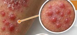
Macule
Flat mole, non-palpable change in skin tone (freckle)

A - asymmetry or symmetric
B - border is defined or undefined
C - color is uniform or different colors
D - diameter/size (less than 5 mm is ideal)
E - elevated or flat, any changes?
F - what does it feel like to pt
What does the ABCDEF model assess for in moles? Highlighted is normal finding
Use sunscreen, no tanning beds, wear hats and sun protection
What are ways that we can promote and primarily prevent melanoma?
Basal cell carcinoma (BCC)
Most common skin cancer, not metastatic
Squamous cell carcinoma (SCC)
2nd most common skin cancer, metastatic
If its unilateral
When is a difference in temperature of skin concerning?
Dorsal
Which part of the hand best palpates temperature?
Dry but not excessively dry
How should the skin feel in terms of moisture?
Diaphoresis
Excessive moisture on skin abnormal with absence of activity, can indicate low sugar
More thin and loses elasticity
As we age, the skin gets…
Acanthosis nigricans
Increased thickness in the skin causing discoloration, due to insulin resistance and NOT poor hygiene
Turgor
Testing hydration, pinching forearm or clavicle and assessing if tenting is or is not present
Clavicle
Where should you assess turgor on an older adult?
Alopecia
Hair loss due to autoimmune disease or anemia, male pattern baldness and patches of balding
Alopecia Areata
Hair loss due to follicles being chronically inflamed, affects any gender
Hirsutism
Too much unwanted hair due to hormonal changes, i.e. women with PCOS have hair on upper lip
Pediculosis
Head lice
Seborrheic dermatitis
Dandruff, common in light skin, cured with certain shampoo
Melanonychia
Dark, longitudinal lines in nails

Beau’s lines
Transverse groove in nails caused by trauma
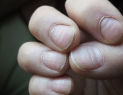
Koilonychia
Spoon nail, typically caused by anemia

Leukonychia
White spots on nails caused by trauma
Clubbing
Angle of 180 degrees or greater at nailbed, sign of chronic hypoxemia
Paronychia
Infected hangnail, redness, swelling, and pus can all be present, often happens with nail biters of frequent exposure to moisture
Onychomycosis
Fungus in the nails, becomes thick, yellow, and brittle

Ingrown toenail
Nail grows laterally into the skin
Acrocyanosis
Blue or gray discoloration of the hands and feet found in newborns, normal
Acrocyanosis, jaundice, melanocytosis, patch and vascular birthmarks
Expected findings in newborns
Melanocytosis
Increased melanocytes in newborns, fades with age
Patches
Large, round/oval birthmarks that are flat and pigmented

Nevus Simplex (Stork bites)
Vascular birthmark, can be caused by dilation or abnormal growth of blood vessels, fades with age, small red or pink spot seen on the back of neck or eyelids, disappears usually with age

Nevus flammeus, infantile hemangioma, cavernous hemangioma
Abnormal findings in newborns
Nevus flammeus (port wine stain)
Large, bluish/purple macule along 5th cranial nerve on face in newborns, does not disappear by itself and laser treatment may be able to remove

Infantile hemangioma (Strawberry hemangioma)
Slightly raised red spot that typically disappears with age in newborns but may require laser treatment
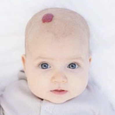
Cavernous hemangioma
More problematic reddish/blue mass of blood vessels located on newborns, may need surgery if it affects eyesight or breathing

Milia (Milk spots)
Small papules on cheeks, nose, chin, and forehead of newborn that should disappear within 3 weeks of birth

Erythema toxicum neonatorum (baby acne)
Common rash located on newborns that goes away within 2 weeks, can be anywhere except palms of hands or soles of feet

Lanugo
Newborn hair that is fine and soft, most common on the back but can also be found on scalp, ears, and shoulders
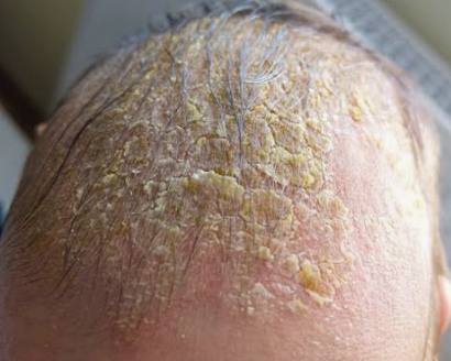
Infantile Seborrheic Dermatitis (Cradle cap)
Crusty, oit scales on scalp in newborns (can also be on ears, nose, and groin), disappears over time
Serous
Vesicles are filled with this type of fluid
Purulent (pus)
Pustules are filled with this type of fluid
Seborrheic Keratoses (SK)
Noncancerous benign growth on older adults that is rough and elevated, typically tan, brown, or black in color, can appear anywhere

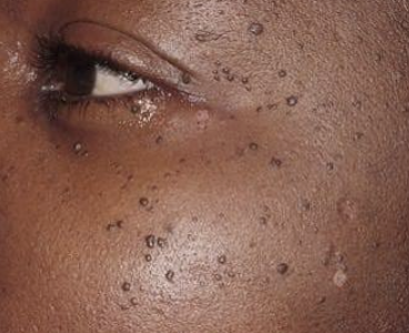
Dermatosis Papulosa Nigra (DPNs)
Variant of SK but is smaller (typically 1-5 mm), not moles, seen in sun exposed areas
Solar lentigo
Flat patches or sun spots on older adults, round or irregular

Acrochordon (Skin tags)
Fleshy, benign lesions in older adults
Multiple bruises in different stages, located in areas that aren’t typically fallen on
When assessing for bruises, what is a sign they are products of abuse?
Skin tears
Risk in older adults with thinner, more fragile skin
Geri sleeves
Sleeves that help to prevent skin tears
Areas with excessive pressure
Where do pressure ulcers happen?
Heels, sacrum, along back, back of the head
Most common areas for pressure ulcers when pt is in supine position
Hip
Most common area for pressure ulcer if patient is laying on the side
Blanch
White spot appears when pressing down on skin
Stage 1
Pressure ulcer that is nonblanchable but skin is still intact
Stage 2
Pressure ulcer that has partial thickness skin loss, shallow and open ulcer, no slough
Stage 3
Pressure ulcer where the subcutaneous fat is visible but no bone, tendon, or muscle is visible yer, presence of purulent drainage (slough)
Stage 4
Pressure ulcer where bone, tendon, and muscle are exposed, slough and escar are present
Kennedy Terminal Ulcer (KTU)
Pressure ulcer that progresses rapidly and is associated with terminally ill patients, typically seen on sacrum
Unstageable
Pressure ulcer that has too much slough or escar present to see into the ulcer
Deep Tissue Injury (DTI)
Pressure ulcer that looks like a bruise but is really deep tissue damage