ANPS 1190 Exam 3
 Call Kai
 Learn
 Practice Test
 Spaced Repetition
 Match
 Flashcards
 Knowt Play
1/269

There's no tags or description
Looks like no tags are added yet.
270 Terms
Skeletal muscles are connected to the CNS by
peripheral nerves
Peripheral nerve structure
collection of axons in the PNS, there is endoneurium, perineurium, and epineurium, and blood vessels
Endoneurium
CT layer surrounding a single axon and its myelin sheath
Peruneurium
CT layer surrounding a fascicle (bundle of axons)
Epineurium
CT layer surrounding an entire nerve
Peripheral nerves carry info
into and out of the CNS

Sensory neurons
some axons in a peripheral nerve belong to sensory neurons, sensory neurons are pseudodiploid in shape, the cell body is outside the CNS is a ganglion, sensory neurons collect info from the periphery and carry it into the CNS, info is collected by a specialized sensory receptor, which stimulates action potentials in the sensory neuron, sensory neurons send signals to the CNS and ultimately the brain through an afferent pathway

Motor neurons
another type of axon in the peripheral nerve belongs to a motor neuron- multipolar in shape, the cell body and dendrites are in the CNS (spinal cord), motor neurons send signals from the CNS to targets in the “periphery” (outside of the CNS), target cells include skeletal muscle, smooth muscle, and glands

Skeletal muscles are innervated by branches of peripheral nerves
somatic sensory and somatic motor axons are bundled together to innervate skeletal muscle, somatic sensory neurons collect info from muscle receptors about ,muscle activity and convey it to the CNS, somatic motor neurons send commands telling the skeletal muscle cells to contract, they release Ach at the neuromuscular junction on individual cells

Cranial and spinal nerves connect the periphery to the CNS
31 pairs of spinal nerves connect the spinal cord to the periphery (limb/trunk region), 12 pairs of cranial nerves connect the brain to the periphery (head/neck)
Spinal nerves connect to the spinal cord through
dorsal and ventral roots

Dorsal roots
axons of sensory neurons enter the spinal cord through dorsal roots, which enters the dorsal portion of the spinal cord
Dorsal root ganglion
cell bodies of sensory neurons are clustered in a dorsal root ganglion which lies just outside the vertebral column (spine)
Ventral root
axons of motor neurons leave the ventral portion of the spinal cord through the ventral root. Cell bodies of motor neurons are in the spinal cord
Spinal nerves
forms when the dorsal root and ventral root merge and thus contain both sensory and motor axons
The CNS has an internal pattern of
gray and white matter, in cross sanctions of the spinal cord, you can see a central area (butterfly shape) surrounded by a white area, this white area is called WHITE MATTER and the darker central area is called GRAY MATTER, although only the spinal cord has a central gray butterfly shape, anywhere we look in the CNS we see areas of gray matter and areas of white matter

Grey matter
dendrites, cell body and terminals have no myelin
White mater
myelinated axons and oligodendrocytes are found in white matter, myelin has a white color in unstained tissue due to its high lipid content
The white mater of the spinal cord contains
tracts, collections of myelinated axons traveling in the longitudinal plane (vertically), there are sensory and motor tracts that are symmetrical
Sensory tracts
some groups of axons carry info up to the brain (Ascending/afferent)
Motor tracts
other groups of axons carry info down from the brain (descending/motor)
The grey matter is divided into function areas
sensory axon synapses in gray matter in the dorsal half of the spinal cord; this gray matter is called the DORSAL HORN which contains small and large neurons, astrocytes, and few axons with oligodendrocytes, motor neurons reside in gray matter in the ventral half of the spinal cord, these are the VENTRAL HORNS, they contain large motor neurons, small local neurons, astrocytes, and few axons with oligodendrocytes, there is an intermediate zone between dorsal and ventral hors where ANS neurons are located

Different regions of the spinal cord have different ratios of
white and gray matter

The CNS is surrounded and protected by the three layers of
Meninges, pia, arachnoid, and dura matter, there is no CT within the CNS to hold tissue together, support for CNS derives from 2 tissue layers outside CNS, dura transitions to epineurium at the spinal nerve
Pia Mater
adhered to surface of brain and spinal cord; it cant be peeled off
Arachnoid mater
transparent, delicate layer external to pia and separated from the pia by a small space this SUBARACHNOID SPACE is filled with a fluid called CSF
Dura mater
is a tough collagen rich tissue layer external to the arachnoid; because of its toughness, it provides the most protection for the CNS tissue
The spinal cord is protected by
bone

There is segmental organization to most of the NS

There are 31 pairs of spinal nerves, so there are 31 spinal segments

Each spinal segment forms a pair of
spinal nerves (right and left)

Each spinal segment innervates a
dermatome and a myotome, each spinal segments innervates skin, muscle and joints that developed adjacent to its early development
Dermatome
the band of skin innervated by a pair of spinal nerves

Myotome
the skeletal muscle cells innervated by a pair of spinal nerves

The adult spinal cord ends at the
L1 vertebra

Cauda equina
the collection of dorsal and ventral roots at the end of the spinal cord, dorsal and ventral roots leaving the lower spinal cord region create a mass of stringy nerves called the cauda equina. Bathed in a large pool; of CSF inside the subarachnoid space, we can collect CSF from here without damaging the spinal cord tissue
Conus medullaris
the end of the spinal cord tapers to a point; this region is called conus medullaris which contains sacral and coccygeal spinal segments
Filum terminale
a strand of pia mater called fiulum terminale extends from the tip of conus medullaris to the bottom of the dura in the sacrum, this helps anchor the spinal cord to the coccyx
Spinal segments and nerves innervate different regions of the body
innervation means to supply with nerves

The cervical and lumbosacral enlargements are their regions of controlling limbs

Most spinal nerves contribute to a nerve plexus
axons from spinal nerves merge with adjacent segments to form a nerve plexus, there are 4 plexuses; cervical, brachial, lumbar and sacral
The cervical plexus
formed from axons in C1-C4 spinal nerves, innervates skin and anterior spine muscles of neck, most important brach is the PHRENIC NERVE from C3-5 levels to the diaphragm
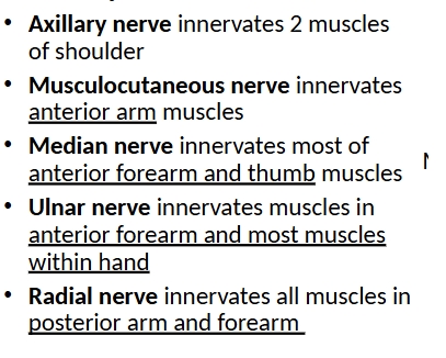
The brachial plexus- formed from axon in C5-T1 spinal nerves, plexus forms under clavicle where it can be damaged by broken bone, branches of plexus innervate skin and muscles of upper limp (exopect tapezius), forms 5 major nerves of limp, axillary, musculocutaneous, median, ulnar, and radial
The lumbar plexus
formed from axons in L1-L4 spinal nerves, plexus forms in abdominal cavity on posterior wall, branches of plexus innervate skin and muscles of anterior and medial thigh and skin of medial leg, femoral nerve, obturator nerve

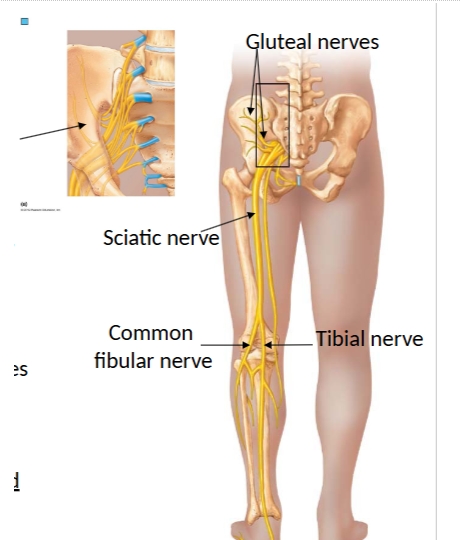
Sacral plexus (lumbosacral plexus)
formed from ventral rami of L4-S3 spinal nerve, plexus forms in pelvis on anterior side of sacrum, branches of plexus innervate skin and muscles of gluteal region, posterior thigh, leg and foot. Gluteal nerves, sciatic nerve: tibial and common fibular nerve
Joints (articulations)
allow for movement in an overwise rigid skeleton, the point of contact between two bones, holding the skeleton together, stability and mobility, mobile joints are the weakest parts of the skeleton
Joints can be classified structurally as
fibrous, cartilaginous, or synovial
Fibrous joints are
bones linked by collagen fibers of CT, no or little movement, dense regular CT, SUTURE JOINTS
Cartilaginous joints
bones linked by cartilage, no or little movement, synchondroses(bones united by hyaline cartilage), symphyses(bones united by fibrocartilage)
Synovial joints
ends of bones not connected by CT, separated by fluid filled joint cavity, movement, components common to all synovial joints- articular cartilage, joint cavity, articular (joint) capsule
Articular cartilage
hyaline cartilage at ends of bones that absorb compression
Joint cavity
potential space containing lubricating synovial fluid
Articular (joint) capsule
two-layer fibrous capsule reinforcing joint- fibrous layer- continuous with bone periosteum, Synovial membrane- inner capsule lining; produce synovial fluid
Ligaments
CT bands connecting bone to bone, limit bone movement in some directions
Tendons
CT from muscle to bone, cross over joints and stabilize, muscle/tendon unit creates movement at joint
Fibrocartilage can provide extra support within a
synovial joint
Articular disc
fibrocartilage disc to absorb joint shock and help joint fit, meniscus in knee
Labrum
fibrocartilage ring that deepens a synovial joint to help keep bones in alignment, found at shoulder and hip joints, acetabular labrum at hip joint
Bursa and tendon sheaths protect
from abrasion at synovial joints
Bursa
CT sac containing synovial fluid, provides padding and reduces friction between two structures that rub at a synovial joint, cynical problems (bursitis- inflammation)
Tendon sheaths
CT sac containing synovial fluid, wraps around tendon at area of abrasion, clinical problems- tendinitis and tenosynovitis
The shape of the bones at the synovial joint dictate the
direction and degree of movement- knee and elbow joints HINGE JOINTS, shoulder and hip joints BALL AND SOCKET JOINTS
Main joints of the knee
a hinge joint between the femur and tibia, there is a separate joint between femur and patella
Knee joint stabilizers
ACL and PCL, MCL and LCL, medial and lateral meniscus cartilages
Anterior cruciate (ACL) and posterior cruciate (PCL) ligaments-
within joint limit anterior-posterior movement of femur over tibia
Medial collateral (MCL) and lateral collateral (LCL) ligaments-
limit side to side movement of femr over tibia
Medial and lateral meniscus cartilages
are c-shaped rings of fibrocartilage helping femoral condyles stay oriented over articular surfaces of tibia
Stabilizing muscles around knee joint
quadriceps group anteriorly (knee extension), hamstring group posteriorly (knee flexion)
Injury to the knee joint; the unhappy triad
injuries to the knee joint often damage more than one stabilizing structure

Injury to the knee joint: Degeneration
without correct alignment of surfaces, cartilage wears away and bone spurs form, inflammation accelerates the process
Shoulder (glenohymeral) joint
a ball and socket joint between the rounded head of Hymers and glenoid fossa of scapula, multiaxial joint with great range of movement (at the expense of stability)
Shoulder joint: Glenoid labrum and glenohumeral ligaments
add stability
Glenoid labrum
a ring of fibrocartilage around edge of glenoid fossa that deepens the cup for the head of the humerus
Glenohymeral ligaments
thickenings of the CT, supporting the position of the head of the humerus
Stabilizing muscles around shoulder joint
4 rotator cuff muscles, muscles acting as the shoulder must cross the shoulder joint, the 4 muscles of the “rotator cuff” surround the joint capsule ad their tendons fuse with the capsule to further stabilize the joint: Supraspinatus, infraspinatus, teres minor, subscapularis, rotator cuff tears destabilize the entire joint and make it more susceptible to injury

Anatomical position and body planes

Movements allowed by synovial joints
ANGULAR MOVEMENTS- increase or decrease the angle between two bones in the sagittal plane, flexion and extension, hypertension is merely extension past anatomical position, can also flex and extend hip, wrist, fingers, toes, knees, elbows
Increase or decrease the angle between two bones in the coronal plane- abduction and adduction- also abduct and adduct hip, fingers, toes, shoulders
Increase or decrease the angle between two bones- circumduction (shoulder and hip joints)
Increase or decrease the angle between two bones in the longitudinal axis- lateral and medial rotation also have medial and lateral rotation at shoulder, often called external and internal rotation
Movements allowed by synovial joints- SPECIAL MOVEMETS
performed by a select few joints, movements within forearm, pronation and supination
Movements at ankle joint dorsiflexion and plantarflexion
Movements within tarsal bones of food inversion and eversion
Movements of jaw or scapula protraction and retraction
Movements of jaw or scapula elevation and depression
Movements of thumb and pinky opposition and reposition

Review of motion terms

Muscle is intimately associated with CT (fascia)
muscle is encased in deep fascia for structural support, force transmission and compartmentalization
Muscle compartments
groups of muscle enveloped in deep fascia; contains muscles that share similar actions
Muscles contract in the direction their muscle cells contract
in each sarcomere the z-lines are pulled toward the center of the sarcomere, a single muscle cell pulls on the CT at each end, in a whole muscle, the orientation of the fascicles tells you the direction of the pull (usually, one end of a muscle movies more easily that the other end)
Origin
attachment on fixed or less moveable bone
Insertion
attachment on the more moveable bone
Fascicle arrangements determine skeletal muscle
shape, function, and overall strength, by identifying the fascicle arrangement, you can infer the direction in which the muscle contracts and the type of movement it produces
The action of a muscle
can be inferred by the position of the muscle relative to the joint it crosses (a muscle that crosses on the anterior side of a joint produces flexion, a muscle that crosses on the posterior side of a joint produces extension)
(a muscle that crosses on the lateral side of a joint produces abduction, a muscle that crosses on the medial side of a joint produces adduction)
Agonist
(prime mover)- muscle that has the major responsibility for producing a specific movement (the biceps in elbow flexion)
Antagonist
muscles that oppose or reverse a specific movement (the triceps to the biceps)
Synergist
assist prime movers by adding extra force or reducing unnecessary movement ( the biceps brachii and brachioradialis muscles)
Fixator
hold a bone (or origin) in place, so that the prime mover has a stable base (at a shoulder joint keep proximal end of biceps stable)
There are four groups of axial muscles
associated with bones of axial skeleton, most move the axial skeleton, some move contents of cavities
Muscle groups of head
Muscles of vertebral column
Muscles of thorax and abdomen
Muscles of the pelvic floor
Muscle groups of head
facial expression, eye movements, jaw movements, tongue, pharynx, plate (speech, swallowing)
Muscles of vertebral column
stabilize, flex, extend, or rotate the vertebral column
Muscles of thorax and abdomen
breathing, core movements
Muscles of the pelvic floor
span pelvic outlets, support pelvic organs
Muscles of facial expression
small, thin, insert into skin of face to change expression, innervated by a cranial nerve, facial nerve (cranial nerve VII)
Muscle groups of the head; Extrinsic eye muscles
(6)- located in the orbit but outside the eyeball, insert into outer surface of eye to move the eyeball, innervated by 3 different cranial nerves
Muscle groups of the head: Muscles of mastication
insert on the mandible (lower jaw), perform chewing movements, innervated by a cranial nerve

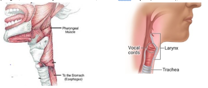
Muscle groups of the head (and neck): Muscles of swallowing a speech
located in the oral cavity and throat, direct movement of food toward esophagus, innervated by a cranial nerves, located in the larynx (voice box), control vocal cords, innervated by a cranial nerve
Muscles of the neck: Head movement
Anterior neck Sternocleidomastoid- muscle, bilateral head flexion, unilateral rotation of head to opposite side (turn head) Posterior neck Trapezius- stabilize, laterally flex, and rotate head

Muscles of the posterior vertebral column: Erector spinae group
prime mover of back extension and maintenance of posture, contribute to lateral bending and rotation of vertebral column, regions in neck assist head extension and rotation, 3 columns (Spinalis group, longissimus group, iliocostalis group)