microbio - streptococcus, enteerococcus, and other Catalase-Negative gram positive cocci
 Call Kai
 Learn
 Practice Test
 Spaced Repetition
 Match
 Flashcards
 Knowt Play
1/27

There's no tags or description
Looks like no tags are added yet.
28 Terms
today’s flow: reduce “jumping” by using one consistent diagnostic path
start broad
GPC
catalase negative
pairs or chains
sort by plate
beta hemolytic
alpha hemolytic
gamma hemolytic/ nonhemolytic
name likely groups
GAS, GBS, C/G, S. pneumoniae, viridans, enterococcus/ group D
confirm
bacitracin, PYR, CAMP, optochin, bile solubility, bile esculin, NaCl
interpret
site + host + clinical significance
general characteristics: what makes these bacteria different from Staphylococci?
microscopy
GPC in pairs or chains
may look more elongated than staph
catalase
catalase NEGATIVE differentiates them from staph
avoid colonies from blood media - hemoglobin can cause FALSE POSITIVES
growth
facultative anaerobes (some are CAPNOPHILIC)
growth is best on enriched media w blood/serum
colony clues
small, transparent colonies are common
hemolysis pattern on BSA drives first major branch of identification
cell wall structure and Lancefield grouping
core cell wall
thick peptidoglycan + teichoic acid + group carbohydrate in many clinically important species
Lancefield groups
serologic groups by C carbohydrate; Group A, B, C, D, F, G, and others
Group A → S. pyogenes
Group B → S. agalactiae
Group D → Enterococcus/ S. gallolyticus (bovis)
NO Lancefield → S. pneumoniae/ viridans
clinical shortcut
group + hemolysis + source often points toward the organism before full biochemical ID

hemolysis
Beta-hemolysis: clear zone around colony
think: GAS, GBS, C/G groups
alpha-hemolysis: green discoloration
think: S. pneumoniae vs Viridans
nonhemolytic: no change in agar
think: Enterococcus/ Group D
alpha prime: tiny intact RBC zone plus wider Beta zone - can be mistaken for beta/alpha mix of both

first pass identification algorithm
GPC
catalase negative
read hemolysis
beta hemolytic: TAXO-A/ PYR/ CAMP/ Hippurate
alpha hemolytic: TAXO-P/ bile solubility
nonhemolytic: bile esculin/ 6.5% NaCl/ PYR
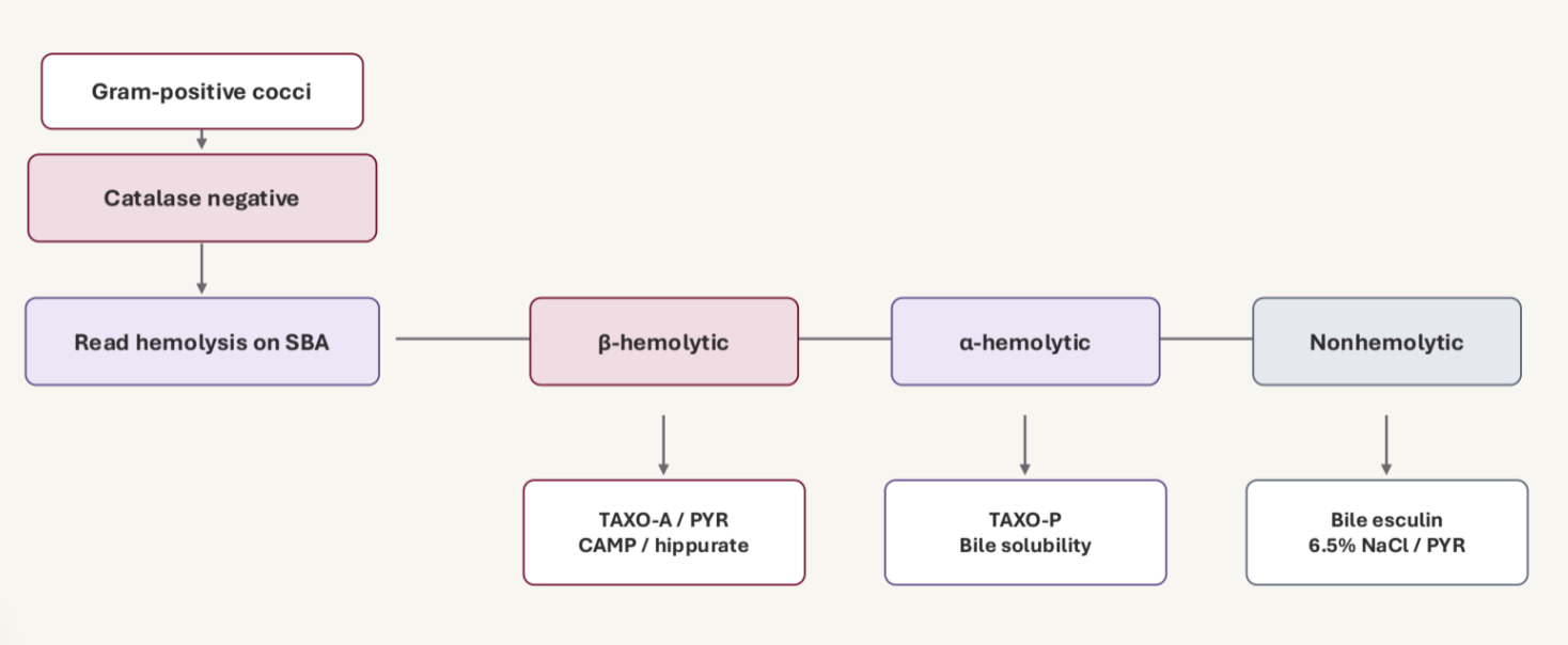
high yield anchors: organism → usual hemolysis → confirmatory clue

Beta hemolytic strep
GROUP A - S. pyogenes
Throat, skin/soft tissue, invasive disease, post strep sequelae
TAXO-A/ PYR/ antigen testing
GROUP B - S. agalactiae
pregnancy/newborn disease; screen vaginal + rectal swabs
CAMP/ hippurate/ NAAT
GROUPS C/G
may cause pharyngitis, impetigo, pyogenic infections; Lancefield typing helps
normally tested w KITS instead of growth
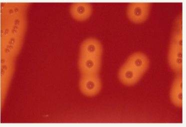
Streptococcus pyogenes (Group A strep)
identity
Lancefield group A
Beta hemolytic
PYR (+)
BACITRACIN SUSCEPTIBLE
common disease
pharyngitis (strep throat)/ tonsilitis
Scarlet fever
impetigo/erysipelas/ cellulitis
serious disease
necrotizing fasciitis
streptococcal TOXIC SHOCK SYNDROME
sequelae: RHEUMATIC FEVER, ACUTE GLOMERULONEPHRITIS

S. pyogenes virulence factors
attach
M protein + lipoteichoic acid + fibronectin-binding protein help adherence to mucosal cells and keratinocytes
avoid phagocytosis
M protein resists phagocytosis
hyaluronic acid capsule MASKS bacterial antigens
damage cells
Streptolysin O is oxygen-labile (easily altered by Oxygen) and immunogenic
streptolysin S is oxygen stable
spread
DNase, streptokinase, and hyaluronidase (spreading factor) help tissue invasion/spread
*antibodies to SLO can be measured in the ASO test to support evidence of recent GAS infection (+ does not mean current infection)
streptococcus pyogenes (GAS) clinical infections - throat
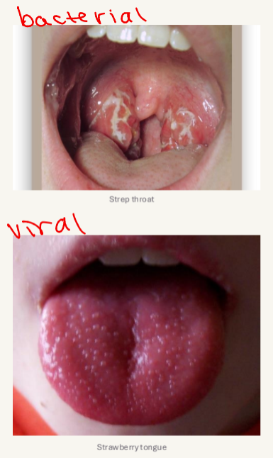
pharyngitis and tonsillitis
primarily in children 5-15 yrs old
spread by droplets and close contact
symptoms: sore throat, fever, malaise, headache, ab pain, nausea, tender cervical lymph nodes
scarlet fever
affects those w strep throat OR STREP SKIN INFECTIONS
streptococcal pyrogenic enterotoxins cause distinctive symptoms
scarlatiniform rash - red w small papules (sandpaper texture)
after rash, skin desquamation (peeling)
strawberry tongue
streptococcus pyogenes (GAS) clinical infections - skin
impetigo
very young CHILDREN
small vesicles → weeping lesions that crust (no actual blister unlike herpes)
erysipelas
in ELDERLY patients
spreading red rash with demarcated but IRREGULAR edge
cellulitis
deep invasion of GAS leading to SEPSIS and GANGRENE (goes deep into tissues)

streptococcus pyogenes (GAS) clinical infections - serious
necrotizing fasciitis (tissue death)
invasive infection characterized by rapidly progressing inflammation and necrosis of the skin, subq fat, and fascia
described as “flesh-eating disease”
life threatening disease
characterized as types 1, 2, 3
type 2 is Group A strep
Streptococcal Toxic Shock Syndrome (TSS)
follows a severe initial strep infection
collapse of entire organ systems - gets access to bloodstream - sepsis can occur

streptococcus pyogenes (GAS) sequelae
rheumatic fever
follows GAS PHARYNGITIS (can ONLY get from throat infection)
fever, inflammation of heart, joints, blood vessels, subq tissues
progressive damage to heart valves
antigenic cross reactivity btwn strep antigens and heart tissue
bacterial enterotoxins
actual bacterial invasion of the heart tissue
acute glomerulonephritis
follows EITHER skin or strep throat infection
malaise, fever, nausea, nephrotic syndrome, hematuria, smoky brown urine
immune complexes deposit in glomeruli causing inflammation and damage

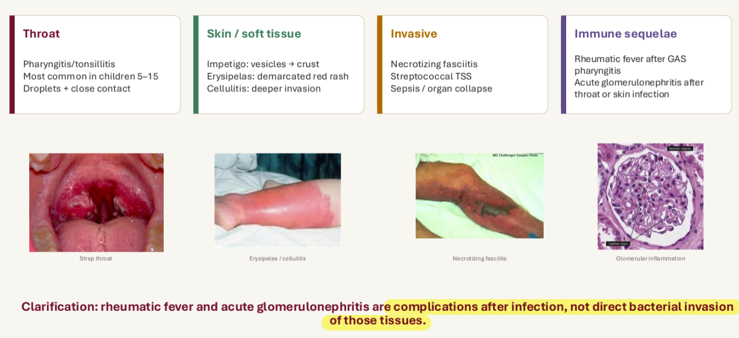
S. pyogenes clinical disease: organize by site and mechanism
rheumatic fever and acute glomerulonephritis are COMPLICATIONS AFTER INFECTION, NOT direct bacterial invasion of those tissues
GAS lab diagnosis
throat specimen → Beta colonies → confirmation
specimen
swab POSTERIOR pharynx + each TONSIL
touch EXUDATE if present
AVOID tongue, uvula, cheeks
culture
SBA - 35C for 18-24 hrs in CO2
if no growth → incubate + 24hrs
can use selective media: SBA containing SMZ, colistin, and polymyxin B
confirmation
Lancefield typing preferred; bacitracin/PYR can support presumptive ID
direct methods
rapid antigen detection tests and PCR are useful for throat swabs
culture remains important when confirmation needed
if kit is negative, still culture to confirm/dbl check

streptococcus agalactiae (GBS) - screening prevents newborn disease
identity
group B antigen
Beta hemolytic
CAMP (+)
Hippurate (+)
reservoir
NORMAL BIOTA of upper respiratory tract, GI tract, and vagina
pregnant women MUST be screened by 7 months (will be checked continuously from 7-9 months/until birth)
risk
major cause of pneumonia, sepsis, and meningitis in NEWBORNS
virulence
capsule containing sialic acid helps inhibit the alternative complement pathway
other products
hemolysins, CAMP factor, neuraminidase, DNases, hyaluronidase, and proteases are useful to know for ID/context
GBS clinical infections
organize by AGE and route
EARLY onset newborn disease
< 7 days old
usually VERTICAL TRANSMISSION during birth (from mom)
associated w/ OBSTETRIC COMPLICATIONS
high mortality rate if not treated quickly
LATE onset newborn disease
7 days to 3 months
primarily MENINGITIS
less tied to obstetric complications
still serious
ADULT disease
postpartum/post-abortion infections
endometriosis, wound infection, endocarditis
elderly or immunodeficient patients
*clinical logic: the same organism may be normal biota in one host but life threatening in another
GBS lab diagnosis
pregnancy screen and confirmation tests
specimen collection
vaginal + rectal swab at 36-27 weeks of pregnancy
enrichment
inoculate in Lim broth, TransVag broth, or StrepB Carrot broth
incubate at 35C, 5% CO2 for 18-24 hrs
then subculture to SBA or chromogenic agar
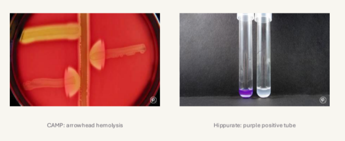
CAMP test
arrowhead of enhanced hemolysis near S. aureus = presumptive GBS
Hippurate
purple after ninhydrin → hippurate positive = GBS
NAATs may also be used, esp when RAPID detection is needed
Lab diagnosis: Beta hemolytic strep
tests for detection/differentiation of GAS and GBS
bacitracin susceptibility (TAXO-A)
CAMP test
sodium Hippurate hydrolysis
PYR (if necessary) - not always accurate
antigenic testing
B-hemolytic strep tests: TAXO-A
TAXO-A
for the presumptive ID of S. pyogenes
disk containing 0.04 IU bacitracin
test
streak 1 colony of B-strep over ½ SBA, add disk onto streaked surface, incubate overnight at 35C in CO2
results
susceptible (ANY zone of inhibition) = GAS
resistant (NO zone of inhibition) = NOT GAS

B-hemolytic strep tests: CAMP
CAMP test (Christine, Atkins, and Munch-Petersen)
for the presumptive ID of Group B Strep
GBS produce beta hemolysis that reacts SYNERGISTICALLY with S. aureus
test
streak vertical straight line of S. aureus, streak B-hemolytic strep to be tested at a right angle (but not touching) , and incubate at 35C overnight in CO2
results
arrowhead of hemolysis @ junction (+) = Group B strep
NO arrowhead of hemolysis @ junction (=) = NOT group B strep

B-hemolytic strep tests: Hippurate
sodium hippurate hydrolysis
for differentiating S. galactiae from other beta strep
S. agalctiae possess hippuricase enzyme
hydrolyzes NaHipp to form sodium benzoate and glycine
test
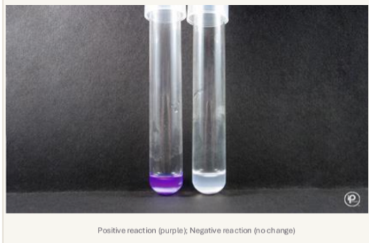
add bacteria to tube of NaHip, incubate overnight at 35C, add 3-2 drops of Nihydrin and incubate at 37C for 10 mins
results
clear turns purple color (+) = GBS
NO color change (=) = other beta strep
B-hemolytic strep tests: PYR
PYR
disk containing PYR substrate (Pyrrolidonyl-α-naphthylamide)
GAS is able to hydrolyze substrate
test
add 1 drop of saline to PYR disk, add 1 colony to disk, incubate RT 5 min, and then add 1 drop of color developer (1 min)
results
color change (red/pink) = positive - GAS
no color change = negative - NOT GAS

other beta hemolytic streptococcal groups: do not stop at beta=GAS/GBS
groups C and G
large colony isolates often S. dysgalactiae subsp. equismillis
infection sites can resemble GAS
S. anginosus group
small colony isolates may be Beta or Alpha or nonhemolytic
frequently linked to abscesses and pyogenic infections
KEY TESTING POINT
Lancefield typing is the best way to separate groups C/G from GAS when morphology overlaps
alpha hemolytic strep: S. pneumoniae vs Viridans strep
S. pneumoniae
pneumococcus
Lancet shaped diplococci
opportunistic pathogen of the RESPIRATORY tract
confirm pneumococcus
optochin SUSCEPTIBLE
bile soluble
quelling reaction can identify capsule
Viridans strep
NORMAL BIOTA of ORAL cavity, URT, GI and female genital tract
opportunistic infection
confirm viridans
optochin RESISTANT
bile insoluble
Lancefield grouping is usually of little value
S. pneumoniae capsule
Identity
alpha hemolytic pneumococcus
part of normal URT flora
lancet shaped diplococci
capsule
abt 90 capsule types
capsule is antigenic and a MAJOR VIRULENCE FACTOR
QUELLUNG RXN
anti-capsular serum causes CAPSULE SWELLING
useful conceptually for capsular typing
other products
hemolysin, IgA protease, neuraminidase, and hyaluronidase also contribute

pneumococcus study anchor
alpha hemolytic
optochin susceptible
bile soluble
capsulated diplococcus
s. pneumoniae clinical infections
pneumococcal lobar pneumonia
common in elderly + underlying disease & can follow another illness as a secondary infection
fluid facilitates spread to adjacent alveoli → reaches fibrous septa that separates major lung lobes
SYMPTOMS: chills, cough, dyspnea, edema, hypoxia
meningitis
occurs in all groups (50% of bacterial meningitis cases in the US)
can follow ear infection or pneumonia
rapid course w HIGH mortality
SYMPTOMS: headache, lethargy, vomiting, fever, nuchal rigidity
COMPLICATIONS: cranial nerve sign, seizures, bacteremia, coma
other infections
most frequently isolated in children under 3 with recurrent ear infections
bacteremia, endocarditis, peritonitis, empyema, and HUS-like complications
prevention/treatments
vaccines area available- routine for children under 5, elderly, and college students
penicillin is drug of choice, though resistance is a concern