RT 110+111 Unit 2: Sacrum/Coccyx and Sacroiliac Joints
 Call Kai
 Learn
 Practice Test
 Spaced Repetition
 Match
 Flashcards
 Knowt Play
1/53

There's no tags or description
Looks like no tags are added yet.
54 Terms
The inferior portion of the lumbar vertebrae formed by the fusion of five sacral vertebral segments is known as the ____.
sacrum

Large wing-like masses lateral to the first sacral segment are the __.
ala

The superior and anterior portion of the sacrum is the ___ while the inferior and posterior portion is the ____.
base; apex

The ____ articulates with the inferior articular processes of the fifth lumbar vertebrae and forms the zygopophyseal joints.
superior articular processes

The anterior ridge of the body of the first sacral segment is the ___.
promontory

Four sets of holes in the sacrum that serve as passageways for sacral nerves and blood vessels are known as ___.
pelvic sacral foramina
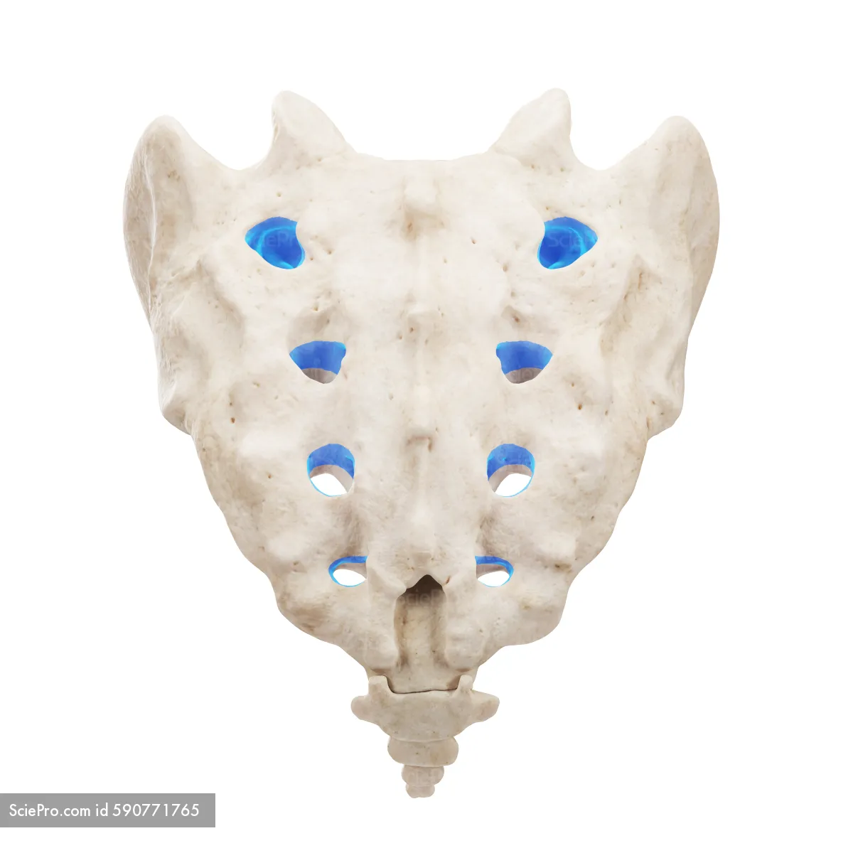
The sacrum articulates with the ilium of the pelvis at the ___ form the sacroiliac joints.
auricular surface

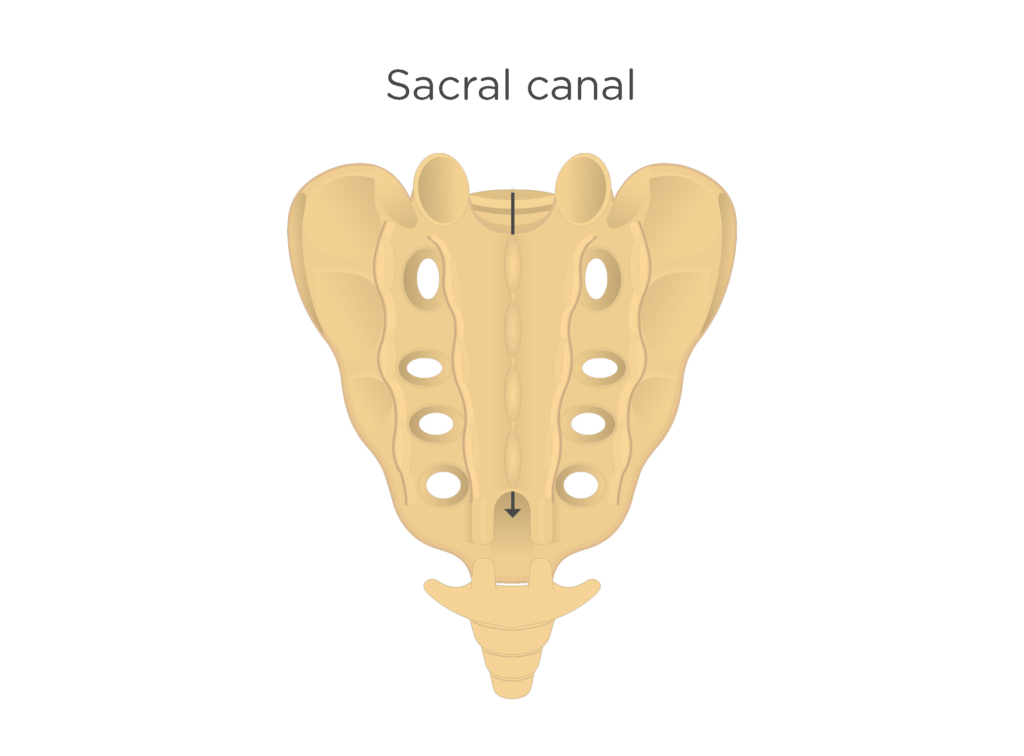
The opening located behind the base of the first sacral segment is the ___.
sacral canal
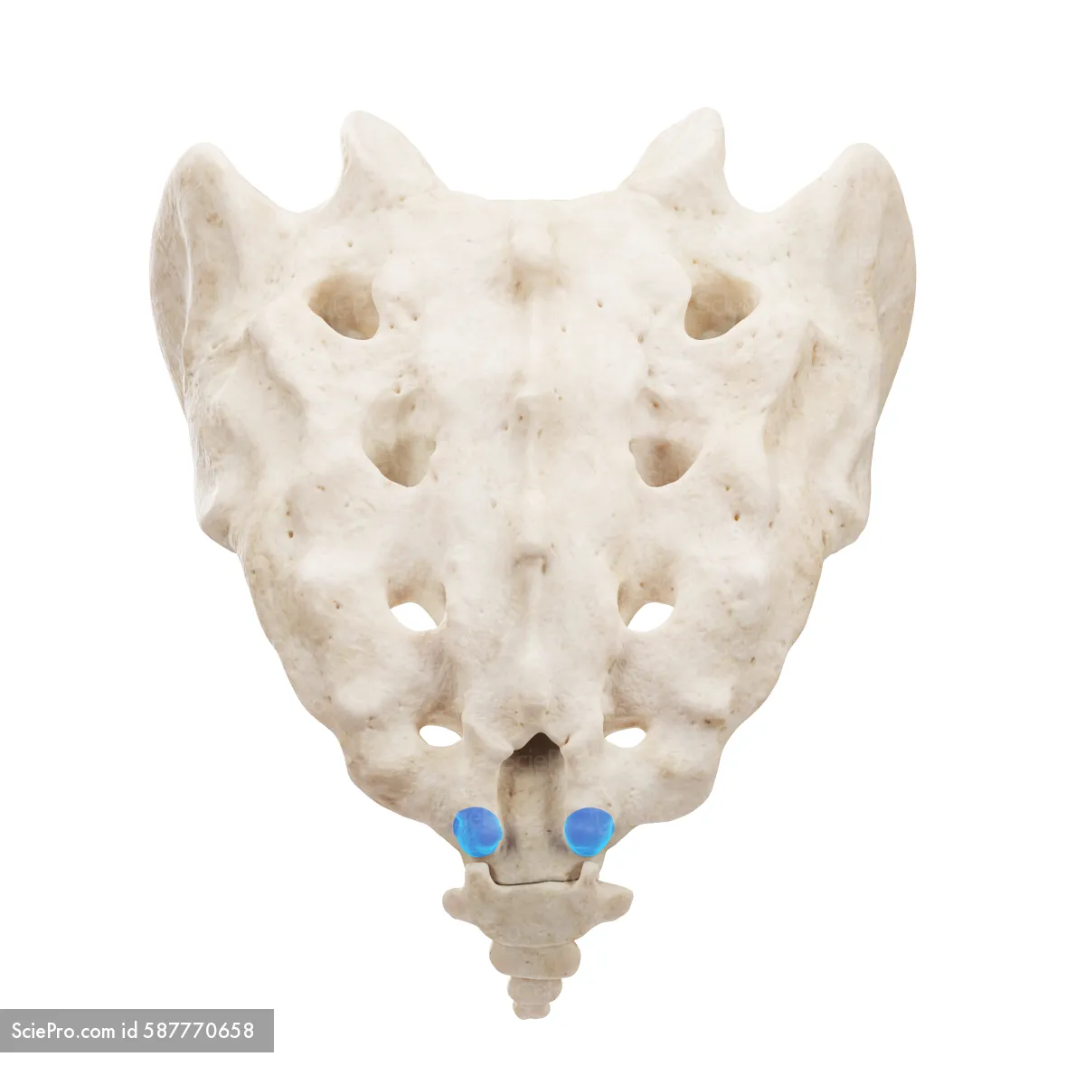
Small bony projections at the inferoposterior aspect of the sacrum that articulate with the horns of the coccyx are the __.
sacral horns
Each sacroiliac joint opens at a ___ oblique angle.
30*

The spinous processes of the sacral vertebrae fuse to form the ___.
medial crest

Three to five segments fuse to form the most distal portion of the vertebral column, which is the ___.
coccyx

The two lateral projections on the most superior segment of the coccyx are the __.
transverse processes

The inferior pointed tip of the coccyx is the ___.
apex

The broader superior portion of the coccyx is the ___.
base

What purpose do the sacroiliac joints serve?
Transfers weight from the spine, pelvis, and lower extremities
T/F: Sacroiliac joints are freely movable synovial joints that disappear or narrow in older adults
False
What are the primary clinical indications visualized in an AP axial projection of the sacrum?
Pathology or fractures in the sacrum
How is the patient positioned for an AP axial projections of the sacrum, coccyx, and sacroiliac joints?
Patient is supine with MSP aligned with the middle of the table
No rotation of the pelvis (ASIS on both sides are at equal distance from the table)
Respiration is suspended to reduce motion

Where is the CR directed for an AP axial projection of the sacrum?
CR is angled 15* cephalic and directed midway between ASIS and symphysis pubis (2” below ASIS/2” above symphysis pubis)

If a patient has a greater posterior curvature, the CR should be angled ___.
20* cephalic
Why is supine preferred over prone for sacrum, coccyx, and sacroiliac joint projections?
Laying supine reduces OID by putting the sacrum and other related structures closer to the IR
What anatomy must be demonstrated to obtain a diagnostic image of the AP axial sacrum?
Sacrum, SI joints, L5-S1 joint spaces, and sacral foramina free of superimposition are visualized

How is no rotation indicated in an AP axial projection of the sacrum?
Alignment of the spinous processes on the lumbar vertebrae, median sagittal crests, and coccyx
What are the optimal exposure factors to demonstrate a diagnostic image of an AP axial sacrum?
40” SID, 75-90 kVp, 14×17” IR portrait, center photocell, grid, collimate to area of interest
If a patient is unable to lay on their back, an PA axial projection of the sacrum can be done prone by angling the tube ___.
15* caudal to the sacrum
What are the primary clinical indications for an AP axial projection of the coccyx?
Pathology or fractures in the coccyx
Where is the CR directed for an AP axial projection of the coccyx?
CR is angled 10* caudal and directed midway between the ASIS and symphysis pubis (2” below ASIS or 2” above symphysis pubis)

If a patient’s coccyx has a greater anterior curve, the CR should be angled ___.
15* caudal to the coccyx
If a patient is unable to lay on their back, a PA axial projection of the coccyx can be done prone by angling the tube ___ to the coccyx.
10* cephalic
How should the coccyx be visualized to produce a diagnostic image of an AP axial coccyx?
Free of superimposition and projected superior to the pubis with segments appearing open
Appears equidistant from the lateral walls of the pelvis opening

What are the optimal exposure factors to produce a diagnostic image of an AP axial coccyx?
40” SID, 75-85 kVp, 14×17” IR portrait, grid, collimate to part
Which projection can be used to visualize pathological conditions or fractures in both the sacrum and coccyx?
Lateral sacrum and coccyx
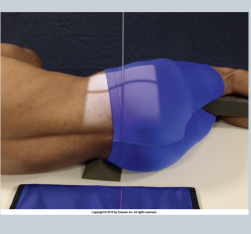
What device is placed behind the patient to reduce scatter and improve image quality in a lateral projection of the sacrum and coccyx?
lead mask
How is the patient positioned for a lateral projection of the sacrum and coccyx?
Patient is in true lateral position
Knees and elbows flexed and placed on top of each other
Shoulders and hips are placed on the same plane
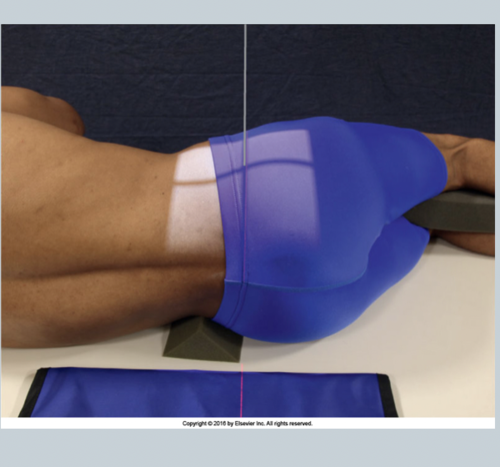
Where is the CR directed for a lateral projection of the sacrum and coccyx?
CR is directed 3 1/2” behind the ASIS for sacrum, 3 1/2” behind and 2” below ASIS to include the coccyx with light skimming patient’s behind
How is no rotation indicated in a lateral projection of the sacrum and coccyx?
Superimposition of the greater sciatic notches and femoral heads
What anatomy must be demonstrated to obtain a diagnostic image of a lateral projection of the sacrum and coccyx?
Sacrum, L5-S1 joint, and coccyx

What are the optimal exposure factors for a lateral projection of the sacrum and coccyx?
40” SID, 85-95 kVp, 14×17” IR portrait, grid, center photocell, lead mask, collimate to part
What are the primary clinical indications for an AP axial projection of the sacroiliac joints (SI joints)?
Pathology, fractures, dislocations, and subluxations
Where is the CR directed for an AP axial projection of the sacroiliac joints?
CR is angled 30*-35* cephalic (30* for males, 35* for females) and directed 2” below ASIS

If a patient is unable to lay on their back, a PA axial projection of the sacroiliac joints can be performed by ___.
Having the patient prone
CR is angled 30*-35* caudal and directed to L4 or slightly above the iliac crest
What anatomy must be demonstrated in an AP axial projection of the sacroiliac joints?
Sacroiliac joints and L5-S1 joint space

How is no rotation indicated in an AP axial projection of the SI joints?
Spinous process of L5 aligned in the center of vertebral body
Symmetric appearance of both alae/wings of the sacrum
How is a patient positioned for a posterior oblique projection of the sacroiliac joints?
Patient is supine with joint of interest aligned to the middle of the table
Side of interest is elevated 25*-30* (LPO for RT SI joint, RPO for LT SI joint)

Where is the CR directed for a posterior oblique SI joint?
CR is directed 1” medial to the upside ASIS of affected side

To demonstrate the inferior part of the SI joint more clearly, the CR may be angled ___.
15*-20* cephalad
What anatomy is demonstrated in a posterior oblique SI joint?
Sacroiliac joint farthest from IR
How is accurate rotation indicated in a posterior oblique SI joint?
No superimposition of the ala of the ilium and sacrum with the open SI joint
What are the optimal exposure factors for a posterior oblique SI joint?
40” SID, 80-95 kVp, 14×17” IR portrait, grid, center photocell, collimate to part
What position can be performed to evaluate the sacroiliac joints if a patient is unable to lay on their back?
Anterior obliques (RAO & LAO)
When peforming anterior obliques to evaluate sacroiliac joints, which side is of interest?
The side of interest is down closest to the table
LAO - LT SI joint
RAO - RT SI joint
How do posterior obliques differ from anterior obliques when looking at SI joints?
Posterior obliques (RPO & LPO) - affected side of interest is up
Anterior obliques (RAO & LAO) - affected side of interest is down
Where is the CR directed for an anterior oblique projection of the sacroiliac joints?
1” medial to the downside ASIS