N245: Alterations in Brain Function
 Call Kai
 Learn
Learn Practice Test
 Spaced Repetition
 Match
Match Flashcards
 Knowt Play
1/63

There's no tags or description
Looks like no tags are added yet.
Name | Mastery | Learn | Test | Matching | Spaced | Call with Kai | Chat |
|---|
No analytics yet
Send a link to your students to track their progress
64 Terms
Cerebral Hypoxia
What: Low levels of oxygen in the brain (well tolerated)
Causes: Reduced atmospheric pressure, CO poisoning, severe anemia, choking, drowning

Cerebral Ischemia
What: Reduced blood flow to the brain
Causes: Cerebral artery occlusion, cardiac arrest, hypotension

Focus Ischemia
What: Reduced blood flow to a specific part of the brain
Causes: Cerebral artery occlusion
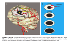
Cerebral infarct
An area of the brain that becomes ischemic and necrotic due to lack of blood supply

Global ischemia
What: Reduced blood flow throughout the brain
Causes: Cardiac arrest, severe hypotension --> shrinking/swelling of neurons
Cerebral Edema
What: Increase in brain fluid --> increased volume of brain tissue

Vasogenic edema
What: A form of cerebral edema
Causes: Hemorrhage, traumatic brain injury, or infections --> leaky capillaries --> increases of extracellular fluid

Cytotoxic edema
What: A form of cerebral edema
Causes: Hypoxic or ischemic injury --> impairment of the Na+ /K+ pumps --> increase of intracellular fluid
S+S: Narrow sulci (depressions in the cerebral cortex), flat gyri (ridges on cerebral cortex)

HACE
What: High altitude cerebral edema
Causes: Climbing to high altitudes quickly
S+S: Extreme fatigue, altered mental status after rapidly ascending to very high altitudes --> death

Consciousness
Being aware of one's self, being aware of and responsive to one's environment

Confusion
Disorientation, impaired ability to think clearly, slightly reduced responses to stimuli

Delirium
Physical restlessness, transient hallucinations, incoherence of thought and speech

Obtundation
Lethargy, very slow responses to stimuli, excessive drowsiness when awake

Stupor
Unresponsiveness, little or no spontaneous activity, patient can be aroused by vigorous and repeated stimuli

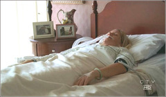
Coma
Deep unconsciousness, complete unresponsiveness, patient cannot be aroused by external stimuli
Glasgow Coma Scale
Assesses patients' coma scale based on eye opening, verbal response, motor response (3 = coma, 15 = full consciousness)

Normal pupillary reflexes
Constrict equally when exposed to light
Pupil characteristics during compression of the oculomotor nerve
Fixed (non-reactive) dilated pupil on the same side as damage

Pupil characteristics during lesions of the brainstem
Fixed (non-reactive) pinpoint pupils in both eyes

Pupil characteristics during irreversible cessation of brain function ("braindeath")
Fixed (non-reactive) dilated pupils in both eyes

Decorticate posturing
Involuntary flexion of arms --> indicates damage to the cerebral hemispheres

Decerebrate posturing
Involuntary extension of arms --> indicates damage to the brainstem

Intracranial Pressure (ICP)
The amount of pressure inside the skull

Intracranial volumes that contribute to the ICP
Brain tissue -- 80% of volume
Blood -- 10% of volume
CSF -- 10% of volume
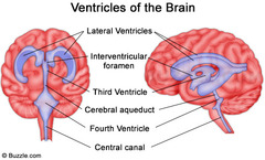
Brain ventricles
A series of cavities in the CNS that contain cerebrospinal fluid
Monro-Kellie hypothesis
Because of limited space in the skull, an increase in any one of components of the skull (brain tissue, blood, CSF) will cause a change in the volume of the others

How do increases in ICP result in brain injury?
1. Physical damage to the neurons --> sensory or motor deficits
2. Compression of blood flow --> ischemia --> hypoxia
3. Herniation/displacements of the brain tissue
Falx cerebri
Septum that separates the two cerebral hemispheres

Tentorium cerebelli
Septum that separates cerebrum from cerebellum

Brain Herniation
Brain shifts from areas of high intracranial pressure to low intracranial pressure
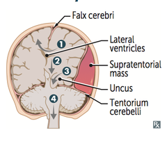
Supratentorial brain herniation
Herniation of cerebral cortex above the tentorium cerebelli

Transtentorial brain herniation
Herniation of the cerebral cortex through the tentorial notch

Tentorial notch
Triangular opening through which the brainstem, blood vessels and oculomotor nerve [CN III] pass
![<p>Triangular opening through which the brainstem, blood vessels and oculomotor nerve [CN III] pass</p>](https://assets.knowt.com/user-attachments/06b96974-b503-4747-83e9-19edba716900.jpg)
Infratentorial brain herniation
Herniation of the cerebellum below the tentorium cerebelli

Cerebrospinal Fluid (CSF)
Clear fluid that circulates in and around the brain and spinal cord; produced in the ventricles

Pathway for CSF
Choroid plexus in ventricles --> subarachnoid space --> arachnoid villi --> heart --> capillaries --> ventricles
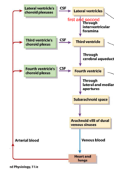
Hydrocephalus
What: Abnormal accumulations of CSF in the ventricles --> enlargement of ventricles
Causes:
1. Overproduction of CSF
2. Impaired reabsorption of CSF
3. Obstruction to the flow of CSF
S+S: Headache, nausea, and vomiting, abnormal eye movements, blindness, weakness, decreased memory

Traumatic Brain Injury
A blow to the head that damages the brain

What is the leading cause of TBIs?
Falls
Focal TBI
Specific, local brain injuries; contusions and hematomas

Global TBI
Widespread, broad brain injuries; concussions
Contusions
What: Bruise on the surface of the brain
Causes: Blunt force to the head --> brain accelerates within the skull --> decelerates abruptly after hitting the inner skull surfaces
S+S: Various neurological defects

Coup injury
Contusions directly under the site of impact with an object

Contrecoup injury
Contusions on the opposite side of the site of impact (whiplash injury)

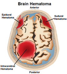
Intracranial Hematomas
What: Abnormal collection of blood in the cranium
Causes: Injury to blood vessels of the brain
Cranial Meninges
Dura mater, arachnoid mater, pia mater

Pia mater
Thin, delicate inner membrane of the meninges

Arachnoid mater
Weblike middle layer of the three meninges

Dura mater
Thick, outermost layer of the meninges

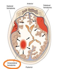
Epidural hematoma
What: Bleeding that develops between the skull and the dura mater
Causes: Severe head trauma --> arterial rupture
S+S: LOC --> Lucid interval of consciousness --> LOC again

Subdural hematoma
What: Bleeding that develops between the dura mater and the arachnoid mater
Causes: Severe head trauma --> venous rupture
S+S: LOC

Intracerebral hematoma
What: Blood clot that develops in the brain tissue
Causes: Severe head trauma, rupture of a cerebral aneurysm
Concussions
What: Mechanical injury --> transient brain dysfunction
Causes: Sports injuries
S+S: Headaches, amnesia, insomnia, confusion

Stroke
What: Non traumatic disruption of blood flow to the brain --> acute neurological deficits; "brain attack"/CVA
S+S: Sensory, motor, verbal, or cognitive deficits depending upon the area of brain affected

Ischemic stroke
A stroke in which the blood flow disruption is caused by a blood clot/blockage

Hemorrhagic stroke
A stroke in which the blood flow disruption is caused by the rupture of a blood vessel

Transient Ischemic Attack
What: Temporary disruption of blood flow to the brain --> brief episode of neurological dysfunction; "mini stroke"

Dementia
What: Deterioration of cognitive function that interferes with social/occupational ADLs
Causes: Drugs, emotional disorders, metabolic disorders, eye/ear problems, normal pressure hydrocephalus, tumors, infections, anemia
S+S: Reduced ability to perceive sensory input, learn and manipulate new information, recognize familiar objects, recall past experiences, solve problems, think abstractly, or make judgments
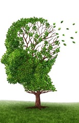
Alzheimer's Disease
What: Most common cause of dementia
Causes: Reduced concentrations of acetylcholine, genetics
S+S: Damage to hippocampus --> inability to store memories

Early stage of Alzheimer's Disease
Memory lapses (denied by the patient but reported by caregivers)
Middle stage of Alzheimer's Disease
Confusion, frustration, anger, social withdrawal, altered sleep patterns, personality and behavioral changes including suspiciousness
Late stage of Alzheimer's Disease
Loss of awareness of recent experiences and surroundings, difficulty in communicating, incontinence, death
Neurofibrillary tangle
Hallmark of Alzheimer's --> defective tau proteins accumulate inside the neurons

Senile (amyloid) plaque
Hallmark of Alzheimer's --> Amyloid plaques form around the outside of the neuron
