PATH 3810 Pathology of the Cardiovascular System
 Call Kai
 Learn
 Practice Test
 Spaced Repetition
 Match
 Flashcards
 Knowt Play
1/132

There's no tags or description
Looks like no tags are added yet.
Name | Mastery | Learn | Test | Matching | Spaced | Call with Kai | Chat |
|---|
No analytics yet
Send a link to your students to track their progress
133 Terms
edema
increased fluid in interstitial space
anasarca
severe and generalized edema with profound subcytaneous tissue swelling
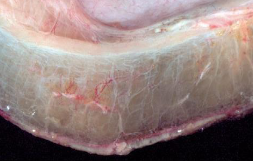
edema of submucosal intestinal tissue

ascites
ascites / hydroperitoneum
edema of the peritoneal cavity

pulmonary edema (bottom) vs normal lung tissue (top)
increased hydrostatic pressure
impaired venous return
arteriolar dilation
category of edema
reduced plasma osmotic pressure (hypoproteinemia)
loss of albumin
category of edema
albumin
serum protein most responsible for maintaining intravascular osmotic pressure
lymphatic obstruction
lymphedema
usually localized
category of edema
sodium water retention
increased salt draws in water and increases hydrostatic pressure
increased renal tubular reabsorption of sodium
category of edema
hyperemia
active process resulting from augmented blood flow due to arteriolar dilation
congestion
passive process resulting from impaired venous return out of tissue
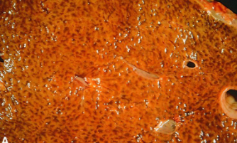
chronic passive congestion of the liver (gross)

chronic passive congestion of the liver (microscopic)
hemorrhage
extravasation of blood from vessels into extracellular space
hemorrhage can be…
…external or confined within a tissue
hematoma
hemorrhage confined within a tissue
petechiae
minute (1-2mm) hemorrhages into the epithelium/underlying tissue
purpura
medium (3-5mm) hemorrhages
ecchymoses
large (1-2cm) subcutaneous hematomas
large accumulation of blood in body cavities
hemothorax
hemopericardium
hemoperitoneum
hemarthrosis

petechiae

intracranial hemorrhage
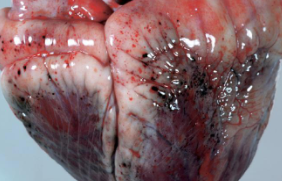
epicardial hemorrhage

ecchymoses

hemorrhage of the leg

gastric hemorrhage
thrombosis
pathologic form of hemostasis
thrombus formation in uninjured vessels or thrombotic occlusion of a vessel after minor injury
three principal influences on thrombus formation
endothelial injury
abnormal blood flow
hypercoagulability
arterial or cardiac thrombi begin at…
…sites of endothelial injury or turbulence
venous thrombi begin at…
…sites of stasis
thrombi are…
…focally attached to the underlying vascular surface and tend to propagate toward the heart
embolus
a mass that is carried by bloodstream to a place distant from point of origin
can be:
broken, free fragment of thrombus (thromboembolism)
solid, liquid, or gaseous mass
fat droplets, air droplets, atherosclerotic debris
vegetations
thrombi on heart valves
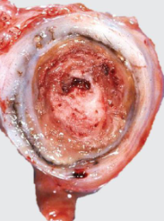
arterial thrombus (gross)

arterial thrombus (microscopic)

venous thrombus

white/pale infarct of the kidney

red/hemorrhagic infarct of the kidney

red infarct of the lung

pale infarct of the spleen
thrombosis outcomes
propagation
embolization
dissolution
organization/recanalization
pulmonary thromboembolism
95% of cases are a venous embolus from deep leg vein thrombus

saddle thrombus in pulmonary artery
systemic thromboembolism
embolism in the arterial circulation
80% arise from intracardial mural thrombi, particularly left ventricular wall infarcts
major sites of embolism are lower extremities and brain
air embolism
may occur during chest wall injury or decompression sickness
amniotic fluid embolism
complication of labor
caused by entry of amniotic fluid into the maternal circulation via tear in the placental membranes and rupture of uterine veins
fat embolism
fat globules may enter bloodstream after long bone fracture or soft tissue trauma
occlude pulmonary and cerebral microvasculature

fat embolus from bone marrow
shock
final common pathway for a number of potentially lethal events
gives rise to systemic hypoperfusion
hypotension, hypoxia, hypoperfusion
cardiogenic shock
results from heart failure
myocardial infarction
ventricular arrythmias
outflow obstruction
hypovolemic shock
results from loss of blood or plasma volume
hemorrhage
extensive burns
septic shock
results from microbial infection
lipid A released by Gram-negative bacteria
neurogenic shock
results from a loss of vascular tone leading to peropheral blood pooling
anaphylactic shock
results from systemic vasodilation and increased vascular permeability mediated by IgE: type I hypersensitivity reaction
shock is characterized by…
…multisystem organ failure due to hypoxic injury
clinical triad of septic shock
hypotension
disseminated intravascular coagulation
metabolic disturbances
blood pressure is a function of…
…cardiac output, peripheral resistance, and hormonal regulation
hypertensive vascular disease
chronic hypertension causes damage to arterioles and endothelium
arteriosclerosis

hyaline arteriosclerosis

hyperplastic arteriosclerosis
atherosclerosis
characterized by intimal lesions called atheromas that protrude into vascular lumina and obstruct blood flow
atheroma
raised lesion with a soft yellow grumous core of cholesterol and covered by a firm white fibrous cap
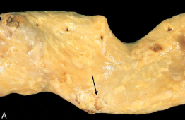
atherosclerosis (gross)

atherosclerosis (gross)

atherosclerosis (microscopic)
aneurysm
localized abnormal dilation of a blood vessel or the heart
true aneurysm
involves all three layers of arterial wall
false aneurysm
breach in the vascular wall leading to extravascular hematoma
dissection
blood enters the wall of the artery as a hematoma separating its layers

aortic aneurysm
aortic dissection
blood separates the laminar planes of the media to form a blood-filled channel within the aortic wall
often secondary to hypertension

pulmonary artery dissection
cystic medial degeneration
loss of smooth muscle cells, fragmentation or mineralization of elastic fibers
predisposes artery to aneurysm or dissection

cystic medial degeneration
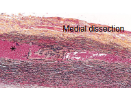
medial dissection
immune-mediated vasculitis
type III hypersensitivity, associated with AgAb complexes
polyarteritis nodosa
systemic
inflammation of small to medium muscular arteries
causes restriction of blood flow

polyarteritis nodosa (gross)

polyarteritis nodosa (microscopic)
shunt
abnormal connection between heart chambers or blood vessels
obstructive congenital heart disease
developmental heart abnormalities that obstruct vascular flow by narrowing of chambers, valves, or major vessels
atresia
complete obstruction of a normal opening
left to right shunt
septal defects & patent ductus arteriosus
atrial septal defects
failure of closure of foramen ovale after birth
patent ductus arteriosus
failure of closure of ductus arteriosus, which allows blood flow from aorta to pulmonary artery

ventricular septal defect: left to right shunt
tetralogy of Fallot
right to left shunt
includes ventricular septal defects, obstruction to right ventricular outflow tract, aorta that overrides the VSD, and right ventricular hypertrophy
congestive heart failure
heart is no longer able to pump blood at rates required for tissues
forward failure of the heart
inadequate cardiac output
backward failure of the heart
increased congestion of venous circulation
left-sided heart failure common causes
systemic hypertension
ischemic heart disease
mitral or aortic valve disease
myocardial disease

myocardial infarction (gross)

necrotic stage of myocardial infarction

acute inflammatory stage of myocardial infarction

subacute inflammatory stage of myocardial infarction

fibrosis stage of myocardial infarction
left-sided heart failure morphology
hypertrophy, dilation, fibrosis, atrial thrombi, cardiomegaly
pulmonary congestion, edema, dyspnea, tachycardia, murmur (third heart sound), fine rales
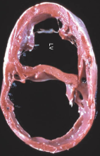
left-sided heart failure