Cardiac Assessment
 Call Kai
 Learn
Learn Practice Test
 Spaced Repetition
 Match
Match Flashcards
 Knowt Play
1/101

There's no tags or description
Looks like no tags are added yet.
Name | Mastery | Learn | Test | Matching | Spaced | Call with Kai | Chat |
|---|
No analytics yet
Send a link to your students to track their progress
102 Terms
What is the role of troponin?
Troponin is a group of regulatory proteins that control the contraction of skeletal and cardiac muscles in response to calcium levels.

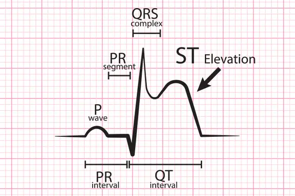
What is a STEMI?
A STEMI (ST-Elevation Myocardial Infarction) is a severe, life-threatening type of heart attack caused by a complete blockage of a major coronary artery.

What is the difference between a STEMI and NSTEMI?
STEMI is a complete blockage of a major coronary artery, whereas an N stemi is an incomplete (partial) blockage of a major coronary artery.

What is a normal troponin level?
6-19 ng/L (nanograms per liter)
The main concept of heart assessment is ____, which is whether the heart can pump blood effectively to the body and whether arteries/veins can move blood where it needs to go.
perfusion

What is the diff betwen ischemia and infarction?
Ischemia = reduced blood/oxygen supply to tissue
Infarction = tissue death after prolonged loss of blood/oxygen.
When seeing elevated troponin levels, that indicates
MI, because troponin is a group of regulatory proteins that control the contraction of skeletal and cardiac muscles in response to calcium levels. If they are elevated, it indicates myocardial injury—meaning heart muscle cells have been damaged or stressed and have leaked the proteins (troponin I or troponin T) into the bloodstream
What is BNP?
Brain Natriuretic Peptide (BNP)
Why does BNP (Brain Natriuretic Peptide) levels rise signficantly after an MI?
BNP is a hormone released by the heart ventricles when they work too hard or stretch under pressure, so higher BNP levels often mean a larger area of heart muscle was damaged during the MI.
What do elevated BNP (brain natriuretic peptide) levels imply?
BNP is a hormone released by the heart ventricles when they work too hard or stretch under pressure, so higher BNP levels often mean a larger area of heart muscle was damaged during the MI.
What is aPTT?
aPTT (Activated Partial Thromboplastin Clotting Time) stands for activated partial thromboplastin time, a blood test that measures how many seconds it takes for your blood to form a clot.
What is Factor Xa?
Factor Xa is an activated blood-clotting enzyme (a serine protease) that plays a critical role in the coagulation cascade by converting prothrombin into thrombin.
What is PT/INR?
A PT/INR (A Prothrombin Time and INR (PT/INR) test is a blood test that measures how many seconds it takes for your blood to form a clot and standardizes the result so doctors everywhere can read it the same way.

What is the difference between a 5-lead and 12-lead ECG?
A 5-lead ECG is used for continuous heart rhythm and rate monitoring, while a 12-lead ECG is used as a diagnostic snapshot to evaluate detailed electrical and structural heart conditions.

What kind of lead ECG would you place to continously monitor heart rhythm and rate?
5-lead ECG
A 5-lead ECG is used for continuous heart rhythm and rate monitoring, while a 12-lead ECG is used as a diagnostic snapshot to evaluate detailed electrical and structural heart conditions.

What is a transthoracic echo?
An ultrasound through the chest that shows structures, valves, wall movement, and ejection fraction (EF).

Name all the diagnostics that can be used to assess the heart.
5 lead EKG
12 lead EKG
Transthoracic Echocardiogram (looks for ejection fraction)
Transesophageal echo (TEE)
Stress Test
Venous/Arterial Ultrasound

What is a transesophageal echo? (TEE)
An ultrasound probe in the esophagus; gives a closer view of posterior heart structures.

What is a stress test?
A cardiac stress test evaluates how well your heart handles physical exertion or simulated stress, helping doctors diagnose coronary artery disease and irregular heart rhythms.
What is a venous/arterial ultrasound?
Venous ultrasound checks for DVT, while an arterial ultrasound/duplex evaluates arterial blood flow, such as PAD.
What does the phrase “Nose to Naval” mean?
“Nose to navel” reminds you that cardiac symptoms can be felt beyond the chest—jaw, neck, arms, upper back, or epigastric area.
What kind of medications can affect your subjective assessment of a patient’s cardiac health?
Blood pressure medications, hyperlipidemia, and erectile dysfunction drugs.
What are the 4 pulses you need to assess for the cardiac assessment?
Rate, rhythm, amplitude, and contour. Compare sides when appropriate.
What is the meaning of each of these pulse strengths?
0+
1+
2+
3+
4+
0+: Absent
1+: Weak/diminished
2+: Expected/normal
3+: Full
4+: Bounding
What is an apical-radial deficit? Why does it happen and what does it imply?
An apical-radial deficit is the difference that occurs when the heart rate counted at the apex of the heart is higher than the pulse rate felt at the wrist.
Definition: It happens when heart contractions are too weak to send a blood wave all the way to the wrist.
The Math: Two workers count beats for one full minute. They subtract the wrist (radial) number from the chest (apical) number.
Normal Result: The number should be zero because every heartbeat should make a pulse at the wrist.
The dorsalis pedis pulse is also known as the
Pedal pulse
What is the equation for pulse pressure?
SBP-DBP (systolic bp-diastolic bp)
Example: For a blood pressure reading of 120/80 mmHg, the pulse pressure is 40 mmHg (120 - 80 = 40).
What is the normal pulse pressure range?
30-40mm Hg
What is MAP?
Mean Arterial Pressure
What is the equation for MAP?
(SBP+2DBP)/3
What is the purpose of measuring pulse pressure and MAP (Mean Arterial Pressure)?
MAP represents the average pressure in a person's arteries during one complete heartbeat cycle. It shows if the blood flow is strong enough to push oxygen and nutrients to vital organs like the brain, heart, and kidneys.
Pulse pressure is the mathematical difference between the top number (systolic) and bottom number (diastolic) of a blood pressure reading (e.g., \(120 - 80 = 40\text{ mmHg}\)).
Detects Vascular Changes: A normal pulse pressure is about 30 to 40 mmHg.
A wide pulse pressure (often over 60–100 mmHg) can mean the arteries have lost their stretch and become stiff (arterial compliance), which is common with aging, high blood pressure, diabetes, or chronic kidney disease.
A narrow pulse pressure (less than 25% of the systolic pressure) can mean the heart is pumping a low volume of blood due to heart failure, severe blood loss, or aortic stenosis.
What is orthostatic blood pressure?
Orthostatic blood pressure measurement checks for a sudden drop in blood pressure when moving from lying down or sitting to standing up, a condition known as orthostatic hypotension.
A patient is lying down and suddenly stands up. Their blood pressure suddenly drops. What condition does this point to?
Orthostatic Hypotension

A patient presents with a blood pressure of 115/78. Is this a normal or elevated blood pressure?

A patient presents with a blood pressure of 128/80. Is this a normal or elevated blood pressure?
Elevated, but not enough for stage 1 hypertension

A patient presents with a blood pressure of 134/89. What stage hypertension is this?
Stage 1
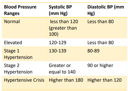
A patient presents with a blood pressure of 143/98. What stage hypertension is this?
Stage 2

A patient presents with a blood pressure of 184/130. What stage hypertension is this?
Hypertensive Crisis
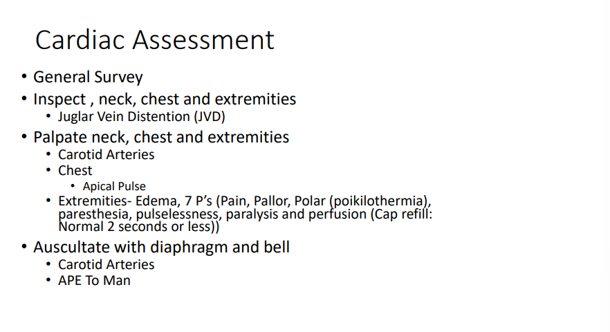
When inspecting the neck, chest, and extremities, you should test for JVD (Jugular Vein Distension). What is JVD and why is it important to look at it? How does it end up looking weird?
Jugular Vein Distension (JVD) is a clinical finding where the jugular veins in the neck become visibly swollen, bulged, or engorged. It serves as an essential, non-invasive indicator of elevated Central Venous Pressure (CVP) and Right Atrial Pressure. Essentially, the jugular vein acts as a built-in "manometer" or pressure gauge for the right side of the heart. When blood cannot flow smoothly into or out of the heart, it backs up into the superior vena cava and spills over into the jugular veins, causing them to bulge.


What is this?
JVD
When palpating the neck, chest, and extremities, what is the 7 P’s?
Assess the 7 P’s for acute limb perfusion concern: pain, pallor, poikilothermia (coolness), paresthesia (abnormal skin sensation, like pins and needles), pulselessness, paralysis, and poor perfusion.
When palpating and assessing the neck, chest, and extremities during a physical or trauma examination, the 7 P’s refer to the mnemonic for a neurovascular assessment. This framework helps healthcare providers identify vascular occlusion, nerve injury, or conditions like compartment syndrome.
When should you use the diaphragm and when should you use the bell when auscultating?
Use the diaphragm for higher-pitched sounds and the bell for lower-pitched sounds.
What is the order of auscultation in the heart and how can you remember it?
APE To Man
Aortic
Pulmonic
Erb’s point
Tricuspid
Mitral
What is the pulse commonly felt during exercise?
3+: Full Volume
You are grading a pulse that is full volume and bounding. What does it mean to bound?
It is a leaping and forceful pulse that quickly disappears
T/F: S1 is closure of the mitral/tricuspid valves; S2 is closure of the aortic/pulmonic valves.
T

S1 is closure of the mitral/tricuspid valves; S2 is closure of the aortic/pulmonic valves. What is S3 and S4? What do they reflect of the heart?
S3: extra sound after S2; can occur with volume overload/heart failure in adults.
S4: extra sound before S1; can reflect a stiff ventricle.

Describe all the locations of the heart when auscultating where S2 is louder than S1/S1 is louder than S2/They’re equal/ etc.
Aortic Area: S2 is louder than S1
Pulmonic Area: S2 is louder than S1
Erb’s Point: S1 and S2 are equal
Tricuspid Area: S1 is louder than S2
Mitral Area: S1 is louder than S2
Which abnormal heart sound sounds like Kentucky?
S3
Which abnormal heart sound sounds like Tennessee?
S4

What does a heart murmur sound like and imply?
Murmur: turbulent blood flow, often from a valve problem.


What does a pericardial friction rub sound like and imply?
Pericardial friction rub: scratchy sound from inflamed pericardial layers.
Edema can be graded on a scale. What does each number on the scale mean?
0+
1+
2+
3+
4+
0+: No pitting edema
1+: Mild pitting edema, 2 mm depression that disappears rapidly
2+: Moderate pitting edema, 4 mm depression that disappears in 10-15 seconds
3+: Moderately severe pitting edema, 6 mm depression that may last more than 1 min
4+: Severe pitting edema, 8 mm depression that can last more than 2 mins
What are the cues for symptomatic bradycardia?
HR < 60
SOB
ALOC
Hypotension
Dizziness
Syncope

A patient has symptomatic bradycardia. What are the actions you should be taking?
Monitor vital signs
12-lead EKG
5-lead EKG (continuous)
Pacemaker
Treat underlying cause

T/F: Caring for a client with a pacemaker can either be temporary or permanent.
T
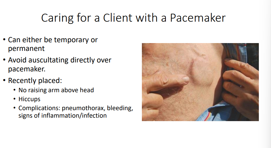
T/F: Caring for a client with a pacemaker can only be permanent.
F, it can be temporary.

Where should you AVOID auscultating if a client has a pacemaker?
Avoid ausculatating directly over the pacemaker.
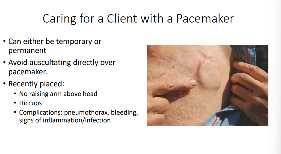
If a pacemaker is recently placed in a client, what are the restrictions for the patient?
Do not raise arm above the head
For the first 4 to 6 weeks, you must avoid raising the arm on the side of the pacemaker above shoulder level. Why it matters: The wires (leads) connecting the pacemaker to your heart tissue need time to secure themselves. Raising your arm too high, reaching behind your back, or lifting heavy objects (usually over 10 pounds) pulls on those areas and can cause lead dislodgement, which requires a repeat procedure to fix.
Frequent or persistent hiccups are an uncommon but important sign to watch out for. Hiccups can indicate phrenic nerve or diaphragmatic stimulation. This means a pacemaker lead may have shifted slightly or its electrical signal is inadvertently triggering the nerve that controls your diaphragm. It can also occasionally point to a lead perforation.
Complications: pneumothorax, bleeding, signs of inflammation/infection
A patient is experiencing left-sided heart failure. Where does this manifest in a patient?
Failure of the left ventricles and atrium mean blood cannot effectively get pumped out to the body, so blood backs up in the lungs, causing crackles, cough, dyspnea, orthopnea, and paroxysmal noctural dyspnea.
A patient is experiencing right-sided heart failure. Where does this manifest in a patient?
Failure of the right atrium and ventricles results in a failure of blood getting pumped out to the lungs, so blood backs up in the body, leading to JVD, dependent edema, weight gain, and ascites.
What can rapid weight gain indicate?
Daily weights are important because rapid gain can mean fluid retention.
A patient presents with new onset chest pain, SOB, diaphoretic, altered HR and BP, and signs and systems of systemic perfusion alteration. They are vomiting, have nausea, and are fatigued. These are cues for what potential disease?
Acute Coronary Syndrome (ACS): Sudden reduced blood flow to the heart
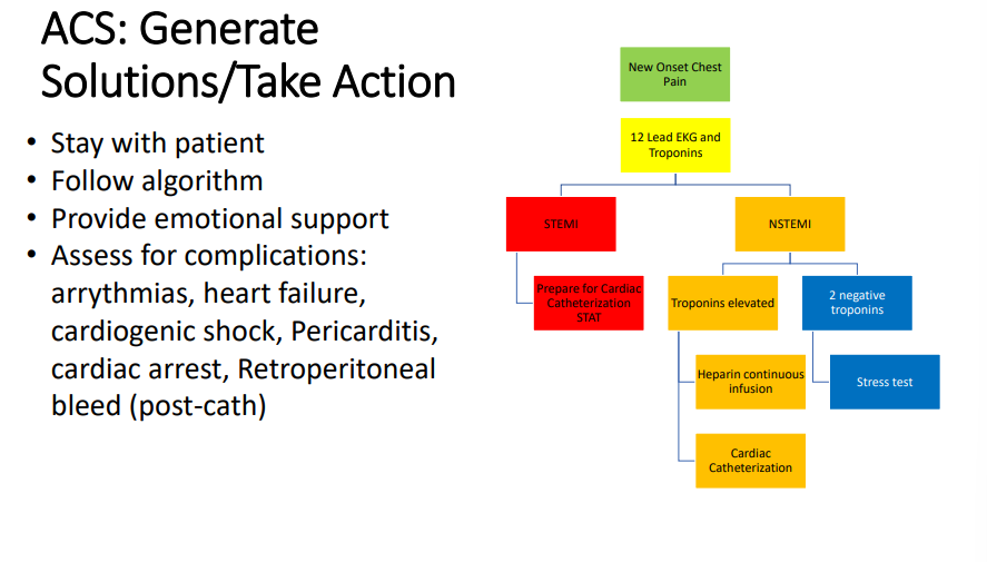
Your patient is experiencing acute coronary syndrome (ACS). You have put a 12 lead EKG and ordered a lab for troponins. Suddenly, a STEMI occurs. What procedure do you need to prepare for STAT?
Cardiac Catherization

Your patient is experiencing acute coronary syndrome (ACS). You have put a 12 lead EKG and ordered a lab for troponins. A NSTEMI occurs. Troponins are elevated. What is the correct procedure to follow?
Heparin Continuous Infusion (A heparin infusion is given to patients with elevated troponin levels when there is a high clinical suspicion or confirmation of an acute coronary syndrome (ACS), such as a Non-ST-elevation myocardial infarction (NSTEMI).
Cardiac Catherization

Your patient is experiencing acute coronary syndrome (ACS). You have put a 12 lead EKG and ordered a lab for troponins. A NSTEMI occurs. 2 negative troponins are present (Two negative troponin tests mean that the levels of cardiac proteins (Troponin I or Troponin T) in your blood are within normal, low, or undetectable ranges, indicating a low likelihood of an acute heart attack (myocardial infarction) or major heart muscle damage at the times the blood samples were drawn). What is the correct procedure to follow?
A stress test.

What is atrial fibrillation with rapid ventricular response (AFib with RVR)?
Atrial fibrillation with rapid ventricular response (AFib with RVR) is an irregular, chaotic heart rhythm where the upper chambers quiver and the lower ventricles beat at a fast rate of over 100 beats per minute.

What are the solutions to a-fib (medications)?
Rate control medication
Antiarrythmic medication
Anticoagulation
Cardioversion
Anticoagulation or TEE
Monitor for signs/symptoms of stroke

What is cardioversion? What is the difference between anticoagulation and TEE cardioversion?
Cardioversion is a medical treatment that uses quick, low-energy electrical shocks or specific medicines to restore a regular heart rhythm when you have a fast or irregular heartbeat.
Conventional Anticoagulation Strategy
Duration: Patients take an oral anticoagulant (such as warfarin or a NOAC) for a minimum of 3 weeks before the procedure and at least 4 weeks after. [1, 2]
Goal: Dissolve or prevent the formation of blood clots in the left atrium or left atrial appendage naturally over time. [1, 2]
Pros: Avoids the need for an invasive diagnostic procedure (TEE). [1]
Cons: Delays the procedure by 3 weeks or more, keeps the patient in atrial fibrillation longer, and carries a cumulative risk of bleeding over the pre-cardioversion window.
TEE-Guided Strategy
Procedure: A specialized ultrasound camera is guided down the esophagus to examine the heart chambers for a thrombus (blood clot). [1, 2]
Timeline: If no clot is found, acute anticoagulation is started (e.g., a dose of a DOAC or heparin), and cardioversion is performed shortly after (often within hours). Anticoagulation is still continued for at least 4 weeks post-procedure. [1, 2]
If a clot is found: Cardioversion is delayed, and the patient is prescribed extended anticoagulation until a repeat TEE proves the clot is gone. [1, 2]
Pros: Shortens the time to cardioversion, reduces overall bleeding complications compared to long pre-treatment, and restores sinus rhythm faster. [1, 2]
Cons: Requires a semi-invasive procedure with mild sedation, increases initial hospital resource/cost use, and does not completely eliminate the risk of post-procedural stroke or atrial stunning. [1, 2, 3]
A patient presents with intermittment claudication (Intermittent claudication is a cramping, aching, or tired pain in the legs that happens during exercise like walking and goes away after a short rest), paresthesia, thin, shiny skin, cool, a loss of hair on LE, and a diminished pulse. What is the possible diagnosis?
Peripheral Arterial Disease (PAD)

What is intermittent claudication?
Intermittent claudication is a cramping, aching, or tired pain in the legs that happens during exercise like walking and goes away after a short rest
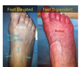
What is the difference between foot elevated and foot dependent PAD? (peripheral arterial disease)
The difference between foot-elevated and foot-dependent states in peripheral arterial disease (PAD) relates to how gravity and position affect blood flow and skin color in the lower extremities.
Elevation: Pallor; Position: The leg or foot is raised above the level of the heart. Because blood cannot easily reach the skin, the foot turns pale, white, or blanched.
Dependent: Rubor; The leg or foot hangs down below the level of the heart (such as dangling off the edge of a bed or chair). Gravity helps pull what little blood is available down into the foot.
Elevated WBC with left shift, positive blood cultres, and elevated lactic acid (if now causing decreased perfusion) points to
inflammation
A pleural friction rub (respiratory) and a pericardial friction rub (cardiac) can be from
inflammation, the body’s natural immune response to tissue damage or foreign organisms/particles.
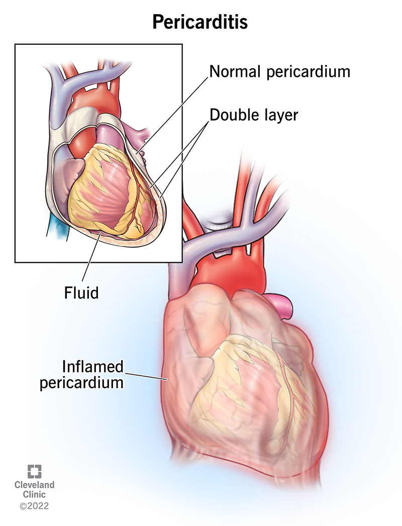
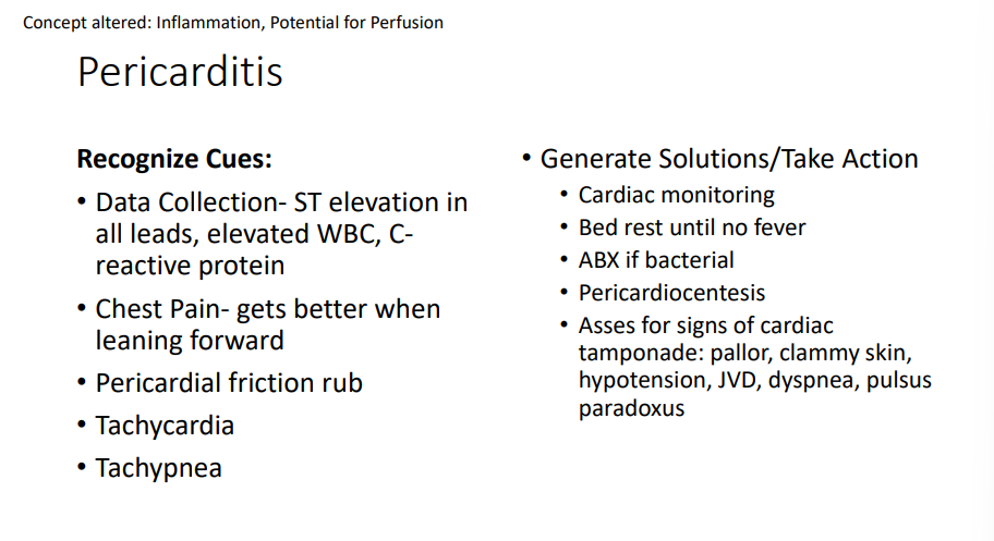
A patient has chest pain that gets better when leaning forward. What condition could this be pointing to?
Pericarditis. Leaning forward eases pericarditis chest pain because the position shifts the heart slightly, which separates the inflamed layers of the heart sac and reduces friction.


What are appropriate actions to take for a patient with pericarditis?
Cardiac Monitoring
Bed rest until no fever
ABX if bacterial
Pericardiocentesis
Assess for signs of cardiac tamponade: pallor, clammy skin, hypotension, JVD, dyspnea, pulsus paradoxus
Cardiac tamponade is a life-threatening medical emergency where excess fluid or blood builds up in the pericardial sac around the heart, compressing the chambers and preventing proper filling.

You are asked to assess for signs of cardiac tamponade. Cardiac tamponade is a life-threatening medical emergency where excess fluid or blood builds up in the pericardial sac around the heart, compressing the chambers and preventing proper filling. What do you need to look for?
pallor, clammy skin, hypotension, JVD, dyspnea, pulsus paradoxus

A patient is educated on the importance of bed rest until they do not have a fever for their condition. What is their likely condition?
Pericarditis.

A patient presents with elevated D-dimer (A D-dimer test is a simple blood test that measures protein fragments left behind when a blood clot dissolves in the body) and a venous ultrasound. What condition does this point to?
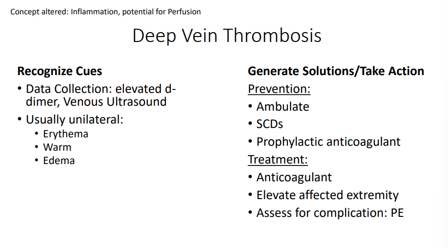
Deep Vein Thrombosis

While up to 50% of DVT cases can be asymptomatic, a symptomatic clot typically presents unilaterally (affecting only one limb, usually the lower leg or thigh). The primary local signs include?
Erythema: Redness or discoloration of the skin over the affected area.
Warmth: The skin feels noticeably warmer to the touch compared to the unaffected limb.
Edema: Unilateral swelling or pitting edema caused by venous congestion.
How can you prevent DVT?
Ambualte the patient
SCDs (Sequential Compression Device)
Prophylactic anticoagulants (medications given in lower doses to prevent harmful blood clots, such as deep vein thrombosis (DVT) and pulmonary embolism (PE), in people at risk.)


What is an SCD?
Sequential Compression Device, Sequential Compression Device, which is a mechanical treatment used primarily for DVT (deep vein thrombosis) prevention rather than an active treatment for an existing clot.
What is the treatment for DVT?
Anticoagulant
Elevate the affected extremity
Assess for complication: PE
T/F: You should NOT elevate the affected extremity for DVT.
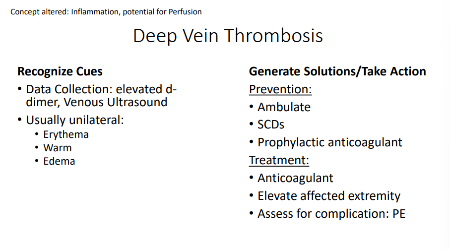
F, you should elevate the affected extremity for DVT to promote venous return to the heart.

DVT can complicate into what condition?
PE

A patient presents with JVD, a wet cough, crackles, increased tactile fremitus, dull percussion tones, and edema. Is this an excess fluid balance or a deficit?
Excess

What is Orthopnea/Paroxysmal Noctural Dyspnea?
Orthopnea and paroxysmal nocturnal dyspnea (PND) are both forms of shortness of breath that happen when lying down, but they differ in when and how they occur.
Orthopnea: Shortness of breath that happens whenever you lie flat, whether you are awake or asleep.
Paroxysmal Nocturnal Dyspnea (PND): Sudden, severe shortness of breath that wakes you up from sleep, usually one to three hours after lying down.

A patient is experincing paroxysmal noctural dyspnea. What could this possibly point to?
Excess fluid balance

You feel a patient’s skin and they have dry mucous membranes and dry skin. They also exhibit tenting. What kind of fluid condition does this indicate?
Fluid deficit

Electrolyte imbalances can cause what symptoms?
Rhythm and heart rate
Nausea/Vomiting
Muscle weakness

What are the cues of hypokalemia?
K<3.5
ABG-metabolic alkalosis
Tachycardia
Rhythm changes
Nausea, diarrhea
Muscle weakness/flaccid paralysis

What are the cues for hyperkalemia?
K>5.5
ABG-metabolic acidosis
Tachycardia
Rhythm changes
Nausea, diarrhea
Muscle weakness/leg cramps

A patient presents with an ABG result of metabolic alkalosis. Would they be experiencing hypo or hyperkalemia?
Hypokalemia

A patient presents with an ABG result of metabolic acidosis. Would they be experiencing hypo or hyperkalemia?
Hyperkalemia

Cardiac monitoring and potassium supplementation is an appropriate action to take when a patient has what condition?
Hypokalemia

Cardiac monitoring, IV calcium gluconate, IV dextrose and IV insulin, albuterol, kayexalate, loop diuretics, and sodium bicarb are appropriate interventions for what condition? Why?
Hyperkalemia
Why Each Intervention Is Used
Cardiac Monitoring:
Why: To continuously watch for dangerous ECG changes (such as peaked T-waves, widened QRS complexes, or heart blocks) and arrhythmias caused by high potassium. [1, 2, 3]
IV Calcium Gluconate:
Why: It stabilizes the cardiac cell membrane (raising the threshold potential) to prevent heart cells from firing uncontrollably, though it does not lower actual potassium levels. [1, 2, 3]
IV Dextrose and IV Insulin:
Why: Insulin stimulates the Na⁺/K⁺-ATPase pump, actively shifting potassium from the bloodstream back into the cells. Dextrose (glucose) is given alongside insulin to prevent dangerous low blood sugar (hypoglycemia). [1, 2]
Albuterol (Nebulized):
Why: As a beta-2 adrenergic agonist, it also activates cellular sodium-potassium pumps to help drive potassium intracellularly, often working synergistically with insulin. [1, 2, 3]
Sodium Bicarbonate:
Why: It helps shift potassium into cells by correcting co-existing severe metabolic acidosis (which trades hydrogen ions out of cells for potassium ions in). [1, 2]
Loop Diuretics (e.g., Furosemide):
Why: They promote the renal (urinary) excretion of potassium out of the body, provided the patient has adequate kidney function and urine output. [1, 2]
Kayexalate (Sodium Polystyrene Sulfonate):
Why: It acts as a gastrointestinal potassium binder that exchanges sodium for potassium in the gut, slowly eliminating potassium via the stool (useful for non-emergent or subacute clearance). [1, 2]

What are the different types of heart failure?
HF with reduced EF (systolic)
HF with preserved EF (diastolic)
Right heart vs left heart failure

What are the appropriate interventions to take for a patient with heart failure?
Daily weights; 3 pounds in a day, 5 pounds in a week
Low sodium diet
Fluid restriction
Administer ordered drugs:
Diuretics
Inotropics
Ace or Arb
Beta blocker
Educate on when to seek medical care

askldjasldjaslkdj
okay

A patient presents with aching, cramping pain. They have present pulses but can be difficult to find due to their edema. Their wounds are superficial, pink, and it is over the inner ankle. The skin is thick and tough. There is a slight brown pigment. What condition is this pointing to?
Peripheral Venous Disease