KN 230 Exam II (Chapters 7, 8, 9)
 Call Kai
 Learn
 Practice Test
 Spaced Repetition
 Match
 Flashcards
 Knowt Play
1/592

There's no tags or description
Looks like no tags are added yet.
593 Terms
Skeleton
The bony framework of the body
Collectively, all the bones of the body
Skeletal system
A living framework that includes the bones of the skeleton as well as cartilage, ligaments, and other connective tissues that stabilize or connect the bones

Bones
The primary organs of the skeletal system
Form the rigid framework of the body
Two types: Compact and spongy
Basic functions: Support and protection, levers for movement, hematopoiesis, storage of mineral and energy reserves
Four classes determined by shape: Long, short, flat, irregular
Coverings and linings: Periosteum and endosteum
Four types of cells: Osteoprogenitor cells, osteoblasts, osteocytes, and osteoclasts
Compact bone
A relatively rigid connective bone tissue that appears white, smooth, and solid
Makes up approximately 80% of the total bone mass
Also called dense or cortical bone
Spongy bone
Located internal to compact bone
Appears porous
Makes up approximately 20% of the total bone mass
Also called cancellous or trabecular bone
Cartilage
A semirigid connective tissue that is more flexible than bone
Three subtypes, but only two that are associated with the skeletal system (hyaline cartilage and fibrocartilage)
Hyaline cartilage
A cartilage subtype associated with the skeletal system
Attaches ribs to the sternum (costal cartilage), covers the ends of some bones (articular cartilage), and is within growth plates (epiphyseal plates)
Provides a model during development for the formation of the fetal skeleton
Fibrocartilage
A cartilage subtype associated with the skeletal system
A weight-bearing cartilage that withstands compression
Forms the intervertebral discs, the pubic symphysis (cartilage between bones of the pelvis), and the cartilage pads of the knee joints (menisci)
Ligament
A band or sheet of dense regular connective tissue that connects bone to bone
Function to stabilize, strengthen, and reinforce most synovial joints
Tendon
A cord of dense regular connective tissue that connects muscle to bone
Are like ligaments, but not part of the synovial joint
Help stabilize joints
Hematopoiesis
The formation and development of blood cells
Occurs in red bone marrow of spongy bone connective tissue
Calcium
An essential mineral for such body functions as muscle contraction, release of neurotransmitter from nerve cells, and blood clotting
Phosphate
A structural component of ATP, other nucleotides, and phospholipids and is an important component of the plasma membrane
Long bones
Bones that are greater in length than width
Have an elongated, cylindrical shaft (diaphysis)
The most common bone shape
Found in the upper limbs (arm, forearm, palm, fingers) and lower limbs (thigh, leg, sole of the foot, toes)
Regions: Diaphysis, epiphysis, metaphysis

Short bones
Bones that have a length nearly equal to their width
Examples: Carpals (wrist bones), tarsals (bones in the foot), sesamoid bones (bones along tendons of some muscles)
Flat bones
Bones that have flat, thin surfaces that may be slightly curved
Provide extensive surface areas for muscle attachment and protect underlying soft tissues
Form the roof of the skull, the scapulae (shoulder blades), the sternum (breastbone), and the ribs

Irregular bones
Bones that have elaborate, sometimes complex shapes and do not fit into any of the other categories
Examples: Vertebrae, hip bones (ossa coxae), several bones in the skull (e.g., ethmoid, sphenoid)
Diaphysis
Elongated, usually cylindrical part of a long bone between its two ends
The shaft of a long bone
Provides for the leverage and major weight support of a long bone
Compact bone composes the more external component, whereas spongy bone extends internally from this compact bone in the form of spicules
Has a cylindrical space within called the medullary (marrow) cavity
Contains red bone marrow in children, which is later replaced by yellow bone marrow in adults
Medullary (marrow) cavity
The cylindrical space (hollow) within the diaphysis
Epiphysis
Expanded, knobby region at the end of a long bone
Two types: Proximal and distal
Composed of an outer, thin layer of compact bone and an inner, more extensive region of spongy bone (resists stress applied from many directions)
Articular cartilage covers its joint surface (helps reduce friction and absorb shock in movable joints)
Proximal epiphysis
The end of the long bone closest to the body trunk
Distal epiphysis
The end of the long bone farthest from the trunk
Articular cartilage
A thin layer of hyaline cartilage covering the joint surface of an epiphysis
Helps reduce friction and absorb shock in movable joints
Metaphysis
Section of a long bone between the epiphysis and the diaphysis
Contains the epiphyseal plate in a growing bone (provides for the continued growth in bone length)
Epiphyseal plate
A thin layer of hyaline cartilage located between a long bone diaphysis and epiphysis
Allows longitudinal growth of the bone
Five zones: Zone of resting cartilage, zone of proliferating cartilage, zone of hypertrophic cartilage, zone of calcified cartilage, and zone of ossification

Epiphyseal line
The remnant of the epiphyseal plate that remains when long bone growth ceases
Thin, defined area of compact bone
Periosteum
A thick, fibrous membrane covering the entire external surface of a bone, except for the articular cartilage on the epiphyses
Consists of two layers: An outer fibrous layer of dense irregular connective tissue and an inner cellular layer primarily composed of bone stem cells and osteoblasts
Anchored to the bone by perforating fibers
Perforating fibers
Numerous collagen fibers that run perpendicular to the diaphysis
Anchors the periosteum to the bone
Also called Sharpey’s fibers
Endosteum
A relatively delicate layer of cells lining the inner surface of bone in the medullary cavity
Composed of a very thin layer of connective tissue containing cells and reticular fibers
Diploë
Central layer of spongy bone between the two layers of compact bone (outer and inner plates) of the flat cranial bones
Nutrient forament
A small opening or hole in the bone where only one nutrient artery enters and one nutrient vein exits the bone
Nerves also enter to innervate the bone as well as the periosteum, endosteum, and marrow cavity
Red bone marrow
Soft connective tissue of bone that contains reticular connective tissue, developing red blood cells, and adipocytes
Primary function: Forms the blood cells of the body
Locations differ in significant ways between children and adults
Also called myeloid tissue
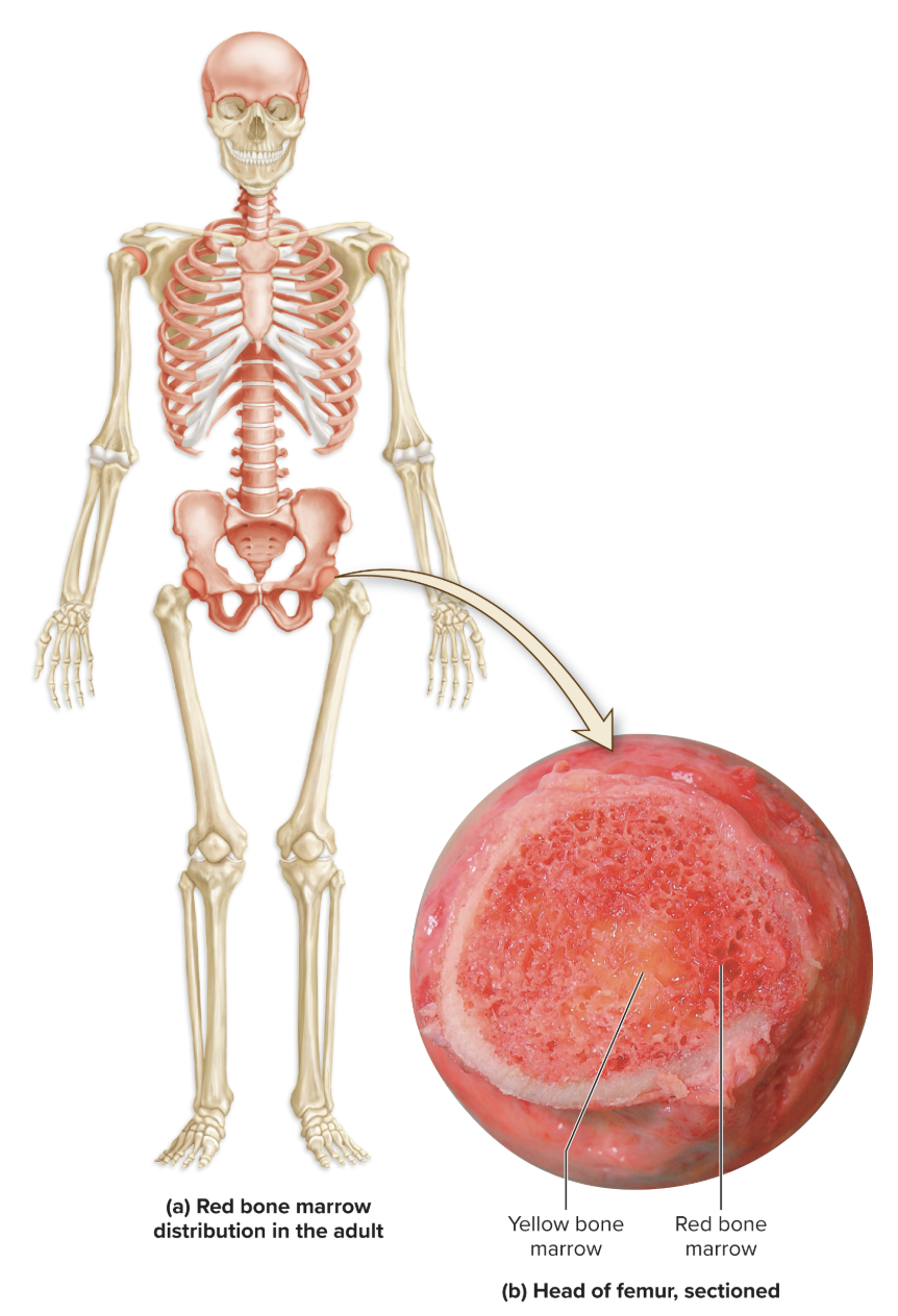
Yellow bone marrow
Soft connective tissue of bone that has fewer developing blood cells and more adipocytes
Primarily within the medullary cavities of long bones and the inner core of most epiphyses as children mature into adults
Anemia
Any condition in which the number of erythrocytes is low or the oxygen-carrying capacity of the blood is reduced
Osteoprogenitor cells
Bone stem cells derived from mesenchyme
Precursor to bone cells
Located in both the periosteum and the endosteum

Osteoblasts
A dividing cell in bone
Formed from osteoprogenitor stem cells
Synthesizes and secretes the initial semisolid organic form of bone matrix called osteoid

Osteocytes
Bone cell in a lacuna within bone matrix
Mature bone cells derived from osteoblasts that have lost their bone-forming ability when the osteocytes become embedded within the calcified osteoid
Maintain the bone matrix and detect mechanical stress on a bone

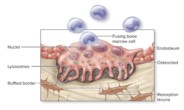
Osteoclasts
Large multinuclear cells that function in the absorption and removal of bone connective tissue
Derived from fused bone marrow cells similar to those that produce monocytes
Exhibit a ruffled border where they contact the bone (increases their surface area exposure to the bone)
Often located within or adjacent to a depression or pit on the bone surface called a resorption lacuna (Howship’s lacuna)
Phagocytic (involved in breaking down bone in a process called bone resorption)
Extracellular matrix
Protein fibers and ground substance in the extracellular space of connective tissue
Has both organic and inorganic components in bone connective tissue
Osteoid
The organic component of the extracellular matrix of bone connective tissue
Composed of both collagen proteins and a semisolid ground substance (proteoglycans and glycoproteins)
Calcification
The process in which structures in the body become hardened as a result of deposited calcium salts
Occurs only in the formation of bone and teeth
Also called mineralization
Bone resorption
A process whereby bone matrix is destroyed by substances released from osteoclasts
These substances are stored within the numerous lysosomes of osteoclasts (proteolytic enzymes and hydrochloric acid (HCl) are released from lysosomes)
Occurs during bone remodeling and when blood calcium levels are low
Osteolysis
The release of the stored calcium and phosphate from the bone matrix
Osteons
The basic functional and structural unit of mature compact bone
Also called Haversian systems
Oriented parallel to the diaphysis of the long bone
Has several components: Central canal, concentric lamellae, osteocytes, lacunae, canaliculi

Central (Haversian) canal
A cylindrical channel at the center of the entire length of the osteon
Blood vessels and nerves that supply the bone extend through
Concentric lamellae
Rings of bone connective tissue that surround the central canal and form the bulk of the osteon
Numbers vary among osteons
Each contains collagen fibers oriented at an angle in one direction (alternating pattern of collagen fiber direction gives strength and resilience to bone)
Lacunae
The small spaces that each house an osteocyte
Canaliculi
Tiny, interconnecting channels within the bone connective tissue that extend from each lacuna, through the lamellae, and connect to other lacunae and the central canal
House osteocyte cytoplasmic projections that permit intercellular contact and communication
Perforating (Volkmann) canals
Canals that resemble central canals in that they also contain blood vessels and nerves, but they run perpendicular to the central canals and help connect multiple central canals within different osteons
Forms a channel for a vascular and innervation connection among the multiple osteons
Circumferential lamellae
Rings of bone immediately internal to the periosteum of the bone (external CL) or immediately external to the endosteum (internal CL)
External circumferential lamellae
Rings of bone immediately internal to the periosteum of the bone
Internal circumferential lamellae
Rings of bone immediately external to the endosteum of the bone
Interstitial lamellae (interstitial systems)
The components of compact bone that are between osteons or the leftover parts of osteons that have been partially reabsorbed
Incomplete and typically have no central canal
Trabeculae
An open lattice of narrow rods and plates of bone in spongy bone
Provides great resistance to stressed applied in many directions by distributing the stress throughout the entire framework
Chondroblasts
A dividing cell in cartilage, which produces the cartilage matrix
Derived from mesenchymal cells
Chondrocytes
Mature, nondividing cartilage cells
Occupy lacunae
Maintain the matrix
Perichondrium
A layer of dense irregular connective tissue around the surface of hyaline cartilage (except articular cartilage)
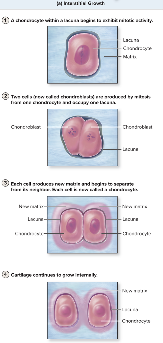
Interstitial growth
An increase in length that occurs within the internal regions of cartilage through the following series of four steps:
Chondrocytes housed within lacunae are stimulated to undergo mitotic cell division.
Following cell division, two cells occupy a single lacuna; they are now called chondroblasts (cells capable of producing cartilage matrix).
As chondroblasts begin to synthesize and secrete new cartilage matrix, they are pushed apart. Each cell now resides in its own lacuna and is called a chondrocyte.
The cartilage continues to grow in the internal regions as chondrocytes continue to produce more matrix.
Appositional growth
An increase in width along the cartilage’s outside edge, or periphery, in the following three steps:
Undifferentiated stem cells at the internal edge of the perichondrium begin to divide.
New undifferentiated stem cells and committed cells that differentiate into chondroblasts are formed. These chondroblasts are located at the periphery of the old cartilage, where they begin to produce and secrete new cartilage matrix.
The chondroblasts, as a result of matrix formation, push apart and become chondrocytes, with each occupying its own lacuna. The cartilage continues to grow at the periphery as chondrocytes continue to produce more matrix.

Ossification
The formation and development of bone connective tissue
Also called osteogenesis
Intramembranous ossification
The formation of bone tissue on a thin layer of mesenchyme (the first type of connective tissue formed in the developing embryo)
Composed of mesenchymal cells and abundant ground substance
Produces the flat bones of the skull, some of the facial bones, the mandible, and the central part of the clavicle
Continues in the following steps:
1. Ossification centers form within thickened regions of mesenchyme beginning at the eighth week of development.
2. Osteoid undergoes calcification.
3. Woven bone and its surrounding periosteum form.
4. Lamellar bone replaces woven bone, as compact bone and spongy bone form.

Woven bone
Primary bone
Newly formed bone connective tissue that is immature and not well-organized during the process of intramembranous ossification
Lamellar bone
Secondary bone
Replaces the trabeculae of woven bone during the process of intramembranous ossification
Endochondral ossification
Bone formation that takes place within hyaline cartilage
Used to form most bones of the body
Takes place in the following steps:
1. The fetal hyaline cartilage model of bone develops.
2. Cartilage calcifies, and a periosteal bone collar forms.
3. The primary ossification center forms in the diaphysis.
4. Secondary ossification centers form in the epiphyses.
5. Bone replaces almost all cartilage, except the articular cartilage and epiphyseal plates.
6. Lengthwise growth continues until the epiphyseal plates ossify and form epiphyseal lines.
Bone remodeling
The constant, dynamic process of continual addition of new bone tissue (bone deposition) and removal of old bone tissue (bone resorption)
Mechanical stress
Occurs in the form of weight-bearing movement and weight-bearing exercise
Also known as mechanical load
Affects the structure of the bone
Detected by osteocytes and communicated to osteoblasts
Growth hormone
Hormone produced by the anterior pituitary gland
Stimulates liver to produce the hormone insulin-like growth factors (IGFs), which causes cartilage proliferation at epiphyseal plate and resulting bone elongation
Thyroid hormone
Hormone that is secreted by the thyroid gland
Stimulates bone growth by stimulating the metabolic rate of osteoblasts
Calcitonin
Hormone that, in childhood, promotes calcium deposition in bone and inhibits osteoclast activity
Appears to have minimal effects in adulthood
Sex hormones (estrogen and testosterone)
Hormones that begin to be secreted in relatively large amounts at puberty
Stimulate osteoblasts
Promote epiphyseal plate growth and closure
Parathryoid hormone
Hormone that increases blood calcium levels by encouraging bone resorption by osteoclasts
Calcitriol
Hormone that increases blood calcium levels by encouraging bone resorption by osteoclasts and stimulating absorption of calcium ions from the small intestine into the blood
Active form of the hormone that regulates blood calcium and is synthesized from vitamin D
Glucocorticoids
A group of steroid hormones that are released from the adrenal cortex and regulate blood glucose levels
Increase bone loss and, in children, impair bone growth in response to chronically high levels
Serotonin
Hormone that inhibits osteoprogenitor cells from differentiating into osteoblasts when there are chronically high levels of serotonin
Osteopenia
Decreased calcification or density of bone
Osteoporosis
Medical condition characterized by decreased bone mass and increased susceptibility to fracture
Fractures
Breaks in bones
Classified in several ways: Avulsion, Colles, comminuted, complete, compound (open), compression, depressed, displaced, epiphyseal, greenstick, hairline, impacted, incomplete, linear, oblique, pathologic, Pott, simple (closed), spiral, stress, transverse
Avulsion fracture
A bone fragment is pulled off or separated from the rest of the bone by a soft tissue attachment
Colles fracture
Fracture of the distal end of the lateral forearm bone (radius)
Produces a “dinner fork” deformity

Comminuted fracture
Bone is splintered into several small pieces between the main parts

Complete fracture
Bone is broken into two or more pieces
Compound (open) fracture
Broken ends of the bone protrude through the skin

Compression fracture
Bone is squashed (may occur in a vertebra during a fall)
Depressed fracture
Broken part of the bone forms a concavity (as in skull fracture)
Displaced fracture
Fractured bone parts are out of anatomic alignment
Epiphyseal fracture
Epiphysis is separated from the diaphysis at the epiphyseal plate
Greenstick fracture
Partial fracture
One side of bone breaks—the other side is bent
More commonly seen in children
Hairline fracture
Fine crack in which sections of bone remain aligned (common in skull)
Impacted fracture
One fragment of bone is firmly driven into the other
Incomplete fracture
Partial fracture extends only partway across the bone
Linear fracture
Fracture is parallel to the long axis of the bone
Oblique fracture
Diagonal fracture is at an angle
Pathologic fracture
Weakening of a bone caused by disease process (e.g., cancer)
Pott fracture
Fracture is at the distal ends of the tibia and fibula

Simple (closed) fracture
Bone does not break through the skin
Spiral fracture
Fracture spirals around axis of long bone
Results from twisting stress

Stress fracture
Thin fractures due to repeated, stressful impact, such as running (these fractures often are difficult to see on x-rays, and a bone scan may be necessary to accurately identify their presence)
Transverse fracture
Fracture is at right angles to the long axis of the bone

Bone fracture repair
A fracture hematoma forms.
A fibrocartilaginous (soft) callus forms.
A bony (hard) callus forms.
The bone is remodeled.
Axial skeleton
The division of the skeletal system that is composed of the bones along the central axis of the body
Includes the bones of the skull (and its associated bones), vertebral column, sternum, and ribs
Forms a framework that supports and protects the organs
Its spongy bone contains hematopoietic tissue that is responsible for blood cell formation