PCS III: Final Objective Structured Clinical Examination (OSCE) Study Material
 Call Kai
 Learn
 Practice Test
 Spaced Repetition
 Match
 Flashcards
 Knowt Play
1/56

There's no tags or description
Looks like no tags are added yet.
57 Terms
symmetry!
What's the first thing to observe upon a live oral exam?
crown
neck
root
What are the three main parts/regions of the tooth?
Start with the muzzle closed. Pull the lips up and examine the incisors and then around the canine teeth and back to the premolars and molar
After observing general symmetry of the head, lips, muzzle, etc what is your next step in the oral exam?
lips and mucosa for gingival redness, masses, swelling, etc.
At the beginning of the oral exam, what soft tissue structures should you be observing after pulling the lips up?
• Remove dental calculus: Supragingival and subgingival
• Evaluate for periodontal disease
• Note oral lesions early
What are three goals of an anesthetized oral exam and dental cleaning?
Tartar removers or calculus removal forceps are made for quick removal of large pieces of calculus
What is this instrument called?
what it do?

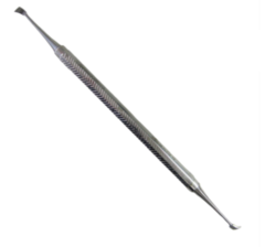
this is a tartar scraper
fine calculus removal
What is this instrument called?
what it do?
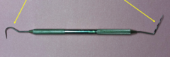
Explorer on left, Periodontal probe on right:
Explorers are used to detect plaque and calculus, explore caries (cavities), and check for pulp exposure
Probing of the gingival sulcus or pockets is performed using a periodontal probe to help measure the gingival sulcus depth
What is this instrument called?
what it do?
dogs: 1-3mm
cats: 0.5mm
What is the normal sulcus depth in dogs?
what about in cats?
This is a scaler
Scalers are for supragingival calculus removal only and come in a variety of sizes and shapes. The have three sharp sides and a sharp tip
What is this instrument called?
what it do?

NO
Can scalers be used subgingivally?
This is a Gracey Curette
Curettes are used for both subgingival and supragingival calculus removal. The Gracey Curette is the most commonly used curette in veterinary medicine
What is this instrument called?
what it do?

Extracting forceps - they are used to grasp the tooth to remove it from the socket.
What is this instrument called?
what it do?
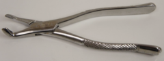
Elevator (these are wing tip elevators)
If the tooth is not loose, elevators are used to loosen and elevate the teeth in their sockets prior to extraction. Intact periodontal ligaments need to be stretched and severed down the entire root in order to remove the tooth
What is this instrument called?
what it do?

This is a luxator!
are also used for tooth extraction, but luxators are thinner and finer than elevators to cut the periodontal ligament. They should not be used in a prying motion like most elevators as the thin head can easily break down in the socket
What is this instrument called?
what it do?

periosteal elevator
is used to elevate the attached gingiva surrounding a tooth from the alveolar bone. This provides exposure of the bone surrounding the tooth root for surgical removal of the tooth and also provides a flap for closure of the socket once the tooth is extracted
What is this instrument called?
what it do?

NO!
- it destroys vital surface structures
- may inhibit culture
do you scrub or prep derm related diseases when you are doing a biopsy?
put them in formalin
what should you do with crusts that you find when performing a biopsy?
- do not scrub or prep
- may be local or general anesthesia dependent upon size and location
- avoid ulcerated areas
biopsy guidelines for derm related diseases
- clip, scrub prep
- may be local or general anesthesia dependent upon size and location
- avoid ulcerated areas
biopsy guidelines for possible cancer cases
~ 4 to 8 mm
what is the typical size of biopsy punch?

the junction of abnormal and normal with a punch. if you want both - take two separate biopsies
what should you not biopsy with a punch biopsy?
NO, you won't get an answer
should you biopsy ulcerated areas?
patient demeanor, depth of the lesion
what is local v. general anesthesia dependent upon for derm cases?
for skin and subQ masses
when do you use incisional biopsys?
- blade incision to minimize crushing of sample
- appositional closure
technique of incisional biospy
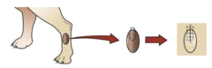
mass is ulcerated or inflamed
incisional biopsy is preferred over needle or punch biopsy when?
subsequent excision in mind
during incisional biopsy, the incision is made with?
complete excision of skin or subQ mass
excisional biopsy
tumor "anatomy"
what must be considered when performing an excisional biopsy?
- Achieve marginal excision if possible
- Best done when benign tumor or ample tissue for wider resection later
tumor "anatomy" when performing excisional biopsy
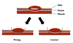
measure and draw with sterile marking pen the margins you wish to obtain
what should you do prior to excisional biopsy?

- Small, superficial, freely movable masses that are easily resectable with minimal morbidity
- Cases that knowledge of histologic type would not alter the type or extent of treatment
Excisional biopsy should be reserved for:
diagnose or stage neoplastic process
lymph node biopsy is used to?
- Complete excision of lymph node
- Incisional biopsy
- Lymph nodes within the abdomen or thoracic should also be sampled
lymph node biopsy
- always submit entire excised tissue
- mark cut borders with india ink
- place sutures for tumor orientation
- communicate with pathologist
what to do with sample after it is removed?
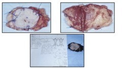
In the event of "dirty" margins it is important to know where the questionable area was located
why is it important to place sutures for tumor orientation?
- skin and subcutis
- SDFT
- DDFT
- carpal sheath
- ALDDFT (check ligament)
- SL
- metacarpus
what structures are in distal horse U/S zone 1A?
- skin and subcutis
- SDFT
- DDFT
- ALDDFT (check ligament)
- SL
- metacarpus
what structures are in distal horse U/S zone 1B?
- skin and subcutis
- SDFT
- DDFT
- ALDDFT (check ligament) joining DDFT
- SL
- metacarpus
what structures are in distal horse U/S zone 2A?
- skin and subcutis
- SDFT
- DDFT
- SL beginning to divide
- metacarpus
what structures are in distal horse U/S zone 2B?
- skin and subcutis
- SDFT
- DDFT
- digital sheath wall
- branches of SL
- metacarpus
what structures are in distal horse U/S zone 3A?
- skin and subcutis
- SDFT
- DDFT
- SDFT ring
- digital sheath wall
- branches of SL
- palmar pouch; metacarpophalangeal joint
- metacarpus
what structures are in distal horse U/S zone 3B?
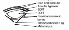
- skin and subcutis
- annular ligament
- SDFT
- DDFT
- proximal sesamoid bones
- intersesamoidean ligament
- metacarpus
what structures are in distal horse U/S zone 3C?
1A
what zone am I in?

1B
what zone am I in?

2A
what zone am I in?

2B
what zone am I in?

3A
what zone am I in?
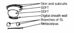
3B
what zone am I in?

3C
what zone am I in?
1A
what zone am I in?

1B
what zone am I in?

2A
what zone am I in?

2B
what zone am I in?

3A
what zone am I in?
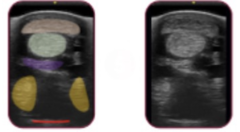
3B
what zone am I in?
