Lymphatic System (Histology)
 Call Kai
 Learn
Learn Practice Test
 Spaced Repetition
 Match
Match Flashcards
 Knowt Play
1/77

There's no tags or description
Looks like no tags are added yet.
Name | Mastery | Learn | Test | Matching | Spaced | Call with Kai | Chat |
|---|
No analytics yet
Send a link to your students to track their progress
78 Terms
Group of cells, vessels, tissues, and organs that monitor body surfaces & internal fluid compartments and react to the presence of potentially harmful substances.
Features of the Lymphatic System
- Collect plasma continuously leaking out from BVs into the interstitial spaces and return this fluid (lymph) to the flood after filtration.
- Regulate both immune and fluid homeostasis
What are the functions of the Lymphatic System?
- Unidirectional (back flow is prevented by valves)
- Movement originates from rhythmic contractions of smooth muscle cells
Describe the flow of lymph
CORRECT: C
A = Hepatic Artery (thick tunica media w/ RBCs in the lumen)
B = Portal Vein (irregularly-shaped lumen with RBCs)
D = Bile Duct (simple cuboidal epithelium)
NOTE: This is a picture of the Portal Triad. C is identified as the lymphatic vessels due to the very thin endothelium w/ lack of RBCs in the lumen.
Identify the lymphatic vessel
A
B
C
D

Lymphocytes
NOTE: Other immune cells also play a role but lymphocytes are the main player.
What is the definitive cell type of the lymphatic system?
Lymphocyte
NOTE: You cannot distinguish whether this is a T cell, B cell, or NK cell without immunohistochemistry to identify cell-surface markers.
Identify this cell

Monocyte
Identify this cell

Monocyte
Identify this cell

Platelet
Identify this cell

Eosinophil
Identify this cell

Neutrophil
Identify this cell

Mast Cell
Identify this cell

(1) Macrophages
(2) Monocytes
(3) Neutrophils, Eosinophils, Basophils
(4) Dendritic cells
(5) Langerhans cells
(6) B cells
NOTE: B cells are both antigen-presenting and antigen-receiving cells. They present antigens to T cells and also receive antigens by either binding antigen to their receptors or through APCs.
List the antigen-presenting cells
Lymphatic Vessel Structures (A - C)
A = endothelium (simple squamous epithelium)
B = basement membrane
C = valve
D = adipocytes
E = connective tissue
Identify the structures

A = Lymphatic Vessel
B = Artery
C = Vein
NOTE: Lymphatic vessels have highly irregular lumen compared to veins and arteries because the endothelium is very thin (to allow greater permeability).
Identify the structures

Lymphocytes
Characteristics:
- 10 microns in diameter
- Slightly larger than an RBC
- Large, prominent nucleus
- Thin peripheral cytoplasm
Identify these cells and describe their characteristics

T Cells:
- Long-life span and is involved in cell-mediated immunity
- Have cell-surface recognition proteins called T-cell receptors
B Cells:
- Have variable lifespans
- Involved in the production of circulating antibodies
NK Cells (Natural Killer):
- Programmed during their development to kill certain virus-infected cells and some types of tumor cells
- Secrete an antiviral agent: INT-alpha
NOTE:
Specific types of lymphocytes cannot be identified under light microscopy and can only be labeled generally as lymphocytes.
You can only identify certain types via immunohistochemistry that would identify cell-surface markers (i.e. CD4 or CD8)
List types of lymphocytes and describe their characteristics
Helper T Cell:
- Express CD4 cell-surface marker and interacts with MHC II
- Subdivided by their ability to secrete cytokines (e.g. TH1 secrete IL-2, IFN-gamma, TNF-alpha)
- Interact with CD8+, NK, & activate macrophages and dendritic cells
NOTE: Helper T cells are the preferential targets of HIV infections.
Describe the characteristic of a Helper T Cell (Lymphocyte)
Cytotoxic T Cell:
- Express CD8 cell-surface marker and interacts with MHC I
- Kills virus-infected cells, cancer-transformed cells, cells infected with intracellular microorganisms & parasites, and transplanted cells (aka transplant rejection)
Describe the characteristic of a Cytotoxic T Cell (Lymphocyte)
Regulatory (Suppressor) T Cell:
- Express CD4+, CD25+, and FOXP3+ cell-surface markers
- Prevent autoimmune response by functionally suppressing an immune response to foreign and self-antigen by influencing the activity of there immune cells
Describe the characteristic of a Regulatory (Suppressor) T Cell (Lymphocyte)
Diffuse lymphatic tissue are cells congregating in the space and are not enclosed by a capsule.
Mucosa-associated lymphoid tissue (MALT) is associated with mucosal lining in the human body
Diffuse lymphatic tissue --> MALT --> BALT
Diffuse lymphatic tissue --> MALT --> GALT
Describe Diffuse Lymphatic Tissue
Gut-Associated Lymphoid Tissue (GALT)
NOTE: The arrow is pointing at an aggregation of lymphatic cells in the extracellular space/lamina propria
Identify this tissue
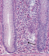
- Alimentary canal (GI)
- Respiratory passages
- Genitourinary tract
Where can diffuse lymphatic tissue be found?
Bronchus-Associated Lymphatic Tissue (BALT)
NOTE: The arrow is pointing to an aggregation of lymphatic cells in the lining of the bronchiole.
Identify this tissue

Secondary Lymphatic Nodule
NOTE: Secondary because it contains a germinal center with actively proliferating B cells (already encountered an Ag) surrounded by a mantle zone or corona
Identify this structure and its features

- Contain mostly B cells
- Found in walls of alimentary canals, respiratory passages, and genitourinary tract
- Most nodules are secondary (contains a germinal center)
Describe the characteristics of Lymphatic Nodules
Peyer's Patch
NOTE: Peyer's Patch is a group of lymphatic nodules, specifically found in the ileum.
Identify the structured pointed at by the arrow

Secondary Lymphatic Nodules
Identify this structures (white box)
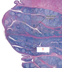
Secondary Lymphatic Nodules (shown in the appendix)
Identify the structured pointed at by the lines
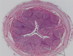
Secondary Lymphatic Nodule
Identify the structure circled in red

F(x):
- traps bacteria and viruses, defends against infection, and provides sites where lymphocytes meet antigens
Function of Tonsils
- Covered by epithelium and have an incomplete capsule
- Most tonsils contain lymphatics nodules but some have diffuse lymphatic tissues
- Located in the oral cavity and posterior roof of the nasopharynx
- Classified as MALT
Features of Tonsils
(1) Pharyngeal Tonsils
- pseudostratified columnar
(2) Palatine Tonsils
- stratified squamous non-keratinized
(3) Lingual Tonsils
- stratified squamous non-keratinized
List the type of tonsils and their epithelium
Pharyngeal Tonsil
- Also called adenoids
- Pseudostratified columnar (resp. epithelium)
- No crypts, only epithelial invaginations/infolds
- Posterior roof of the nasopharynx
- Thin and incomplete capsule
Identify this structure and describe its features

Pharyngeal Tonsil
Identify this structure
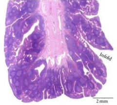
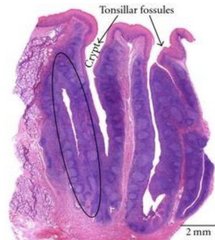
Palatine Tonsil
- Paired tonsils on the posterolateral walls of the oral cavity
- Stratified squamous non-keratinized
- Deep and branched crypts
- Thick and incomplete capsule
Identify this structure and describe its features

Lingual Tonsil
- Paired tonsils on the posterior floor of the mouth
- Stratified squamous non-keratinized
- Non-branched crypts (not as deep as palatine's)
- No capsule
Identify this structure and describe its features

Palatine Tonsil
Identify this structure
Lymph Node
A = Hilum
B = Medulla
C = Cortex
D = Paracortex (Deep Cortex)
E = Capsule
Identify the overall structure and its components (in letters)

F(x):
- primary site where T and B lymphocytes undergo antigen-dependent proliferation and differentiation into effector lymphocytes (i.e. plasma cells) and memory B cells and T cells
Function of the Lymph Node
- Afferent lymphatic vessels: toward the lymph node
- Efferent lymphatic vessels: away from the lymph node
- Encapsulated
- Trabeculae: invaginations from the capsule which separates lymphatic nodules
- Cortex: peripheral region with rows of lymphatic nodules (B cells only)
- Medulla: center area with lymphocytes, plasma cells, macrophages, and dendritic cells, in which bacteria and Ag can be trapped (mostly B cells)
- Paracortex: b/w cortex and medulla, contains HEV (high endothelial venules) (T cells only)
Features of the Lymph Node
Lymph Node
Identify this structure

Lymph Node
Identify this structure

Lymph Node
NOTE: (for reference)
SCS = Sub-capsular space
T = Trabeculae
M = Medulla
MS = Medullary sinus
MC = Medullary cord
Identify this structure
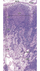
Lymph Node
Identify this structure

High Endothelial Venules (HEV) in the paracortex of a lymph node
Identify this structure and where it is located

High Endothelial Venules (HEV) in the paracortex of a lymph node
Identify this structure and where it is located

High Endothelial Venules (HEV) in the paracortex of a lymph node
Identify this structure and where it is located

F(x):
- an immune component (white pulp) to activate lymphocytes and promote Ab production by plasma cells
- filtration of blood and destruction of aged RBCs (red pulp)
- reservoir for RBCs and platelets
Functions of the Spleen
- Encapsulated (most dense CT capsule compared to all encapsulated lymphatic organs)
- Non-lobulated but still has discontinuous trabeculae
- White Pulp: highest concentration of WBCs; also called splenic/lymphatic nodules
- Red Pulp: highest concentration of RBCs
Features of the Spleen
Spleen
A = Capsule
B = White pulp (splenic/lymphatic nodule)
C = Red pulp
D = Trabeculae
Identify the overall structure and its components (in letters)

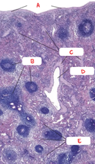
Spleen
A = Capsule
B = White pulp (splenic/lymphatic nodule)
C = Trabeculae
D = Trabecular vein
E = Red pulp
Identify the overall structure and its components (in letters)
- Comprised of lymphatic nodules (considered as splenic nodules)
- Nodules w/ germinal centers are secondary nodules where B cells actively differentiate into lymphoblasts and lymphocytes
- Consists of cylindrical mass of lymphocytes arranged around a central artery that constitutes the periarterial lymphatic sheath (PALS)
Features of White Pulp
Periarterial Lymphatic Sheath
- Diffuse lymphoid tissue surrounding a central artery
- Comprised of concentrated T cells
Features of PALS
Spleen
A = Marginal zone
B = Germinal center
C = Mantle zone
D = PALS (High T cell concentration)
E = Primary follicle
F = Central artery
Identify the overall structure and its components (in letters)

- Contains large numbers of RBCs to be filtered and degraded; also blood storage
- Consists of splenic sinuses separated by splenic cords (cords of Billroth)
NOTE: Splenic cord is a framework of reticular tissue that contains B cells, T cells, plasma cells, macrophages, and other blood cells
Features of White Pulp
Splenic Nodule w/ Germinal Center
NOTE: Also pictured is the central artery of the spleen.
Identify this structure
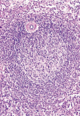
Splenic Nodule w/ Germinal Center
NOTE: Also pictured is the central artery of the spleen. This is a reticular stain to show the reticular fibers of the splenic nodule.
Identify this structure

F(x):
- give rise to hematopoietic stem cells that are the precursor to lymphocytes
Function of the Bone Marrow
- B and T cells are the main cell types located in lymphoid organs.
- Each is programmed to respond to a particular determinant.
- Each naïve B or T cell is relatively short-lived unless activated by Ag it recognizes.
- Both give rise to memory cells and effector cells if they interact with an Ag (Ag-dependent).
Common Features of B Cells & T Cells
- B cell Ag recognition is mediated by Ig molecules in their surface membranes
- T cell Ag recognition is mediated by TCR and activation requires presentation of Ag w/ an MHC molecule from an APC
- Activated B cells differentiates into Ab-secreting plasma cells (humoral immune response)
- Activated T cells differentiates either into helper T cells, cytotoxic T cells, or suppressor T cells (cell-mediated immune response)
Differential Features of B Cells & T Cells
Bone Marrow
Identify the overall structure (disregard the arrows)

- Encapsulated
- NO LYMPHATIC AFFERENT VESSELS
- NO LYMPHATIC NODULES (no B cells, only T cells in the thymus)
- Trabeculae: keeps structures lobulated (thymic lobules)
- Thymic parenchyma contains developing T lymphocytes in an extensive meshwork formed by epithelioreticular cells (6 types)
Each lobule is organized into:
- Cortex: densely-packed T cells; Types I, II, III epithelioreticular cells
- Medulla: loosely-packed T cells; Types IV, V, VI epithelioreticular cells; contains Hassall's corpuscles (Type VI epithelioreticular cells)
Features of the Thymus
Thymus
Identify this structure

Thymus
Identify this structure
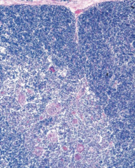
Hassall's Corpuscle (in Thymic Medulla)
NOTE: The tiny arrows are pointing at Type V Epithelioreticular cells.
Identify this structure

In the Thymic Cortex:
- Type I: separates thymic parenchyma from CT
- Type II: thymic cell education
- Type III: create functional junction complexes separating the cortex and medulla
NOTE: Also in the cortex are macrophages for phagocytosis of T cells that do not fulfill thymic education requirement.
In the Thymic Medulla:
- Type IV: create functional junction complexes separating the cortex and medulla
- Type V: located throughout the medulla
- Type VI: forms the Hassall's corpuscles
List the types of Epithelioreticular Cells and their functions
Hassall's Corpuscle (in Thymic Medulla)
Identify this structure

Thymus
NOTE: The arrow is pointing at the Hassall's Corpuscle
Identify the overall structure

Appendicitis
- May be triggered by bacterial & viral inf
- Results in hyperplasia of lymphatic nodules, neutrophil infiltration, increase number of lymphocytes, & obstruction of the lumen of appendix
What clinical correlation is this?

HIV infection (of the lymph node)
- Leads to progressive decline in CD4+ T cells
- Primarily infects CD4+ as well as macrophages and dendritic cells (APCs)
NOTE: The lymph node is in the early stage of HIV infection, which shows large, irregular lymphatic nodules and an increased number of macrophages in the GC.
What clinical correlation is this?

Lymphoid tissues of the nasopharynx, which includes the palatine tonsils, lingual tonsils, and pharyngeal tonsil (adenoid), which may be an extranodal site of lymphoma development
Describe the Waldeyer's Ring
Lymphagioma
- Congenital malformation of the lymphatic system that involves the skin and subcutaneous tissues
- Often occurs in the head and neck
- Characterized by multiple clusters of translucent vesicles that contain lymph fluid
- Appears as a raised, soft, spongy, and peckish-white or red lesions
A 42 y/o woman visited her dermatologist due to the appearance of multiple spongy and red lesions in her head and neck. A biopsy was obtained for pathological examination. What clinical correlation is this most likely?

Diffuse Large B-cell Lymphoma
- Most common non-Hodgkin lymphoma
- Fast-growing and often symptomatic mass at a nodal or extranodal site
- Most common extranodal site is the GI, but could also affect the skin, soft tissues, Waldeyer ring, lung, spleen, & kidneys
- Pt may experience fever, weight loss, and drenching night sweats
- Histology: large tumor cells with large nuclei, open chromatin, and prominent nucleoli. Tumor grows in a diffuse patter
What clinical correlation is this?

Hodgkin's Lymphoma
- Malignant lymphoid cancer characterized by painless enlargement of lymph nodes, spleen, and liver
- Pt may experience fever, night sweats, unexpected weight loss, and fatigue
- Characteristic Reed-Sternberg cells ("owl's eyes)
What clinical correlation is this?
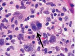
Hodgkin's Lymphoma
What clinical correlation is this?

Hodgkin's Lymphoma
What clinical correlation is this?

Hodgkin's Lymphoma
What clinical correlation is this?
