Nephrotic Syndrome
 Call Kai
 Learn
 Practice Test
 Spaced Repetition
 Match
 Flashcards
 Knowt Play
1/34

There's no tags or description
Looks like no tags are added yet.
35 Terms
Findings that raise suspicion for glomerular disease in general
Hematuria (dysmorphic RBCs, RBC casts) and/or proteinuria, Dec GFR
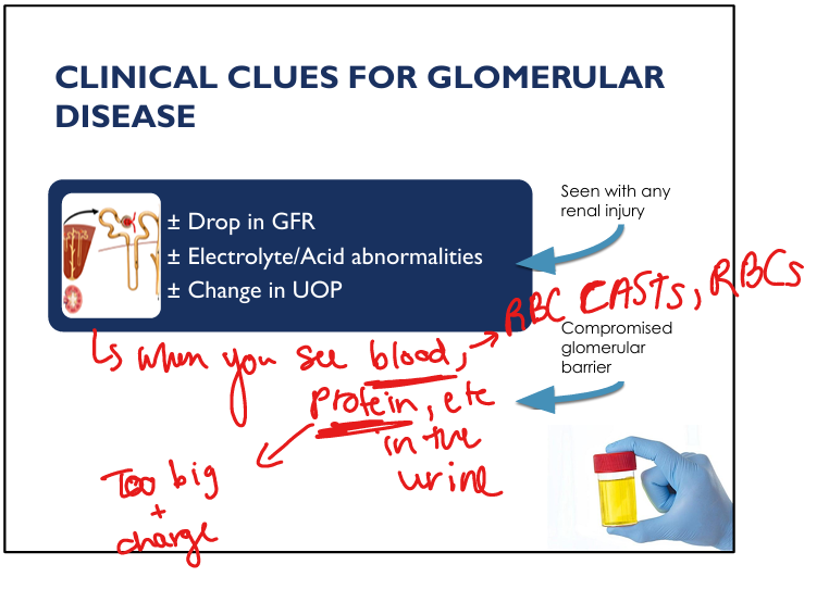
Why dysmorphic RBCs suggest glomerular disease
RBCs squeezed through damaged filtration barrier → irregular shapes.

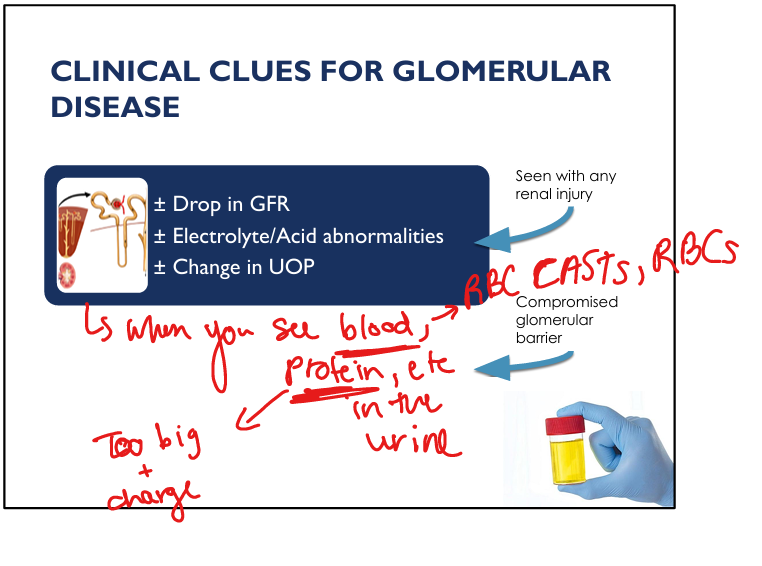
Why RBC casts indicate glomerular disease
Casts form only when bleeding originates in the nephron → nephritic process.
Which barrier prevents albumin filtration
Negative charge of GBM + slit diaphragm size restriction.

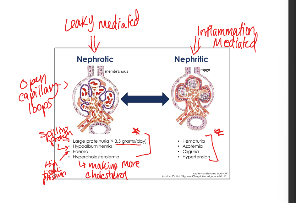
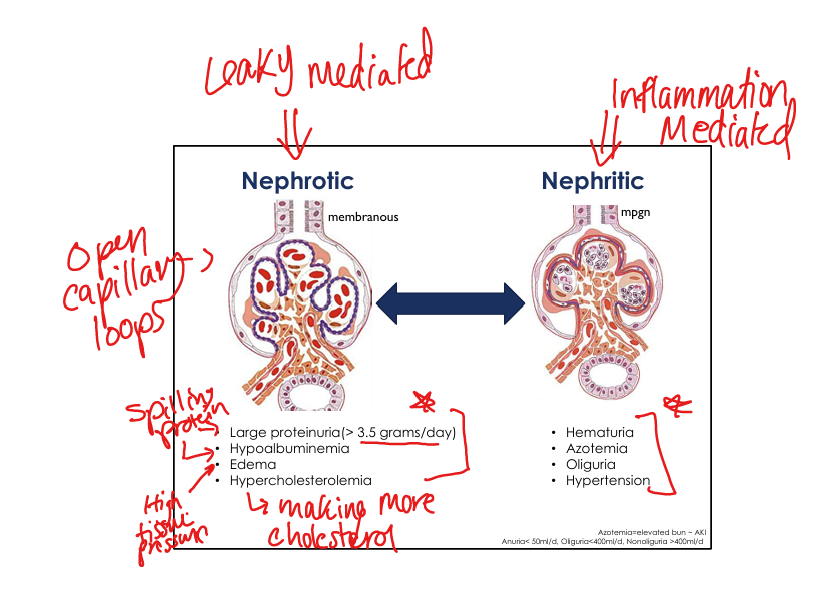
Nephrotic vs nephritic
Nephrotic = proteinuria, leaky
Nephritic= hematuria, inflammation

Classic nephrotic syndrome features
Proteinuria >3.5 g/day, hypoalbuminemia, edema, hyperlipidemia.
Why nephrotic syndrome causes edema
Tubular sodium retention + low oncotic pressure from hypoalbuminemia.

Why nephrotic syndrome causes hyperlipidemia
Liver increases lipoprotein synthesis in response to low oncotic pressure(due to low albumin leaked out).

Oval fat bodies significance
Lipid‑laden tubular cells seen in nephrotic syndrome.

Complication: hypercoagulability in nephrotic syndrome MOA
Loss of ATIII and fibrinolytic proteins → ↑ thrombosis risk (DVT and renal vein thrombosis)

Complication: infection risk in nephrotic syndrome
Loss of IgG and complement proteins → ↑ susceptibility.

Complication: hypovitaminosis D
PT dysfunction→Loss of vitamin D–binding protein in urine.

Normal daily protein excretion? Proteinuria definition?
<150 mg/day in adults. Proteinuria= >150mg or 3.5g/day

Nephrotic Syndrome 3 Pathology Diseases
Minimal Change Disease, Membranous Glomerulopathy, Focal and Segmental Glomerulosclerosis

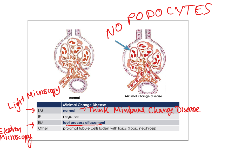
Minimal Change Disease hallmark (view from: LM, IF, EM)
NO Podocyte foot process(damage to podocytes), look normal on light microscopy, Infrared Fluoroscopy is negative.
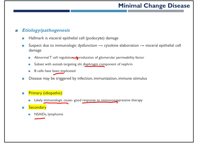

Minimal Change Disease triggers
Infection, immunization, NSAIDs, lymphoma. → they can cause immunologic dysfunction

Minimal Change Disease Clinical presentation + treatment
Most common in kids»sudden onset of massive proteinuria following infection. Tx with Corticosteroids (high response rate).

Focal & Segmental Glomerulosclerosis (FSGS) hallmark (view from: LM, IF, EM)
Focal and segmental sclerosis and hyalinosis on LM; Not an immune complex disease but slight focal IgM on IF; On electron microscope: Loss of Podocytes


Focal & Segmental Glomerulosclerosis (FSGS) genetic associations (1 in particular)
APOL1 variants. NPHS2, TRPC6, ACTN4,

Primary Focal & Segmental Glomerulosclerosis (FSGS) recurrence after transplant
Can recur within 24 hours → circulating permeability factor.
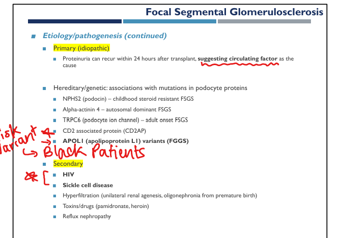

Secondary Focal & Segmental Glomerulosclerosis (FSGS) causes
HIV, sickle cell, heroin, reflux nephropathy, hyperfiltration.

Focal & Segmental Glomerulosclerosis (FSGS) treatment
ACE/ARB + immunosuppression for primary disease.

Membranous nephropathy hallmark (view from: LM, IF, EM)
LM: diffuse capillary wall + GBM thickening + “spike and dome” on silver stain
IF: Immune deposits in GBM
EM: No podocytes, subepithelial immune deposits

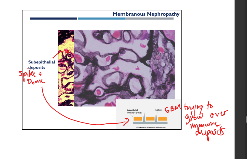

Membranous nephropathy primary antigen
PLA2R autoantibody (adult primary membranous).
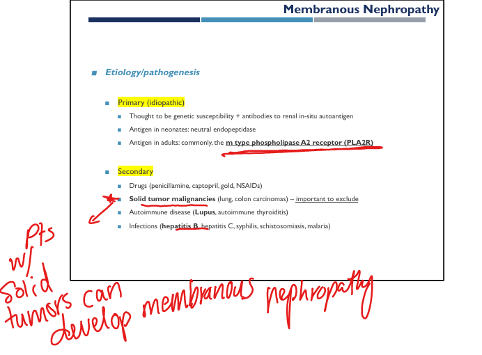
Secondary membranous nephropathy causes
Cancer (lung/colon), hepatitis B/C, lupus, NSAIDs, syphilis.

Diabetic nephropathy hallmark lesions (view from: LM, IF, EM)
LM: GBM thickening, mesangial expansion, Kimmelstiel‑Wilson nodules, afferent+efferent arteriolar hyalinosis
EM: No podocytes, GBM thickening

Diabetic nephropathy pathogenesis
Advanced Glycosylation End-products(AGEs) → basement membrane glycation → hyalinosis → hyperfiltration injury→glomerulosclerosis

Amyloidosis renal hallmark
Congo red positivity with apple‑green birefringence and cotton candy deposition in mesangium; randomly arranged fibrils


Nephrotic syndrome: mechanism of protein leakage
Loss of podocyte slit diaphragm integrity → albumin filtration.
Subepithelial deposits clinical correlate
Nephrotic syndrome (non‑inflammatory, leaky barrier).
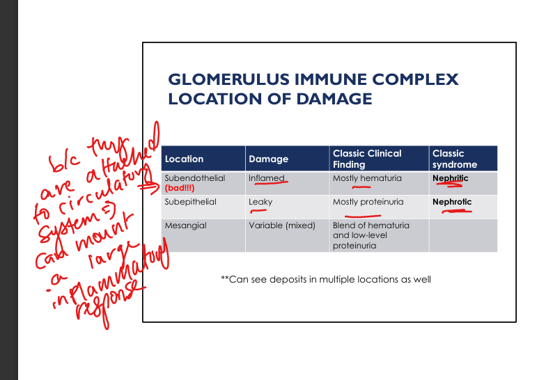
Subendothelial deposits clinical correlate
Nephritic syndrome (inflammatory, hematuria).
Mesangial deposits clinical correlate
Mixed nephritic/nephrotic features.
Why nephrotic urine sodium is low
Tubules retain sodium to compensate for low oncotic pressure → edema formation.

Classic Nephritic Changes
Hematuria, Azotemia(high nitrogen→inc BUN), Oliguria(little peeing), Hypertension

HIV Associated Nephropathy
Caused by direct infection of glomerular/tubular cells with HIV. Tx with Anti retroviral therapy

