Occular symmetry
 Call Kai
 Learn
 Practice Test
 Spaced Repetition
 Match
 Flashcards
 Knowt Play
1/37

There's no tags or description
Looks like no tags are added yet.
38 Terms
initial examination
Observe patient prior to restraint (from a distance):
• Differences in symmetry
• Presence of discharge (eye or nose)
• Presence of anatomical defects
how should eyes look?
nice and open, symmetrical, look from front and side
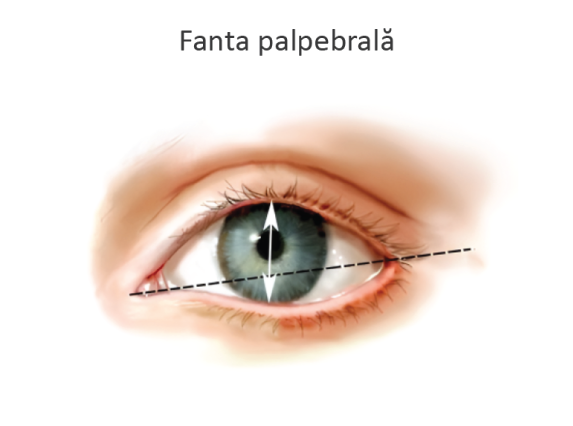
palpebral fissure
space between upper and lower eyelid
uniform/equal bilateral width
muscles of the eyelid
orbicularis oculi - ocludes palpebral fissure
levator mm -
if problem with nervous structure or sympathetic muscles are affected to the point eye becomes droopy
orbital ligaments
enculeations

retractor bulbi muscle
retracts eye
defensively to pull eye into cavity so its protected

afferent
trigeminal nerve (sensory- ophtalmic)
efferent
facial n (motor)
1YO squinting OS
reduced palpebral fissure size (caused by pain/blepharospasm)
tearing
epiphoria
clean up/flush out
photophobia
light hurts/sensitivity
complete ophtalmic examination
fluorescein stain
water based dye (exposed stroma/epithelium is broken, it loves water)
intraocular pressure assessment
distichia
abnormal eyelashed that originate from the eyelid marigin
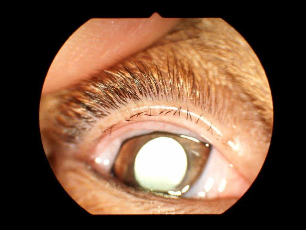
ectopic cilia
abnormal eyelashes that emerge through the conjunctiva
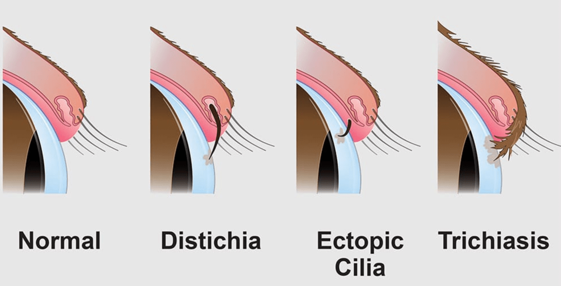
trichiasis
when normal hair touches the eye
prominent rolls of skin
entropion- eyelid margin rolls in
long facial hair

trauma
chemical, mechanical, blunt or sharp trauma
primary entropion
inherited
due to long eyelid length or excessive folds
chow chow, shar pei, rottweiler, lab, etc.

systemic disease process
illness can cause loss of retrobulbar volume - sunken eyes bc not support eyelids and causes entropion, etc.
loss of muscle, fat, dehydration
temporalis and masseter mm atrophied
lagophtalmos
facial n (CN VII) (efferent) or trigeminal n (CN V) (trigeminal) dysfunction
incomplete closure of the eyelid
ptosis
drooping of the eyelid
loss of sympathetic innervation to eyelid muscles (sympathetic)
usually not painful
miosis, 3rd eyelid elevation
congenital defect
smaller than normal eye
microphtalmos or anophtalmos
rupture of eye (volume inside of eye reduces)
adnexa
group of tissue around the eye (periocular) that protect, support, and assist in the function of the globe
globe
the eye and its direct anatomical structures
organized into coats or tunics (fibrous, nervous, uveal)
tapetum lucidum
shiny layer on caudal aspect of globe
more sensitivity at night, helps catch prey

loss of normal retroillumination (reflection of light from the back of the eye)
if the opacity involves the cornea or anterior chamber=blocks ability to see the iris
corneal opacity identification
gray corneal opacity=scar
inactive lesion, non-painful distinct borders
flat

blue corneal opacity
edema
active process
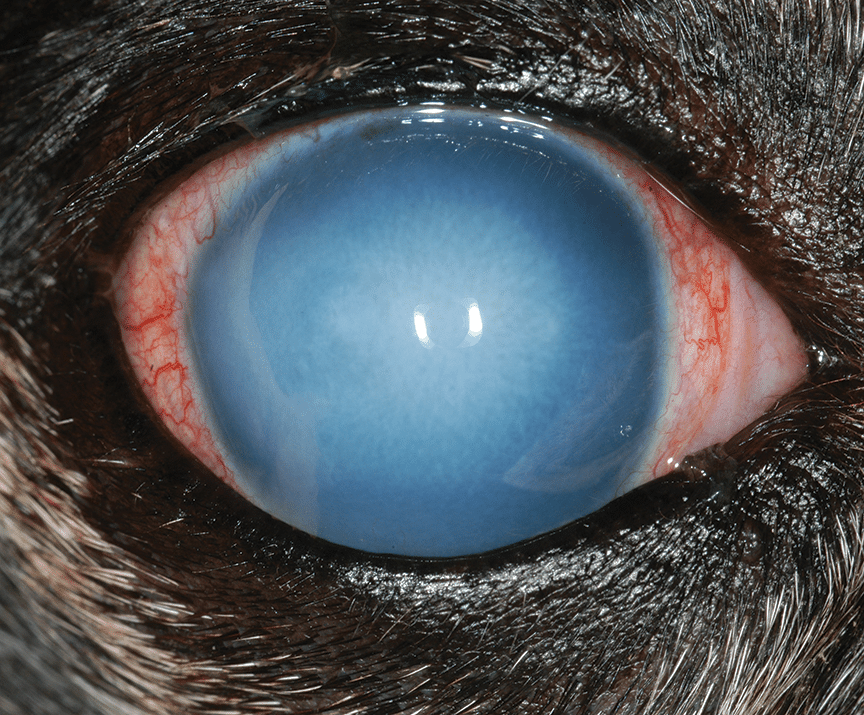
focal pattern
associated with epithelial injury such as an ulcer
confirm by fluorescein stain uptake
diffuse pattern
associated with endothelial disease such as that seen with uveitiis (tipically fluorescein negative)
fluorescein stain does not correspond to region of edema
hydrophobic
do not replicate/non mitotically active monolayer of cells (need transplant, will not have vision otherwise)

stroma
90% of overall corneal thickness aprox
organized in lamellae for clarity
will bind water (hydrophilic)- results in corneal edema

endothelial degeneration
in humans we do corneal transplant
age related
cant see

mucopurulent
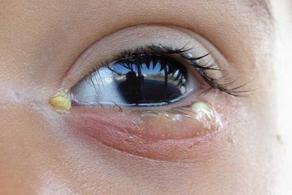
serosanguineous

schirmer tear test
keep it in place 1 min
keratoconjunctivitis sicca
adherent, tacky mucopurulent ocular discharge
dry, lackluster corneal surface

Jones test
patent nasolacrimal duct = positive - fluorescein dye goes through nostril
nasolacrimal flush
if neg jones test » canulate that duct and get fluid to flush (in SA)
nasolacrimal duct flush in LA
IV sedation
topical cetacaine spray
lubricant
nose to eye vs eye to nose (in SA)
slaine eye wash