Evaluation & Management of Small Animal Thoracic Trauma
 Call Kai
 Learn
 Practice Test
 Spaced Repetition
 Match
 Flashcards
 Knowt Play
1/29

There's no tags or description
Looks like no tags are added yet.
30 Terms
How do we define and classify trauma?
A trauma - is a physical injury which is classified based on the organ system
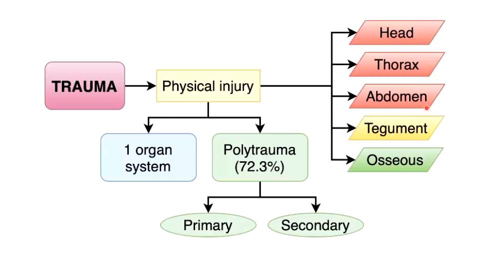
What are the potential consequences of trauma?

Describe how a trauma is evaluated in an emergency setting?
• Triage
• Primary Survey
• Scoring systems
ATT (Animal Trauma Triage) score
mGCS (modified Glasgow Coma Scale)
• Secondary Survey
How is effective circulating volume different from total blood volume?
Effective circulating volume (ECV)—often called effective circulating blood volume—is the amount of blood that is actively perfusing (flowing through) the tissues and organs.
Unlike total blood volume, ECV is not a measurable physical space. Instead, it is a functional concept representing the fluid that is in the arterial system and actively maintaining blood pressure.
Death tends to be associated with what three main traumas?
Thoracic Trauma
Abdominal Trauma
CNS Trauma
What is TRIAGE?
Triage: RANK and MANAGE injuries based on their THREAT TO LIFE
What is apnoea and dyspnoea?
Apnoea is a temporary stop in breathing, while dyspnoea is the medical term for shortness of breath or difficult, uncomfortable breathing.
What are the two main causes of thoracic trauma?
Penetrating - something communicating between outside and inside of the chest
Blunt - any traumatic event damaging internal structures of the chest without opening communication with the “outside”

What are the main features of pulmonary contusions?
• Pulmonary contusions (44-88% of trauma events) - extremely common
- Compression/Decompression injury following blunt trauma - impact forces acting like lungs on an acordian, causing rupture and hemorrhage
- Pulmonary interstitial & alveolar haemorrhage/oedema
- Ventilation/Perfusion (V/Q) mismatch / hypoxaemia - gas exchange function altered leading to hypoxemia and shock since inflammatory response takes a bit to develop
Progressive deterioration over several hours
- 34-57% dogs / 17% cats in RTA
High index of suspicion in trauma patients
Concomitant injuries
How do we assess/diagnose pulmonary contusions?
POCUS -
C-lines: irregular pulmonary-pleural line and indicate the presence of lung consolidation
CT
Gold standard but expensive and patient must be anesthetized
X-Ray
If done immediately after injury might miss since pulmonary conditions take a few days to develop
What are the main features of pneumothorax?
• Pneumothorax (7-47% of trauma events)
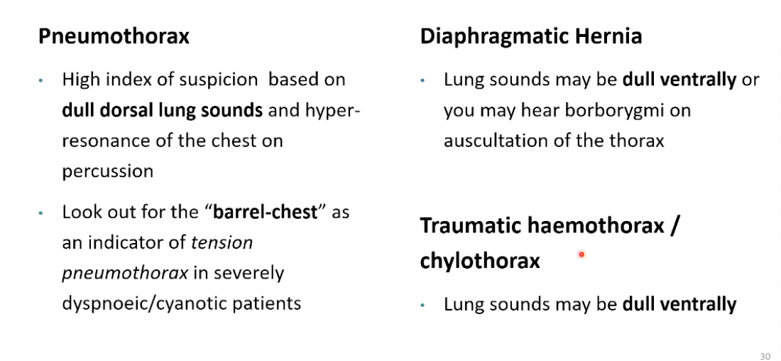
- Accumulation of air within the pleural space, compressed lungs do not participate in gas exchange
Atelectasis of lungs
No participation in gas exchange
Classified in two ways:
- Open
E.g. Rib fractures, penetrating injuries
- Closed
E.g. ruptures alveolus, bronchus, trachea, oesophagus...
How do we assess/diagnose pneomothorax?
X-Ray
Pockets of air surrounding the lungs where parenchyma should be
Absence of Glide Sign - ultrasound
Expected in pneumothorax
What are the main features of a tension pneumothorax?
• Tension Pneumothorax
- The site of air-leakage acts as a one-way valve, with every breath, more leakage of air into pleural space, and next inspiration increases more
- Pleural pressure increases during each inspiration until is is greater than Ptm
→ Atelectasis → life-threatening hypoxaemia
Poor venous return
Cardiovascular collapse
Shock
What are the main features of diaphragmatic hernias/ruptures?
• Diaphragmatic hernia/Rapture (2- 6%)
- Often concurrently with trauma caudal to the diaphragm
Sudden increase in abdominal pressure with an open glottis is thought to result in a tear to the diaphragm, displaces organs to chest cavity which compresses the lungs
- Herniated organs within the pleural space contributes to hypoxaemia
- Reduced venous return may contribute to cardiovascular signs.
What are the main features of fractured ribs?
• Fractured ribs (9-14% of trauma events)
- Often associated with pulmonary contusions & pleural space disease
- Pain associated hypoventilation
- "Flail chest" - two or more adjacent rib segments fragmented creating a window, acts independently of the rest of the chest (Dorsal and ventral)
Rarely as a result of RTA
Fracture (dorsally and ventrally) of 2 or more adjacent rib segments
Paradoxical chest motion
How do we assess thoracic trauma?
• Assessment of thoracic trauma
- Physical Examination
Breathing pattern
Auscultation
Percussion
- Advanced assessment
• Point of Care Ultrasound (POCUS)
• Diagnostic/Therapeutic thoracocentesis
• Thoracic Imaging: radiography vs. CT
• ECG
What sort of findings will you find after doing the physical exam characteristic of the different thoracic traumas?
Describe the different appearance of the lung on the POCUS of lungs.

Describe management of a pneumothorax.
• Pneumothorax
- Goal
Re-expansion of the collapsed lung
Improvement in venous return & cardiac output
- Thoracocentesis / Thoracostomy tube placement
Intermittent or continuous pleural drainage may be required
Consider induction-intubation in rapidly deteriorating patients and commence IPPV
Potential need for exploratory thoracotomy if ongoing/unresolved pneumothorax
Describe management of pulmonary contusions.
- Supportive management
- 02 supplementation
Intubation and ventilation may be indicated in severe cases
Avoiding over-zealous fluid therapy
Lesions typically worsen over 24- 48h and improve over 3-10 days
Describe management practice for:
Diaphragmatic Hernia/Rupture
Fractured Ribs
Pleural / Pericardial Effusion

What are the most common head traumas and TBI’s occurring after an injury.
• Injury to the CNS is common following RTA
- Traumatic Brain Injury (TBI)
• 25% of dogs involved in RTA
• Spinal Cord Injury (SCI)
- May have concurrent skeletal and soft tissue trauma affecting the head
• Fractured mandible
• Orbital fractures
• Soft palate trauma
What is the difference between a primary and secondary injury?
• Primary Injury: immediate
• Secondary injury (hours to days after trauma)
Describe the most common primary head injuries following trauma.
• Primary Injury: immediate
• Concussion (no histologic lesion) - temporary failure of function of the brain
• Contusion (parenchymal haemorrhage & oedema)
"coup" same side of injury, or "contrecoup" injury, opposite side injury
• Laceration resulting in haematoma formation & brain compression
Axial (within the brain parenchyma)
Extra-axial (epidural, subdural, subarachnoid)
Describe the most common secondary head injuries following trauma.
Systemic - anything in body affecting brain
Intracranial - occurred as consequence of the injury

What occurs if trauma raises ICP?
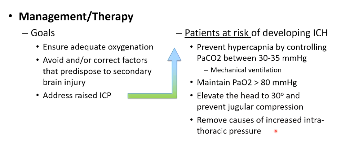
Raised Intracranial Pressure (ICP)
• The volume of the intracranial contents exceeds compensatory mechanisms, haematoma increases volume beyond what the skull can contain
• Results in brain herniation if not identified and treated
Transtentorial (cerebrum herniating caudally)
Transforminal (cerebellum herniates caudally through the foramen magnum)
When should you consider ICH as a consequence of head trauma?
Consider ICH (Intracranial Hypertension) if you observe deterioration in neurological function
Deterioration in mentation, brain- stem function, postural changes (decerebrate rigidity)
Development of abnormal. respiratory patterns
Systemic hypertension and bradycardia: "Cushing reflex"
How are severity of TBI’s assessed using the Glasgow coma scale?
- Glasgow Coma Scale (modified) mGCS
• Level of consciousness
• Brain-stem reflexes (pupil size, PLRs and eye movement)
• Limb movements
• Respiratory pattern
• Blood pressure and heart rate
Lower the score → the worst outcome

What are the goals of management and therapy regarding head traumas and TBIs?
What does management look like for skeletal traumas?
• Mandibular symphyseal fractures are relatively common in cats following RTA
Consider the nutritional management in these cases particularly as concurrent facial and nasal trauma frequently affects appetite in cats
Consider placement of an oesophagostomy tube
Decision making regarding the timing of fracture repair should take into account the clinical status of the patient