APR Nervous System
 Call Kai
 Learn
Learn Practice Test
 Spaced Repetition
 Match
Match Flashcards
 Knowt Play
1/37

There's no tags or description
Looks like no tags are added yet.
Name | Mastery | Learn | Test | Matching | Spaced | Call with Kai | Chat |
|---|
No analytics yet
Send a link to your students to track their progress
38 Terms

Cauda equina
Location: vertebral canal inferior to L2 vertebral level
Description:
large bundle of dorsal & ventral roots for nerves below L2
Latin for “horse tail”

Conus medullaris
Location: upper lumbar vertebral canal
Description: tapered inferior end of spinal cord (spinal cord w nerves ends at L2)

Dorsal root ganglion (spinal ganglion)
Location: intervertebral foramen
Description: contains afferent/sensory neurons

Dura mater
Location: surrounds brain and spinal cord (protection)
Description: tough, dense, irregular connective tissue
Cranial dura mater has periosteal (outer) and meningeal (inner) parts
Cranial dura mater has dural venous sinuses

Spinal cord
Location: vertebral canal
Description: ends at L2 in adults
Composed of inner gray matter and outer white matter
Divided into cervical, thoracic, lumbar, sacral, and coccygeal regions
Comments:
Continuous w/ brain at foramen magnum
31 pairs of spinal nerves

Ventral ramus
Location: lateral to vertebral foramen
Cervical VR enters cervical
Lumbar VR enters branchial
Sacral VR enters lumbosacral plexuses
Thoracic VR becomes intercostal and thoracoabdominal nerves
Description: motor and sensory components
Distributed primarily to skin, limb muscles, anterolateral trunk and neck
Comment: all VR receives gray ramus communicans from sympathetic trunk
VRs from T1 to L2 receives white ramus communicans from sympathetic trunk

Dorsal root of Cervical region
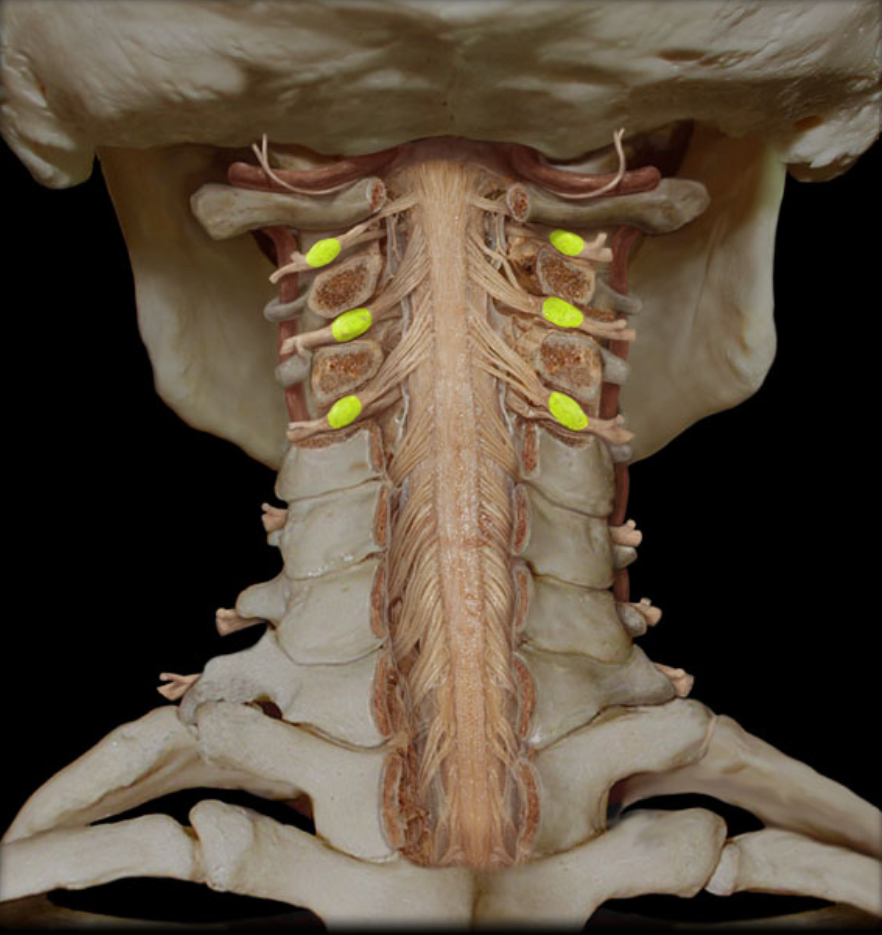
Dorsal root ganglion of Cervical region

Dura mater of Cervical region

Ventral ramus of Cervical region

Ventral root of Cervical region

Dorsal root ganglion of Thoracic region

Dura mater of Thoracic region

Ventral ramus of Thoracic region

Cauda equina of Lumbar region

Conus medullaris of Lumbar region

Dorsal root ganglion of Lumbar region
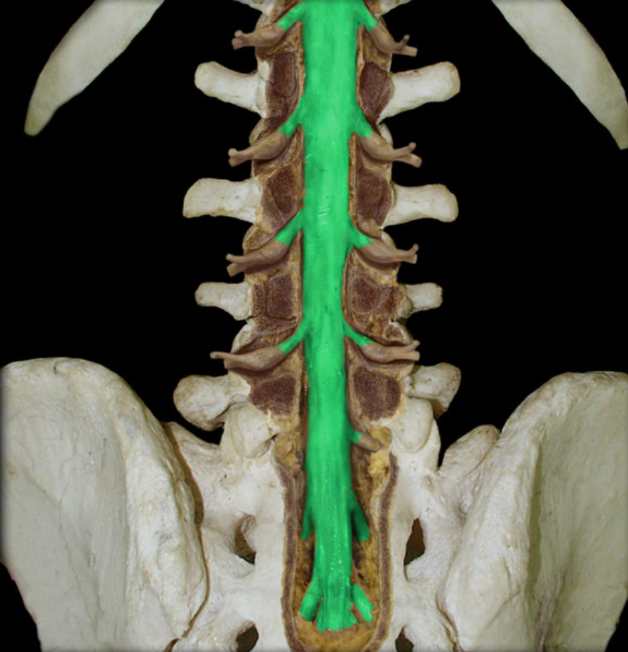
Dura mater of Lumbar region

Ventral ramus of Lumbar region

Dorsal root of Spinal Nerve

Dorsal root ganglion of Spinal Nerve

Dura mater of Spinal Nerve

Epidural space of Spinal Nerve
Location: space external to dura mater
Description:
Contains loose connective tissues, fat, and venous plexus in spine
In cranial, potential space

Gray matter
Description: unmyelinated nervous tissue shaped like letter “H”
Ventral horns house motor neurons
Dorsal horns house sensory neurons

Spinal cord

Ventral ramus of Spinal Nerve

Ventral root of Spinal Nerve

White matter
Description: ascending and descending tracts of myelinated axons

Accessory n. (CN XI)
Location: vertebral canal, posterior cranial fossa, neck
Motor
Cranial: joins vagus nerve (CN X) to distribute to palate muscles, pharynx, and larynx
Spinal: trapezius and sternocleidoomastoid
CNS Connection
Cranial: medulla oblongata
Spinal: ventral horn of C1-C4
Cranial foramina
foramen magnum in spinal root
jugular foramen
Comments
Cranial and spinal roots unite in jugular foramen to form accessory nerve
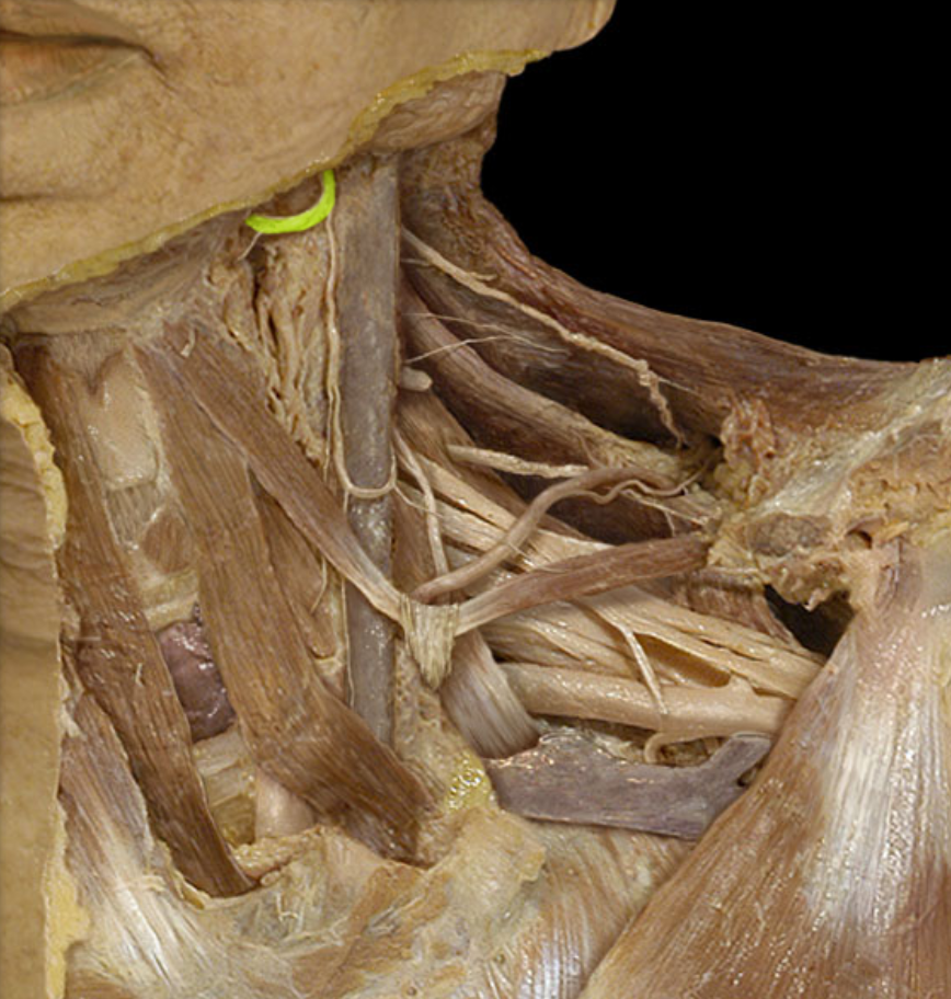
Hypoglossal n. (CN XII)
Location: posterior cranial fossa, neck, oral cavity
Motor: genioglossus, hyoglossus, styloglossus, intrinsic muscles of tongue
CNS Connection: medulla oblongata
Cranial foramina: hypoglossal canal
Comment:
Hypoglossal nerve innervates all tongue muscles except palatoglossus (vagus nerve)
Intrinsic tongue muscles originate and insert within tongue

Vagus n. (CN X)
Location: posterior cranial fossa, head, neck, thorax, abdomen
Composition: Motor, general sensation, special sensation, parasympathetic
Motor: muscles of palate, pharynx, and larynx
General sensation:
Thoracic and abdominal viscera
Epiglottis and laryngopharynx
External acoustic meatus
Special sensation: taste from epiglottis and surrounding region
Parasympathetic:
Mucous glands and smooth muscles of respiratory and digestive systems in neck (larynx and pharynx), thorax, and abdomen
Cardiac muscle
CNS Connection: all medulla oblongata
Motor: nucleus ambiguus
General: spinal nucleus of trigeminal nerve
Special: nucleus of solitary tract
Parasympathetic: dorsal nucleus of vagus nerve
Cranial foramina: Jugular foramen
Comment:
General sensation from thoracic and abdominal viscera only involves stretch
General sensation from epiglottis and laryngopharynx involves pain, touch, and temperature
Vagus nerve is the only cranial nerve that extends beyond head and neck
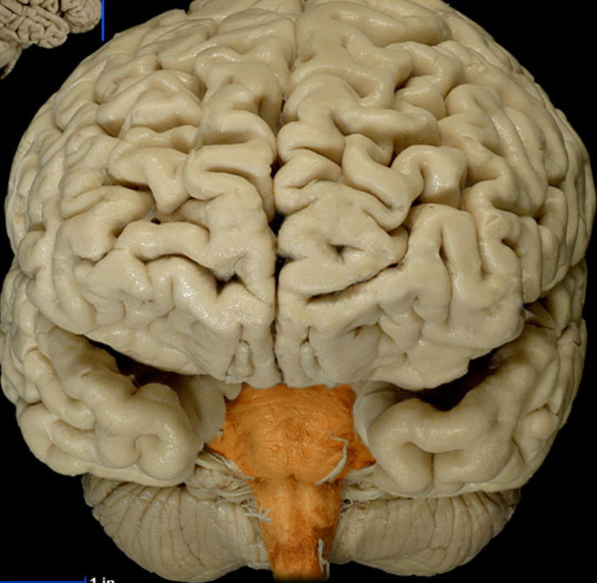
Brainstem
Location: caudal portion of brain
Description: midbrain, pons, and medulla oblongata

Cerebellum
Location: dorsal to brainstem
Description:
Many lobes with highly folded cortex
Attached to pons via cerebellar peduncles
Functions:
Coordinates complex movements
Monitor muscles to ensure fluid movements
Comment:
cerebellar cortex has folds called folia
White matter resembles branching trees and is called arbor vitae
Influence motor function through thalamus and motor cortex

Cerebrum
Location: rostral portion of brain (towards the nose)
Description:
2 cerebral hemispheres separated by longitudinal fissure
Hemispheres are connected by corpus callosum
Cerebral cortex is the gray matter
Each hemisphere has a core of white matter
Gray matter can include basal nuclei

Corpus Callosum
Forms floor of longitudinal fissure

Frontal Lobe
Location: anterior portion of cerebral hemisphere
Description:
Extends from anterior pole of brain to central sulcus
Contains precentral gyrus
Function:
Controls voluntary motor activity
Higher mental processing
Emotional behavior
Speech output (Broca’s area in left hemisphere)

Hippocampus
Location: deep in temporal lobe
Description:
Major output via fornix
Part of limbic system
Function:
Learning and memory processing
Emotional behavior

Insular Lobe
Location: Deep in each lateral sulcus
Description: Cerebral lobe
Function:
Understanding spoken language
Perception of taste and smell
Integrates information from visceral receptors